Fig 1.

Fig 1.
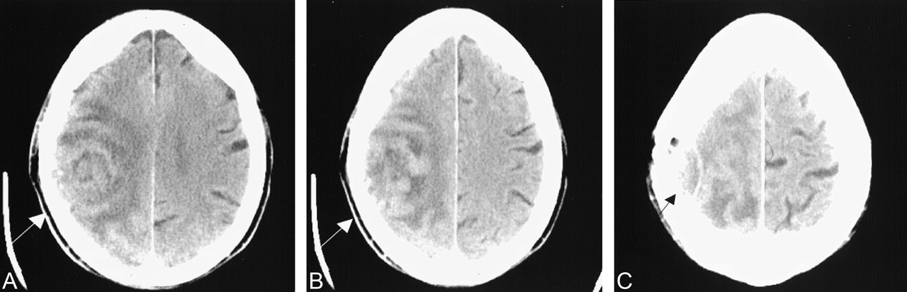
CT scans.
A and B, Nonenhanced scans obtained at slightly different heights show a rounded heterogeneous mass in the right frontoparietal region (arrow).
C, Higher section shows inward dural displacement (arrow) and overlying calvarial destruction, suggesting extra-axial extension of the mass.


