
Abstract
Summary: We report a case of cerebral hyperperfusion syndrome complicated by a fatal intracranial hemorrhage as a consequence of endovascular recanalization of an occluded left subclavian artery. This entity is thought to be secondary to the failure of the normal autoregulatory mechanism controlling cerebral blood flow after recanalization. Patients at risk for this relatively rare complication are not easily identified. Further investigation is warranted to identify patients at risk for this complication and to determine optimal periprocedural management to decrease risk.
Cerebral hyperperfusion syndrome was first recognized as a rare and sometimes fatal complication following carotid endarterectomy (1–6). With the advent of carotid artery stent placement, the incidence of this complication may possibly be seen in a comparatively larger percentage of patients (7). Until recently, no reported data implicating hyperperfusion syndrome as a consequence of endovascular treatment of subclavian stenosis were available. One uncomplicated case was identified after such an intervention to treat an occluded left subclavian artery with angioplasty and stent placement (8). At our institution, a case of cerebral hyperperfusion syndrome complicated by a deadly intracranial hemorrhage was documented subsequent to the recanalization of an occluded left subclavian artery.
Case Report
A 66-year-old white female patient presented for angiographic evaluation and possible endovascular therapy subsequent to a workup for vertigo, blurred vision, and near syncope with orthostasis. The patient reportedly experienced these symptoms for approximately 3 months. In addition, the patient demonstrated signs of left-arm claudication. Her medical history was pertinent for severe peripheral vascular disease and coronary artery disease, diabetes mellitus, and a 125-pack-per-year smoking history. The patient was status-post right femoral popliteal bypass. Physical examination revealed bilateral carotid bruit, absent left radial and brachial pulses in addition to severe manifestations of peripheral vascular disease in the right lower extremity. Neurologically, the patient’s gait was found to be grossly intact, although she experienced weakness most pronounced at the left upper extremity. Workup at another institution revealed near occlusion of both carotid arteries and complete occlusion of the left subclavian artery on MR angiograms and duplex sonograms. This was noted proximally. A CT scan of the head obtained before the planned procedure showed only mild cerebral atrophy and white matter disease.
Occlusion of the left subclavian artery was confirmed at diagnostic angiography. A stump was visualized at the proximal aspect of this vessel (Fig 1A). Angiography of the left common carotid artery demonstrated very slow flow as well as occlusion of the left external carotid artery. An ostial stenosis of 95% was noted at the left internal carotid artery. Evaluation of the innominate artery showed moderate stenosis at its ostium. Sixty percent to 70% stenosis of the right common carotid artery was present. The right internal carotid artery showed 50% stenosis. The right subclavian artery demonstrated 65% stenosis. Multiple branches of this vessel were enlarged, crossing the midline to reconstitute an enlarged left vertebral artery. In addition, the right vertebral artery was occluded; however, reconstitution via muscular branches of the right subclavian artery was seen. Opacification of this vessel was followed by visualization of unremarkable basilar and bilateral posterior cerebral arteries. Filling of the distal left vertebral artery, suggestive of a steal phenomenon, was also seen.
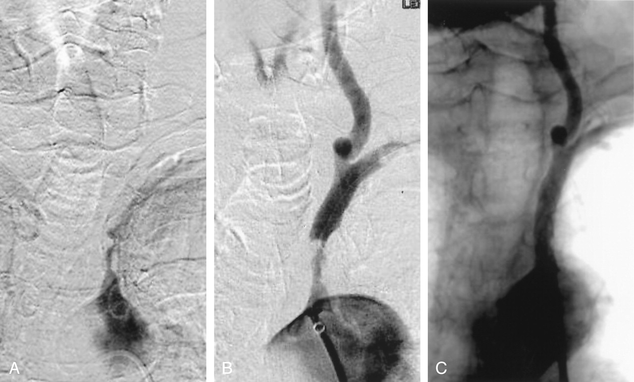
A, Occlusion of the left subclavian artery.
B, Appearance of the left subclavian artery after predilatation with 4 × 40 mm balloon. Anterograde opacification of the left vertebral artery.
C, Control angiography post stent placement (two Medtronic Arterial Vascular Engineering [AVE] stents, 6 × 16 and 6 × 10 mm).
At the time of stent placement, the proximal left subclavian artery occlusion was crossed by using an 0.038-inch glidewire, guided by a 5F 100-cm Vitek catheter introduced via the right common femoral. The 5F catheter was then advanced across the occluded segment of the left subclavian artery into the axillary artery. The 0.038-inch glide wire was changed for 0.035-inch stiff exchange wire and a 6F 90-cm arterial sheath was then introduced into the ostium of the left subclavian artery followed by the injection of 4000 U heparin. The occlusion was then predilated by using a 4.5/40 Trac four balloon at 10 atm for 180 seconds (Fig 1B). A 6.0/16-mm bridge stent was subsequently deployed within the proximal left subclavian artery, effectively eliminating the occlusion (Fig 1C). Approximately 1 minute after the stent was deployed, the patient experienced a short episode of hypotension, which returned to baseline without treatment after approximately 2 minutes. An interval of hypertension during the procedure was never reported.
Approximately 1 hour after the procedure, the patient experienced a severe right-sided headache. A noncontrast CT of the head was obtained. During this examination, the patient became comatose and underwent respiratory arrest. An acute hemorrhage centered at the right thalamic nucleus was noted with extension into the lateral ventricles and the subarachnoid space. Severe dilatation of the lateral ventricles was also present (Fig 2). The patient expired 8 days later.

A–F, CT scan of the head 1 hour after the left subclavian artery recanalization. Large hematoma in the right thalamus with rupture into the third ventricle and spilling of the blood through the 4th ventricle into the subarachnoid space.
Discussion
In our review of the literature, we did not find a case of cerebral hyperperfusion syndrome complicated by intracerebral and subarachnoid hemorrhage subsequent to the endovascular recanalization of a subclavian artery. In 1975, Sundt et al first described the entity of “hyperperfusion syndrome” in five patients who suffered seizures during the early postoperative period subsequent to carotid endarterectomy (9). They described this complication to include atypical migraines, transient focal seizure activity, and intracranial hemorrhage (1). Today this syndrome continues to plague a relatively small number of patients who undergo endovascular recanalization therapies. The onset of this syndrome is typically marked by headaches, occasionally with lateralizing neurologic signs, focal seizures that can generalize to grand mal, and, in the event of brain hemorrhage, loss of consciousness. Its cause is thought to be the failure of the normal autoregulatory mechanism controlling cerebral blood flow after recanalization. With gradually decreasing perfusion pressures due to chronic stenosis, it is postulated that the cerebral arterioles become maximally dilated and lose their ability to constrict after normal perfusion pressure is reinstated (6, 10). This can lead to hyperemia of the brain tissue, swelling with increase of intracranial pressure, temporary loss of neuronal function and rarely to the rupture of the arterioles with resulting brain hematoma (9, 11, 12).
Although we do not have pathologic evidence to support hyperperfusion injury as cause of our patient’s ultimately fatal hemorrhage, cerebral infarction has been excluded by the absence of periprocedural symptoms and vascular occlusions on the post-stent placement control angiography. Other causes of intracranial hemorrhage following revascularization techniques, such as periprocedural hypertension or anticoagulant use (7, 13), are unlikely because the patient never became hypertensive during the procedure nor in the postprocedural period, and antiplatelet medications were not used.
Patients at risk for cerebral hyperperfusion syndrome are not easily identified. This presents a predicament in the treatment of stenotic or occlusive atherosclerotic disease. It is thought that preprocedural risk factors include hypertension, high-grade stenosis, or occlusion of the treated vessel and chronic ischemic, angiopathic changes of the brain tissue. Until now, these risks have not been applied to the subclavian artery recanalization techniques, as it is an uncommon procedure. The optimal parameters for treatment remain unknown. Further investigation is warranted to identify patients at risk for this complication and to determine optimal periprocedural management to decrease risk.
References
- Received March 21, 2004.
- Accepted after revision May 13, 2004.
- Copyright © American Society of Neuroradiology
In this issue


Jump to section
Related Articles
Cited By...
- No citing articles found.