Advertisement

Main menu

  • Home
  • Content
    • Current Issue
    • Accepted Manuscripts
    • Article Preview
    • Past Issue Archive
    • Video Articles
    • AJNR Case Collection
    • Case of the Week Archive
    • Case of the Month Archive
    • Classic Case Archive
  • Special Collections
    • AJNR Awards
    • ASNR Foundation Special Collection
    • Most Impactful AJNR Articles
    • Photon-Counting CT
    • Spinal CSF Leak Articles (Jan 2020-June 2024)
  • Multimedia
    • AJNR Podcasts
    • AJNR SCANtastic
    • Trainee Corner
    • MRI Safety Corner
    • Imaging Protocols
  • For Authors
    • Submit a Manuscript
    • Submit a Video Article
    • Submit an eLetter to the Editor/Response
    • Manuscript Submission Guidelines
    • Statistical Tips
    • Fast Publishing of Accepted Manuscripts
    • Graphical Abstract Preparation
    • Imaging Protocol Submission
    • Author Policies
  • About Us
    • About AJNR
    • Editorial Board
    • Editorial Board Alumni
  • More
    • Become a Reviewer/Academy of Reviewers
    • Subscribers
    • Permissions
    • Alerts
    • Feedback
    • Advertisers
    • ASNR Home

User menu

  • Alerts
  • Log in

Search

  • Advanced search
American Journal of Neuroradiology

American Journal of Neuroradiology

ASHNR American Society of Functional Neuroradiology ASHNR American Society of Pediatric Neuroradiology ASSR
  • Alerts
  • Log in

Advanced Search

  • Home
  • Content
    • Current Issue
    • Accepted Manuscripts
    • Article Preview
    • Past Issue Archive
    • Video Articles
    • AJNR Case Collection
    • Case of the Week Archive
    • Case of the Month Archive
    • Classic Case Archive
  • Special Collections
    • AJNR Awards
    • ASNR Foundation Special Collection
    • Most Impactful AJNR Articles
    • Photon-Counting CT
    • Spinal CSF Leak Articles (Jan 2020-June 2024)
  • Multimedia
    • AJNR Podcasts
    • AJNR SCANtastic
    • Trainee Corner
    • MRI Safety Corner
    • Imaging Protocols
  • For Authors
    • Submit a Manuscript
    • Submit a Video Article
    • Submit an eLetter to the Editor/Response
    • Manuscript Submission Guidelines
    • Statistical Tips
    • Fast Publishing of Accepted Manuscripts
    • Graphical Abstract Preparation
    • Imaging Protocol Submission
    • Author Policies
  • About Us
    • About AJNR
    • Editorial Board
    • Editorial Board Alumni
  • More
    • Become a Reviewer/Academy of Reviewers
    • Subscribers
    • Permissions
    • Alerts
    • Feedback
    • Advertisers
    • ASNR Home
  • Follow AJNR on Twitter
  • Visit AJNR on Facebook
  • Follow AJNR on Instagram
  • Join AJNR on LinkedIn
  • RSS Feeds

AJNR is seeking candidates for the AJNR Podcast Editor. Read the position description.

Research ArticleSPINE

Experimental Autoimmune Encephalomyelitis in the Rat Spinal Cord: Lesion Detection with High-Resolution MR Microscopy at 17.6 T

Andreas Steinbrecher, Thomas Weber, Thomas Neuberger, André M. Mueller, Xiomara Pedré, Gerhard Giegerich, Ulrich Bogdahn, Peter Jakob, Axel Haase and Cornelius Faber
American Journal of Neuroradiology January 2005, 26 (1) 19-25;
Andreas Steinbrecher
  • Find this author on Google Scholar
  • Find this author on PubMed
  • Search for this author on this site
Thomas Weber
  • Find this author on Google Scholar
  • Find this author on PubMed
  • Search for this author on this site
Thomas Neuberger
  • Find this author on Google Scholar
  • Find this author on PubMed
  • Search for this author on this site
André M. Mueller
  • Find this author on Google Scholar
  • Find this author on PubMed
  • Search for this author on this site
Xiomara Pedré
  • Find this author on Google Scholar
  • Find this author on PubMed
  • Search for this author on this site
Gerhard Giegerich
  • Find this author on Google Scholar
  • Find this author on PubMed
  • Search for this author on this site
Ulrich Bogdahn
  • Find this author on Google Scholar
  • Find this author on PubMed
  • Search for this author on this site
Peter Jakob
  • Find this author on Google Scholar
  • Find this author on PubMed
  • Search for this author on this site
Axel Haase
  • Find this author on Google Scholar
  • Find this author on PubMed
  • Search for this author on this site
Cornelius Faber
  • Find this author on Google Scholar
  • Find this author on PubMed
  • Search for this author on this site
  • Article
  • Figures & Data
  • Info & Metrics
  • Responses
  • References
  • PDF
Loading

Article Figures & Data

Figures

  • Fig 1.
    • Download figure
    • Open in new tab
    • Download powerpoint
    Fig 1.

    Sagittal MR images of rat spinal cord. Arrowheads indicate the cord surface; asterisks, gray matter; white circle, white matter; and arrows, lesions.

    A, Thoracic cord in a control rat.

    B–D, Lumbar (B), thoracic (C), and cervical (D) sections in a rat with grade 4 EAE.

  • Fig 2.
    • Download figure
    • Open in new tab
    • Download powerpoint
    Fig 2.

    Axial MR and histopathologic images of rat spinal cord.

    A, Lumbar cord in a control rat. Letter A indicates the corticospinal tract; B, gracile fasciculus; C, lateral funiculus; and D, anterior/ventral funiculus.

    B–D, Lumbar portion (L3) in a rat with grade 4 EAE. Multiple lesions (1–6) can be identified in the anterior and lateral parts of the white matter. Histologic section (LFB stain) in B shows an inflammatory infiltrate in the gray matter (arrow). Corresponding MR images in C and D, spaced 150 μm, account for the slightly different orientation of the axial plane introduced by histologic processing. Lesions 1–6 in A are shown.

  • Fig 3.
    • Download figure
    • Open in new tab
    • Download powerpoint
    Fig 3.

    Lesion correlation in the ventral and lateral columns. Bar in A–D= 250 μm.

    A and D, Details of the MR image in Figure 2D show a large lesion (1) in the ventral part of the cord (A) and two lesions (2 and 3) in the lateral part (D).

    B, C, E, and F, Corresponding histologic sections stained for HE (B and E) and LFB (C and F) show the lesions depicted in A and Figure 2B. Note the fingerlike protrusion of lesion 1 (arrowhead) in A and C.

  • Fig 4.
    • Download figure
    • Open in new tab
    • Download powerpoint
    Fig 4.

    Lesion correlation in the dorsal column. Bar in A = 250 μm.

    A, Detail of the MR image in Figure 2B shows only two small lesions (arrows).

    B and C, Corresponding histologic sections stained for HE (B) and LFB (C) show. only a few inflammatory cells (arrows).
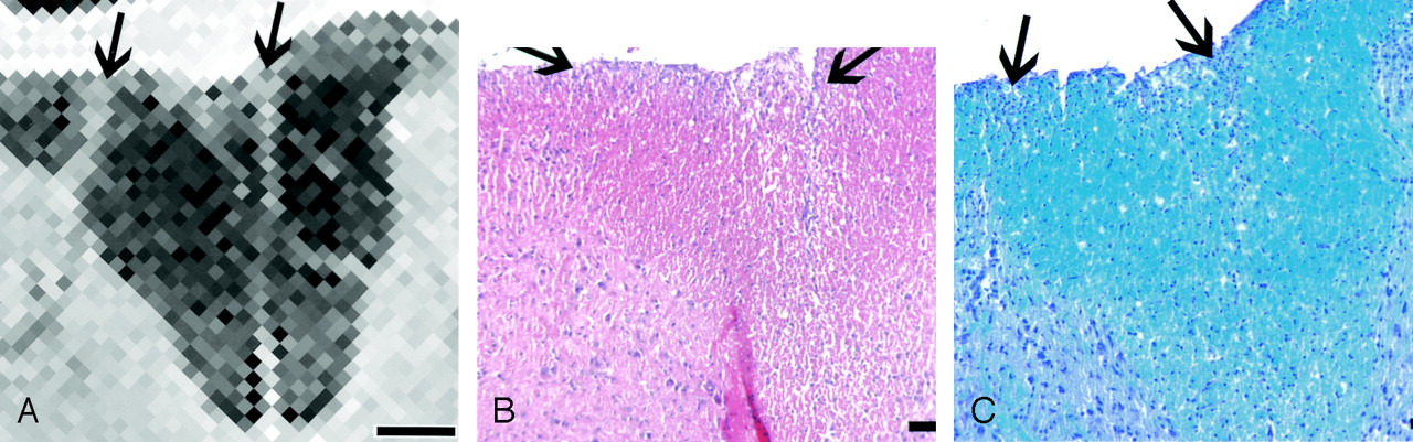
  • Fig 5.
    • Download figure
    • Open in new tab
    • Download powerpoint
    Fig 5.

    Lesion load quantification. 3D visualization of the thoracic spinal cord in the animal with grade 4 EAE. Gray matter is green; white matter lesions are blue (arrow); 1 is ventral horn; 2 is dorsal horn. Lesion load in the depicted cord is 16% of the white matter. Note the relative paucity of lesions in the dorsal part versus the ventral and lateral parts of the cord.

    A, Ventrolateral view.

    B, Dorsolateral view.

PreviousNext
Back to top

In this issue

American Journal of Neuroradiology: 26 (1)
American Journal of Neuroradiology
Vol. 26, Issue 1
1 Jan 2005
  • Table of Contents
  • Index by author
Advertisement
Print
Download PDF
Email Article

Thank you for your interest in spreading the word on American Journal of Neuroradiology.

NOTE: We only request your email address so that the person you are recommending the page to knows that you wanted them to see it, and that it is not junk mail. We do not capture any email address.

Enter multiple addresses on separate lines or separate them with commas.
Experimental Autoimmune Encephalomyelitis in the Rat Spinal Cord: Lesion Detection with High-Resolution MR Microscopy at 17.6 T
(Your Name) has sent you a message from American Journal of Neuroradiology
(Your Name) thought you would like to see the American Journal of Neuroradiology web site.
CAPTCHA
This question is for testing whether or not you are a human visitor and to prevent automated spam submissions.
Cite this article
Andreas Steinbrecher, Thomas Weber, Thomas Neuberger, André M. Mueller, Xiomara Pedré, Gerhard Giegerich, Ulrich Bogdahn, Peter Jakob, Axel Haase, Cornelius Faber
Experimental Autoimmune Encephalomyelitis in the Rat Spinal Cord: Lesion Detection with High-Resolution MR Microscopy at 17.6 T
American Journal of Neuroradiology Jan 2005, 26 (1) 19-25;

Citation Manager Formats

  • BibTeX
  • Bookends
  • EasyBib
  • EndNote (tagged)
  • EndNote 8 (xml)
  • Medlars
  • Mendeley
  • Papers
  • RefWorks Tagged
  • Ref Manager
  • RIS
  • Zotero
0 Responses
Respond to this article
Share
Bookmark this article
Experimental Autoimmune Encephalomyelitis in the Rat Spinal Cord: Lesion Detection with High-Resolution MR Microscopy at 17.6 T
Andreas Steinbrecher, Thomas Weber, Thomas Neuberger, André M. Mueller, Xiomara Pedré, Gerhard Giegerich, Ulrich Bogdahn, Peter Jakob, Axel Haase, Cornelius Faber
American Journal of Neuroradiology Jan 2005, 26 (1) 19-25;
del.icio.us logo Twitter logo Facebook logo Mendeley logo
  • Tweet Widget
  • Facebook Like
  • Google Plus One

Jump to section

  • Article
    • Abstract
    • Methods
    • Results
    • Discussion
    • Conclusion
    • Footnotes
    • References
  • Figures & Data
  • Info & Metrics
  • Responses
  • References
  • PDF

Related Articles

  • No related articles found.
  • PubMed
  • Google Scholar

Cited By...

  • Single cell-resolution in situ sequencing elucidates spatial dynamics of multiple sclerosis lesion and disease evolution
  • Crossref
  • Google Scholar

This article has not yet been cited by articles in journals that are participating in Crossref Cited-by Linking.

More in this TOC Section

  • Bern Score Validity for SIH
  • MP2RAGE 7T in MS Lesions of the Cervical Spine
  • Resisted Inspiration for CSF-Venous Fistula
Show more Spine

Similar Articles

Advertisement

Indexed Content

  • Current Issue
  • Accepted Manuscripts
  • Article Preview
  • Past Issues
  • Editorials
  • Editor's Choice
  • Fellows' Journal Club
  • Letters to the Editor
  • Video Articles

Cases

  • Case Collection
  • Archive - Case of the Week
  • Archive - Case of the Month
  • Archive - Classic Case

Special Collections

  • AJNR Awards
  • ASNR Foundation Special Collection
  • Most Impactful AJNR Articles
  • Photon-Counting CT
  • Spinal CSF Leak Articles (Jan 2020-June 2024)

More from AJNR

  • Trainee Corner
  • Imaging Protocols
  • MRI Safety Corner

Multimedia

  • AJNR Podcasts
  • AJNR Scantastics

Resources

  • Turnaround Time
  • Submit a Manuscript
  • Submit a Video Article
  • Submit an eLetter to the Editor/Response
  • Manuscript Submission Guidelines
  • Statistical Tips
  • Fast Publishing of Accepted Manuscripts
  • Graphical Abstract Preparation
  • Imaging Protocol Submission
  • Evidence-Based Medicine Level Guide
  • Publishing Checklists
  • Author Policies
  • Become a Reviewer/Academy of Reviewers
  • News and Updates

About Us

  • About AJNR
  • Editorial Board
  • Editorial Board Alumni
  • Alerts
  • Permissions
  • Not an AJNR Subscriber? Join Now
  • Advertise with Us
  • Librarian Resources
  • Feedback
  • Terms and Conditions
  • AJNR Editorial Board Alumni

American Society of Neuroradiology

  • Not an ASNR Member? Join Now

© 2025 by the American Society of Neuroradiology All rights, including for text and data mining, AI training, and similar technologies, are reserved.
Print ISSN: 0195-6108 Online ISSN: 1936-959X

Powered by HighWire