Advertisement

Main menu

  • Home
  • Content
    • Current Issue
    • Accepted Manuscripts
    • Article Preview
    • Past Issue Archive
    • Video Articles
    • AJNR Case Collection
    • Case of the Week Archive
    • Case of the Month Archive
    • Classic Case Archive
  • Special Collections
    • AJNR Awards
    • ASNR Foundation Special Collection
    • Most Impactful AJNR Articles
    • Photon-Counting CT
    • Spinal CSF Leak Articles (Jan 2020-June 2024)
  • Multimedia
    • AJNR Podcasts
    • AJNR SCANtastic
    • Trainee Corner
    • MRI Safety Corner
    • Imaging Protocols
  • For Authors
    • Submit a Manuscript
    • Submit a Video Article
    • Submit an eLetter to the Editor/Response
    • Manuscript Submission Guidelines
    • Statistical Tips
    • Fast Publishing of Accepted Manuscripts
    • Graphical Abstract Preparation
    • Imaging Protocol Submission
    • Author Policies
  • About Us
    • About AJNR
    • Editorial Board
    • Editorial Board Alumni
  • More
    • Become a Reviewer/Academy of Reviewers
    • Subscribers
    • Permissions
    • Alerts
    • Feedback
    • Advertisers
    • ASNR Home

User menu

  • Alerts
  • Log in

Search

  • Advanced search
American Journal of Neuroradiology

American Journal of Neuroradiology

ASHNR American Society of Functional Neuroradiology ASHNR American Society of Pediatric Neuroradiology ASSR
  • Alerts
  • Log in

Advanced Search

  • Home
  • Content
    • Current Issue
    • Accepted Manuscripts
    • Article Preview
    • Past Issue Archive
    • Video Articles
    • AJNR Case Collection
    • Case of the Week Archive
    • Case of the Month Archive
    • Classic Case Archive
  • Special Collections
    • AJNR Awards
    • ASNR Foundation Special Collection
    • Most Impactful AJNR Articles
    • Photon-Counting CT
    • Spinal CSF Leak Articles (Jan 2020-June 2024)
  • Multimedia
    • AJNR Podcasts
    • AJNR SCANtastic
    • Trainee Corner
    • MRI Safety Corner
    • Imaging Protocols
  • For Authors
    • Submit a Manuscript
    • Submit a Video Article
    • Submit an eLetter to the Editor/Response
    • Manuscript Submission Guidelines
    • Statistical Tips
    • Fast Publishing of Accepted Manuscripts
    • Graphical Abstract Preparation
    • Imaging Protocol Submission
    • Author Policies
  • About Us
    • About AJNR
    • Editorial Board
    • Editorial Board Alumni
  • More
    • Become a Reviewer/Academy of Reviewers
    • Subscribers
    • Permissions
    • Alerts
    • Feedback
    • Advertisers
    • ASNR Home
  • Follow AJNR on Twitter
  • Visit AJNR on Facebook
  • Follow AJNR on Instagram
  • Join AJNR on LinkedIn
  • RSS Feeds

AJNR is seeking candidates for the AJNR Podcast Editor. Read the position description.

Research ArticleSPINE

Apparent Diffusion Coefficients in Spinal Cord Transplants and Surrounding White Matter Correlate with Degree of Axonal Dieback After Injury in Rats

Eric D. Schwartz, Chih-Liang Chin, Jed S. Shumsky, Abbas F. Jawad, B. Kooper Brown, Suzanne Wehrli, Alan Tessler, Marion Murray and David B. Hackney
American Journal of Neuroradiology January 2005, 26 (1) 7-18;
Eric D. Schwartz
  • Find this author on Google Scholar
  • Find this author on PubMed
  • Search for this author on this site
Chih-Liang Chin
  • Find this author on Google Scholar
  • Find this author on PubMed
  • Search for this author on this site
Jed S. Shumsky
  • Find this author on Google Scholar
  • Find this author on PubMed
  • Search for this author on this site
Abbas F. Jawad
  • Find this author on Google Scholar
  • Find this author on PubMed
  • Search for this author on this site
B. Kooper Brown
  • Find this author on Google Scholar
  • Find this author on PubMed
  • Search for this author on this site
Suzanne Wehrli
  • Find this author on Google Scholar
  • Find this author on PubMed
  • Search for this author on this site
Alan Tessler
  • Find this author on Google Scholar
  • Find this author on PubMed
  • Search for this author on this site
Marion Murray
  • Find this author on Google Scholar
  • Find this author on PubMed
  • Search for this author on this site
David B. Hackney
  • Find this author on Google Scholar
  • Find this author on PubMed
  • Search for this author on this site
  • Article
  • Figures & Data
  • Info & Metrics
  • Responses
  • References
  • PDF
Loading

Article Figures & Data

Figures

  • Tables
  • Fig 1.
    • Download figure
    • Open in new tab
    • Download powerpoint
    Fig 1.

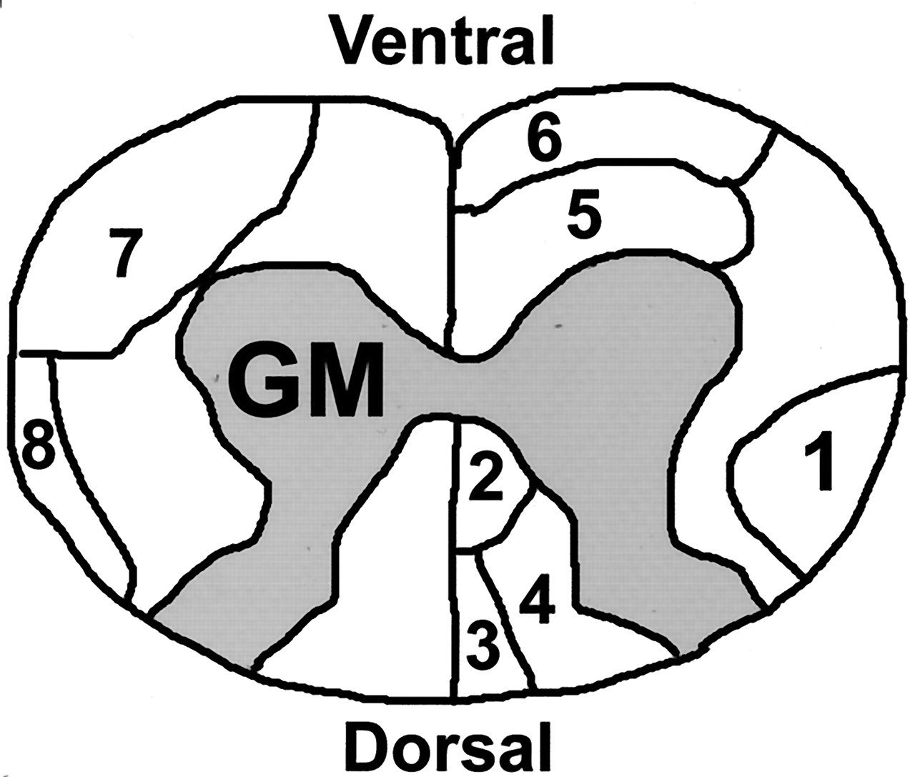
    Schematic of rat cervical spinal cord. The labeled tracts in the white matter are RST (1), dorsal corticospinal tract (2), fasciculus gracilis (3), fasciculus cuneatus (4), reticulospinal tract (5), vestibulospinal tract (6), spinothalamic tract (7), dorsal spinocerebellar tract (8). GM indicates gray matter.

  • Fig 2.
    • Download figure
    • Open in new tab
    • Download powerpoint
    Fig 2.

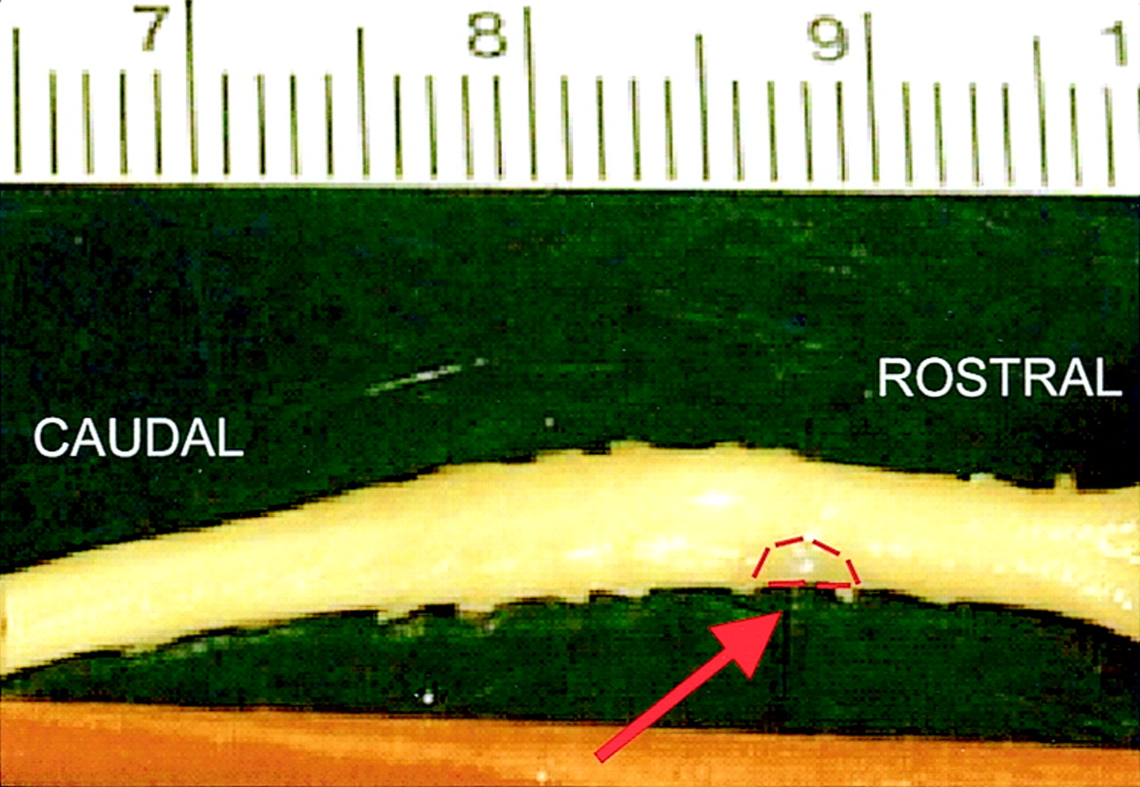
    Picture of cervical spinal cord after partial right-sided hemisection and transplantation with unmodified fibroblasts (arrow, transplant outlined in red).

  • Fig 3.
    • Download figure
    • Open in new tab
    • Download powerpoint
    Fig 3.

    Spinal cord images obtained 2 mm rostral to a right-sided partial hemisection and transplant of unmodified fibroblasts.

    A, T2-weighted (b = 0) MR image used as a guide to draw an ROI in the right DLF. An ROI in the surrounding buffer was also created to account for slight differences in temperatures.

    B and C, Corresponding tADC (B) and lADC (C) maps show atrophy in the right DLF (arrow) compared with the contralateral side (arrowhead). Spinal cord injury is expected to increase transverse water diffusion and decrease longitudinal water diffusion in injured white matter; these findings are reflected by a subtle increase in signal intensity in the right DLF on the tADC map (B) and a decrease in signal intensity in the right DLF on the lADC map (C) compared with the signal intensity of the undamaged left DLF.

  • Fig 4.
    • Download figure
    • Open in new tab
    • Download powerpoint
    Fig 4.

    Spinal cord images obtained at the level of right-sided partial hemisection and transplant of BDNF-expressing fibroblasts.

    A, T2-weighted (b = 0) image used as a guide to draw ROIs in the transplant (TP), bilateral corticospinal tracts (CST), and left DLF.

    B and C, Corresponding tADC (B) and lADC (C) maps. Analysis of the transplant showed diffusion to be close to isotropic.

  • Fig 5.
    • Download figure
    • Open in new tab
    • Download powerpoint
    Fig 5.

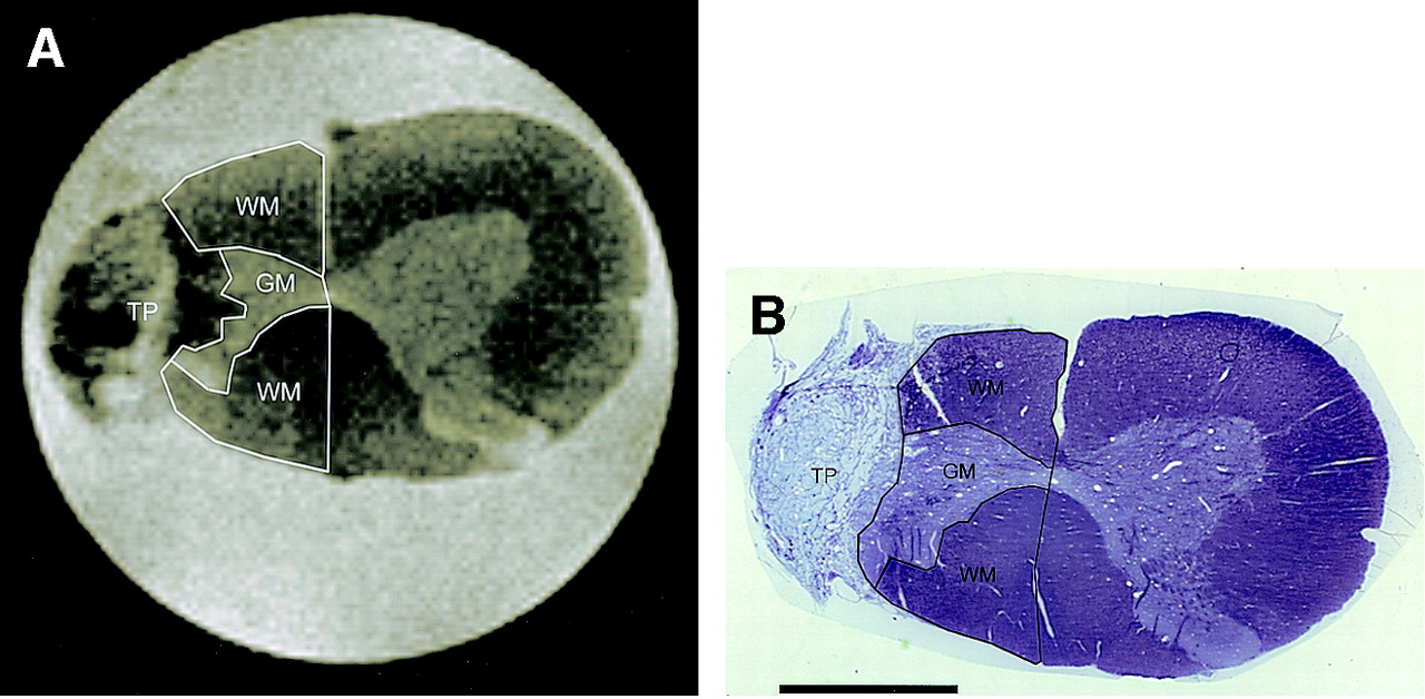
    MR image and corresponding histologic image of spinal cord at level of right-sided partial hemisection and unmodified fibroblast transplant.

    A, T2-weighted (b = 0) image with remaining right-sided white matter (WM) and remaining gray matter (GM) outlined. TP indicates transplant.

    B, Photomicrograph of corresponding histologic section (toluidine blue stain, original magnification ×16, bar = 1.0 mm), also with white matter (WM) and gray matter (GM) outlined, demonstrates close correspondence to MR image in A. TP indicates transplant

  • Fig 6.
    • Download figure
    • Open in new tab
    • Download powerpoint
    Fig 6.

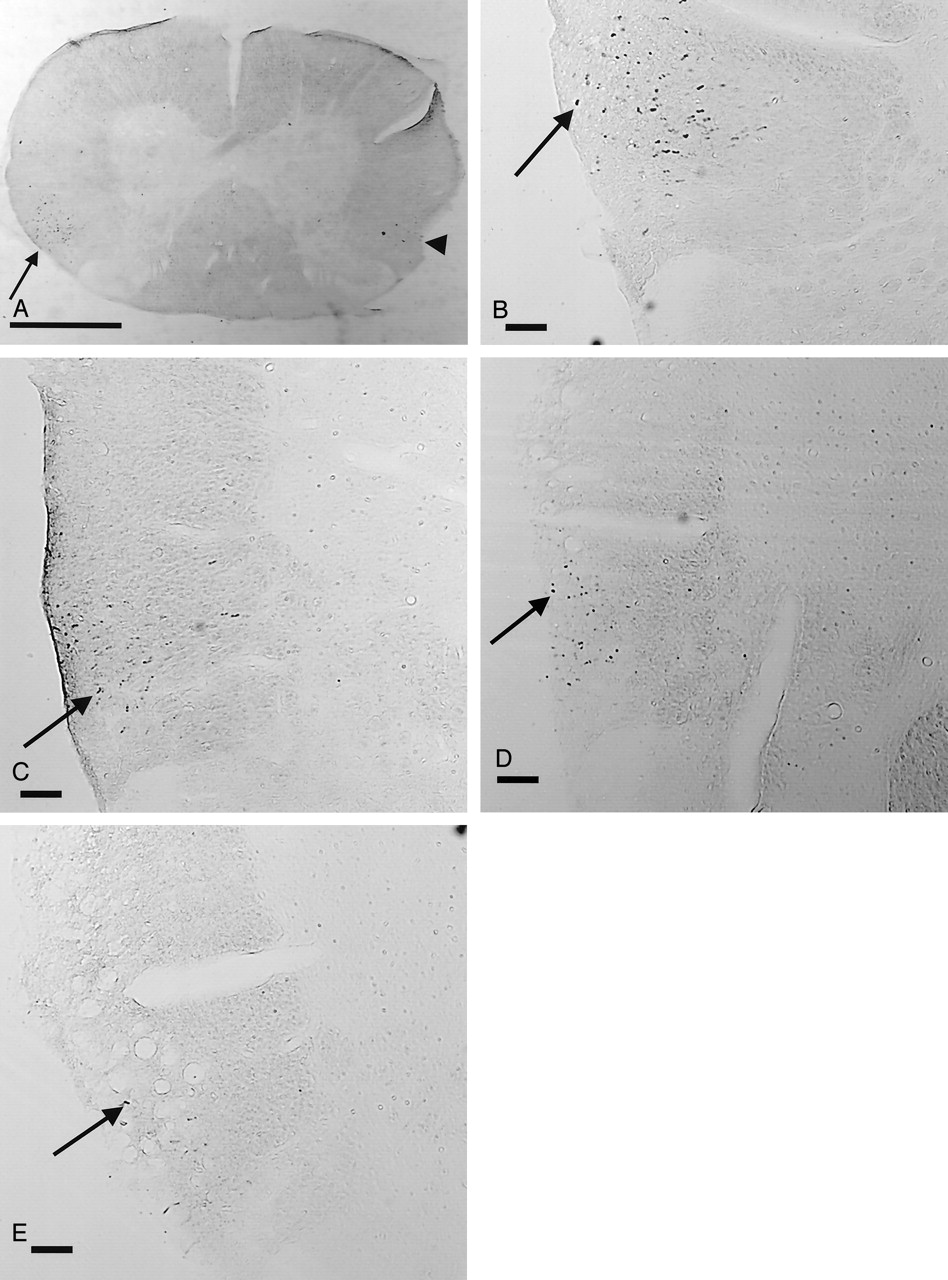
    Histologic images of BDA anterograde of labeled axons in the right RST.

    A and B, Normal images at magnification X16 (A, bar = 1.0 mm) and at magnification X50 (B, bar = 0.1 mm) show numerous labeled axons in the right RST (arrow). There are a few labeled axons on the contralateral side (arrowhead in A), as approximately 10% of RST axons do not cross the midline. For orientation, a slit was made in the ventral left white matter before sectioning.

    C–E, Histologic images of the right RST in an animal with an unmodified fibroblast graft at magnification X50 (bar = 0.1 mm) obtained at 3.5 mm rostral (C), 1.5 mm rostral (D), and 1.0 mm rostral (E) to the transplant. Note that the number of labeled axons (arrow) decreases as images are obtained closer to the transplant.

  • Fig 7.
    • Download figure
    • Open in new tab
    • Download powerpoint
    Fig 7.

    RST axonal dieback. Graph indicates increasing percentage loss of axons as counts are obtained closer to the grafts. Axon count at 4.0 mm rostral to graft is reference value (0% loss). Note that the normal animal (▴) has a small loss of axons due to the normal exiting of RST axons in the cervical spinal cord.

  • Fig 8.
    • Download figure
    • Open in new tab
    • Download powerpoint
    Fig 8.

    A–C, Graphs of ADC values and anisotropy index (AI) in the right DLF both rostral and caudal to the transplants. * indicates both animals with unmodified fibroblasts and those with BDNF-expressing fibroblasts were significantly different from normal controls; #, unmodified fibroblasts significantly different from normal controls; ∼, BDNF-expressing fibroblasts significantly different from normal controls; +, unmodified fibroblasts significantly different from BDNF-expressing fibroblasts.

    A, The tADC values in the right DLF become significantly elevated in white matter adjacent to the grafts, both rostral and caudal. These values become closer to normal as measurements are obtained further from injury site.

    B, The lADC values in the right DLF become significantly decreased in white matter adjacent to the grafts compared with those of normal controls, both rostral and caudal. These values become closer to normal as measurements are obtained further from injury site.

    C, Anisotropy index (AI) values become significantly elevated (decreased anisotropy) in white matter adjacent to the grafts compared with those in normal controls, both rostral and caudal. These values become closer to normal as measurements are obtained further from injury site.

  • Fig 9.
    • Download figure
    • Open in new tab
    • Download powerpoint
    Fig 9.

    Scatterplots of ADC values and anisotropy index versus axonal dieback

    A and B, Scatterplots of tADC and lADC versus percentage axonal dieback show that as axonal loss increases closer to the injury site, tADC increases and lADC decreases.

    C, Scatterplot of anisotropy index (tADC/lADC ratio) versus percentage axonal dieback shows that the changes in ADC values seen with increasing axonal loss produce an increasing anisotropy index, indicating decreasing anisotropic water diffusion.

  • Fig 10.
    • Download figure
    • Open in new tab
    • Download powerpoint
    Fig 10.

    Recovery of function in the affected right hindlimb occurred in all animals over time as measured with a modified BBB scale. At 12 weeks, the animals with BDNF-expressing fibroblast grafts had significantly better scores than those of animals with unmodified fibroblast grafts (+ indicates P < .05). The difference at 12 weeks is small, however, and likely not functionally significant.

Tables

  • Figures
    • View popup
    TABLE 1:

    Measurement of ADC values in the BDNF-expressing fibroblast transplants and the unmodified fibroblast transplants

    ADC MeasureBDNF-Expressing (n = 5)Unmodified (n = 6)
    tADC (10 −4 mm 2/s)*5.13 ± 0.15.64 ± 0.1
    lADC (10 −4 mm 2/s)*6.14 ± 0.16.95 ± 0.1
    AI (tADC/lADC ratio)0.84 ± 0.10.81 ± 0.1
    • Note.—AI indicates anisotropy index. All scores are mean ± standard error.

    • * Indicates significant difference ( P < .05) between transplant types.

    • View popup
    TABLE 2:

    MR imaging and histologic measurements of preserved right-sided tissue

    Preserved Right-Sided TissueMR Imaging MeasurementsHistologic Measurements
    BDNF-Expressing Fibroblasts (n = 5)Unmodified Fibroblasts (n = 6)Normal Control (n = 3)BDNF-Expressing Fibroblasts (n = 5)Unmodified Fibroblasts (n = 6)Normal Control (n = 3)
    White matter (mm 2)0.73 ± 0.090.86 ± 0.093.00 ± 0.120.70 ± 0.090.87 ± 0.082.56 ± 0.11
    Gray matter (mm 2)0.10 ± 0.040.14 ± 0.041.06 ± 0.50.20 ± 0.080.40 ± 0.070.96 ± 0.10
    • Note.—All scores are mean ± standard error.

PreviousNext
Back to top

In this issue

American Journal of Neuroradiology: 26 (1)
American Journal of Neuroradiology
Vol. 26, Issue 1
1 Jan 2005
  • Table of Contents
  • Index by author
Advertisement
Print
Download PDF
Email Article

Thank you for your interest in spreading the word on American Journal of Neuroradiology.

NOTE: We only request your email address so that the person you are recommending the page to knows that you wanted them to see it, and that it is not junk mail. We do not capture any email address.

Enter multiple addresses on separate lines or separate them with commas.
Apparent Diffusion Coefficients in Spinal Cord Transplants and Surrounding White Matter Correlate with Degree of Axonal Dieback After Injury in Rats
(Your Name) has sent you a message from American Journal of Neuroradiology
(Your Name) thought you would like to see the American Journal of Neuroradiology web site.
CAPTCHA
This question is for testing whether or not you are a human visitor and to prevent automated spam submissions.
Cite this article
Eric D. Schwartz, Chih-Liang Chin, Jed S. Shumsky, Abbas F. Jawad, B. Kooper Brown, Suzanne Wehrli, Alan Tessler, Marion Murray, David B. Hackney
Apparent Diffusion Coefficients in Spinal Cord Transplants and Surrounding White Matter Correlate with Degree of Axonal Dieback After Injury in Rats
American Journal of Neuroradiology Jan 2005, 26 (1) 7-18;

Citation Manager Formats

  • BibTeX
  • Bookends
  • EasyBib
  • EndNote (tagged)
  • EndNote 8 (xml)
  • Medlars
  • Mendeley
  • Papers
  • RefWorks Tagged
  • Ref Manager
  • RIS
  • Zotero
0 Responses
Respond to this article
Share
Bookmark this article
Apparent Diffusion Coefficients in Spinal Cord Transplants and Surrounding White Matter Correlate with Degree of Axonal Dieback After Injury in Rats
Eric D. Schwartz, Chih-Liang Chin, Jed S. Shumsky, Abbas F. Jawad, B. Kooper Brown, Suzanne Wehrli, Alan Tessler, Marion Murray, David B. Hackney
American Journal of Neuroradiology Jan 2005, 26 (1) 7-18;
del.icio.us logo Twitter logo Facebook logo Mendeley logo
  • Tweet Widget
  • Facebook Like
  • Google Plus One

Jump to section

  • Article
    • Abstract
    • Methods
    • Results
    • Discussion
    • Methodological Considerations
    • Conclusion
    • Acknowledgments
    • Footnotes
    • References
  • Figures & Data
  • Info & Metrics
  • Responses
  • References
  • PDF

Related Articles

  • No related articles found.
  • PubMed
  • Google Scholar

Cited By...

  • Growing Minds, Integrating Senses: Neural and Computational Insights into Age-related Changes in Audio-Visual and Tactile-Visual Learning in Children
  • Another Barrier to Regeneration in the CNS: Activated Macrophages Induce Extensive Retraction of Dystrophic Axons through Direct Physical Interactions
  • Crossref
  • Google Scholar

This article has not yet been cited by articles in journals that are participating in Crossref Cited-by Linking.

More in this TOC Section

  • Bern Score Validity for SIH
  • MP2RAGE 7T in MS Lesions of the Cervical Spine
  • Deep Learning for STIR Spine MRI Quality
Show more Spine

Similar Articles

Advertisement

Indexed Content

  • Current Issue
  • Accepted Manuscripts
  • Article Preview
  • Past Issues
  • Editorials
  • Editor's Choice
  • Fellows' Journal Club
  • Letters to the Editor
  • Video Articles

Cases

  • Case Collection
  • Archive - Case of the Week
  • Archive - Case of the Month
  • Archive - Classic Case

Special Collections

  • AJNR Awards
  • ASNR Foundation Special Collection
  • Most Impactful AJNR Articles
  • Photon-Counting CT
  • Spinal CSF Leak Articles (Jan 2020-June 2024)

More from AJNR

  • Trainee Corner
  • Imaging Protocols
  • MRI Safety Corner

Multimedia

  • AJNR Podcasts
  • AJNR Scantastics

Resources

  • Turnaround Time
  • Submit a Manuscript
  • Submit a Video Article
  • Submit an eLetter to the Editor/Response
  • Manuscript Submission Guidelines
  • Statistical Tips
  • Fast Publishing of Accepted Manuscripts
  • Graphical Abstract Preparation
  • Imaging Protocol Submission
  • Evidence-Based Medicine Level Guide
  • Publishing Checklists
  • Author Policies
  • Become a Reviewer/Academy of Reviewers
  • News and Updates

About Us

  • About AJNR
  • Editorial Board
  • Editorial Board Alumni
  • Alerts
  • Permissions
  • Not an AJNR Subscriber? Join Now
  • Advertise with Us
  • Librarian Resources
  • Feedback
  • Terms and Conditions
  • AJNR Editorial Board Alumni

American Society of Neuroradiology

  • Not an ASNR Member? Join Now

© 2025 by the American Society of Neuroradiology All rights, including for text and data mining, AI training, and similar technologies, are reserved.
Print ISSN: 0195-6108 Online ISSN: 1936-959X

Powered by HighWire