Advertisement

Main menu

  • Home
  • Content
    • Current Issue
    • Accepted Manuscripts
    • Article Preview
    • Past Issue Archive
    • Video Articles
    • AJNR Case Collection
    • Case of the Week Archive
    • Case of the Month Archive
    • Classic Case Archive
  • Special Collections
    • AJNR Awards
    • ASNR Foundation Special Collection
    • Most Impactful AJNR Articles
    • Photon-Counting CT
    • Spinal CSF Leak Articles (Jan 2020-June 2024)
  • Multimedia
    • AJNR Podcasts
    • AJNR SCANtastic
    • Trainee Corner
    • MRI Safety Corner
    • Imaging Protocols
  • For Authors
    • Submit a Manuscript
    • Submit a Video Article
    • Submit an eLetter to the Editor/Response
    • Manuscript Submission Guidelines
    • Statistical Tips
    • Fast Publishing of Accepted Manuscripts
    • Graphical Abstract Preparation
    • Imaging Protocol Submission
    • Author Policies
  • About Us
    • About AJNR
    • Editorial Board
    • Editorial Board Alumni
  • More
    • Become a Reviewer/Academy of Reviewers
    • Subscribers
    • Permissions
    • Alerts
    • Feedback
    • Advertisers
    • ASNR Home

User menu

  • Alerts
  • Log in

Search

  • Advanced search
American Journal of Neuroradiology

American Journal of Neuroradiology

ASHNR American Society of Functional Neuroradiology ASHNR American Society of Pediatric Neuroradiology ASSR
  • Alerts
  • Log in

Advanced Search

  • Home
  • Content
    • Current Issue
    • Accepted Manuscripts
    • Article Preview
    • Past Issue Archive
    • Video Articles
    • AJNR Case Collection
    • Case of the Week Archive
    • Case of the Month Archive
    • Classic Case Archive
  • Special Collections
    • AJNR Awards
    • ASNR Foundation Special Collection
    • Most Impactful AJNR Articles
    • Photon-Counting CT
    • Spinal CSF Leak Articles (Jan 2020-June 2024)
  • Multimedia
    • AJNR Podcasts
    • AJNR SCANtastic
    • Trainee Corner
    • MRI Safety Corner
    • Imaging Protocols
  • For Authors
    • Submit a Manuscript
    • Submit a Video Article
    • Submit an eLetter to the Editor/Response
    • Manuscript Submission Guidelines
    • Statistical Tips
    • Fast Publishing of Accepted Manuscripts
    • Graphical Abstract Preparation
    • Imaging Protocol Submission
    • Author Policies
  • About Us
    • About AJNR
    • Editorial Board
    • Editorial Board Alumni
  • More
    • Become a Reviewer/Academy of Reviewers
    • Subscribers
    • Permissions
    • Alerts
    • Feedback
    • Advertisers
    • ASNR Home
  • Follow AJNR on Twitter
  • Visit AJNR on Facebook
  • Follow AJNR on Instagram
  • Join AJNR on LinkedIn
  • RSS Feeds

AJNR is seeking candidates for the AJNR Podcast Editor. Read the position description.

Research ArticleBRAIN

Pyogenic Brain Abscess: Findings from In Vivo 1.5-T and 11.7-T In Vitro Proton MR Spectroscopy

Ping H. Lai, Kun T. Li, Shu S. Hsu, Chia C. Hsiao, Chi W. Yip, S. Ding, Lee R. Yeh and Huay B. Pan
American Journal of Neuroradiology February 2005, 26 (2) 279-288;
Ping H. Lai
  • Find this author on Google Scholar
  • Find this author on PubMed
  • Search for this author on this site
Kun T. Li
  • Find this author on Google Scholar
  • Find this author on PubMed
  • Search for this author on this site
Shu S. Hsu
  • Find this author on Google Scholar
  • Find this author on PubMed
  • Search for this author on this site
Chia C. Hsiao
  • Find this author on Google Scholar
  • Find this author on PubMed
  • Search for this author on this site
Chi W. Yip
  • Find this author on Google Scholar
  • Find this author on PubMed
  • Search for this author on this site
S. Ding
  • Find this author on Google Scholar
  • Find this author on PubMed
  • Search for this author on this site
Lee R. Yeh
  • Find this author on Google Scholar
  • Find this author on PubMed
  • Search for this author on this site
Huay B. Pan
  • Find this author on Google Scholar
  • Find this author on PubMed
  • Search for this author on this site
  • Article
  • Figures & Data
  • Info & Metrics
  • Responses
  • References
  • PDF
Loading

Article Figures & Data

Figures

  • Tables
  • Fig 1.
    • Download figure
    • Open in new tab
    • Download powerpoint
    Fig 1.

    Patient 6. A 43-year-old man with surgically proved pyogenic brain abscess in the right basal ganglion secondary to Eubacterium lentum (obligate anaerobe) infection.

    A, Axial contrast-enhanced T1-weighted MR image (500/30) shows a ring-shaped cystic lesion and surrounding edema. The 2 x 2 x 2-cm voxel (box) in the center of the lesion represents the 1H MR spectroscopic volume of interest.

    B and C, In vivo 1H MR spectra (TR/TE 1600/270 [B] and 1600/135 [C]) from the abscess cavity show spectral pattern A, which represents succinate (Succ), acetate (Ac), alanine (Ala), lactate (Lac), and amino acids (AA). At a TE of 135 ms (C), the phase reversal resonances are well depicted at 1.5, 1.3, and 0.9 ppm, which confirms the assignment to alanine, lactate, and amino acids, respectively.

    D and E, One-dimensional, in vitro, single-pulse (D) and spin-echo (TE = 135 ms; E) spectra of the pus removed from the abscess cavity show the prominent resonance for acetate (Ac), as well as signals for succinate (Succ), alanine (Ala), lactate (Lac), amino acids (AA) at 0.9 and 3.75 ppm, leucine (leu), lysine (lys) at 1.73 and 3.0 ppm, glutamate/glutamine (Glx) at 2.09–2.36 ppm, glycine (gly) at 3.55 ppm, and taurine (Tau) at 3.24 and 3.42 ppm. Alanine is equally prominent as lactate in vitro, compared with the in vivo study. Note the phase reversal at 1.5, 1.3, and 0.9 ppm, suggesting alanine, lactate, and amino acids, respectively, in the spin-echo (135 ms) spectrum (E).

    F, In vitro 2D COSY spectrum of the pus obtained from the abscess cavity assigns leucine (L), isoleucine (I), valine (V), lipids (Lip), and lysine (Lys) unambiguously. COSY spectrum shows the J-couplings between amino acids and other metabolites, as well as the difference between the coupling values of amino acids and other metabolites. The individual amino acids (e.g., leucine, isoleucine, and valine) can be identified only through 2D COSY. Ala indicates alanine; Glx, glutamate/glutamine; Lac, lactate; Tau, Taurine.

  • Fig 2.
    • Download figure
    • Open in new tab
    • Download powerpoint
    Fig 2.

    Patient 14. A 78-year-old woman with surgically proved pyogenic brain abscess and ventriculitis in the right temporal region secondary to Pseudomonas aeruginosa (aerobe) infection.

    A, Axial contrast-enhanced T1-weighted MR image (500/30) shows a regular thin-walled ring-enhanced cystic lesion and adjacent temporal horn ventricular enhancement and surrounding edema. The 2 x 2 x 2-cm voxel (box) in the center of the lesion represents the 1H MR spectroscopic volume of interest.

    B and C, In vivo 1H spectra (TR/TE 1600/270 [B] and 1600/135 [C]) from the abscess cavity show spectral pattern B with only two resonance peaks identified; however, the phase reversal of the amino acid (AA) signal at 0.9 ppm is depicted on the MR spectrum obtained with a 135-msec TE (C), which is indicative of a pyogenic brain abscess. Lac inidcates lactate.

    D and E, One-dimensional, in vitro, single-pulse (D) and spin-echo (TE = 135 ms; E) spectra of the pus removed from the abscess cavity show the signals for lactate (Lac), amino acids (AA), alanine (Ala), glutamate/glutamine (Glx), leucine (leu), and lysine (lys). Alanine is present and more prominent in vitro, compared with the in vivo study. Note the phase reversal at 1.5, 1.3, and 0.9 ppm, suggesting alanine, lactate, and amino acids, respectively, in the spin-echo (135 ms) spectrum.

    F, In vitro 2D COSY spectrum of the pus obtained from the abscess cavity assigns leucine (L), isoleucine (I), valine (V), lipids (Lip), and lysine (Lys) unambiguously. The individual amino acids (e.g., leucine, isoleucine, and valine) can be identified only through 2D COSY. Ala indicates alanine; Glx, glutamate/glutamine; Lip, lipid; Lys, lysine; Tau, Taurine.
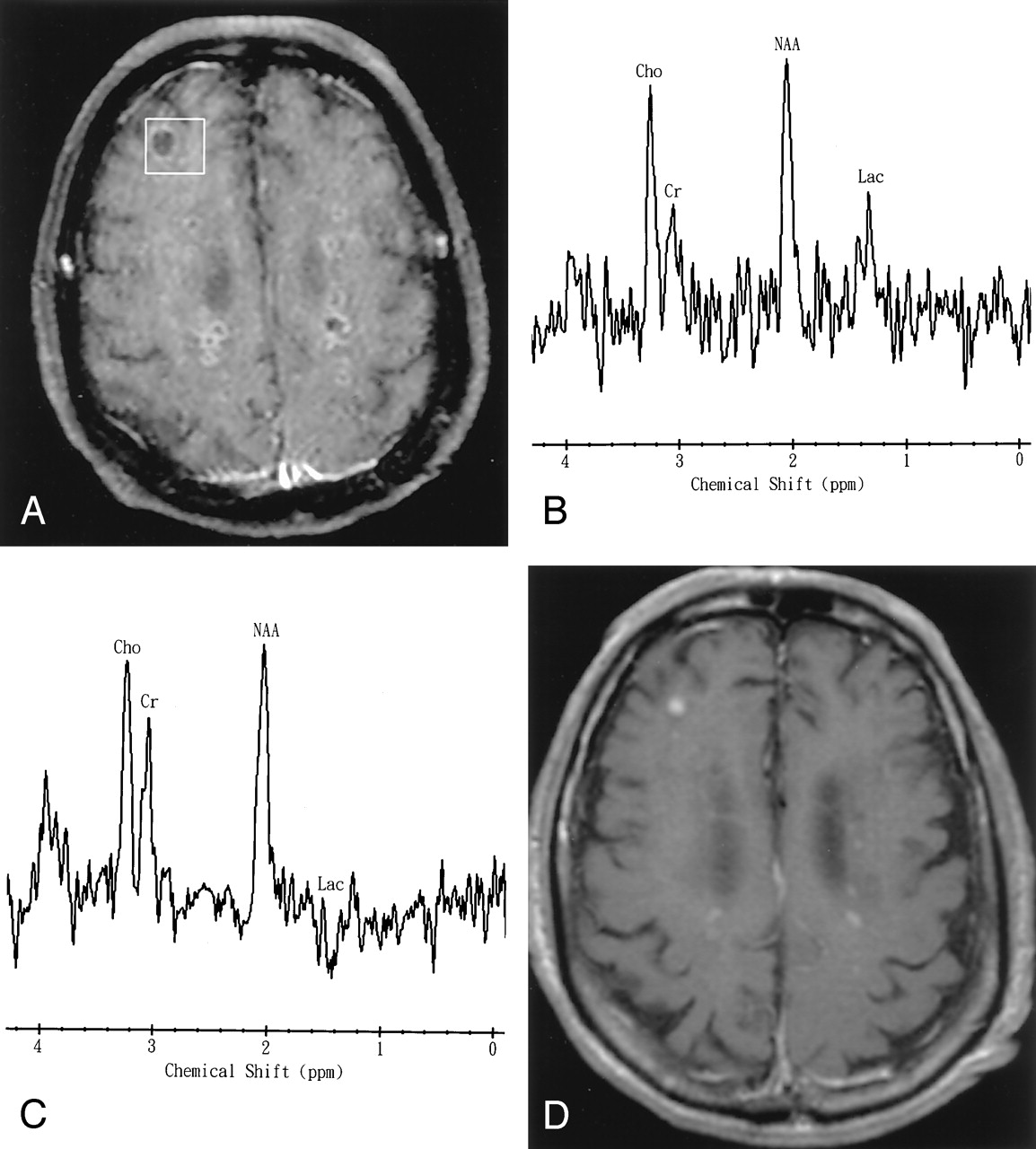
  • Fig 3.
    • Download figure
    • Open in new tab
    • Download powerpoint
    Fig 3.

    Patient 15. A 72-year-old man with Streptococcus mitis (aerobe) bacteremia, infective endocarditis, and multiple miliary pyogenic brain abscesses.

    A, Axial contrast-enhanced T1-weighted MR image shows numerous miliary nodules with ring enhancement. The 2 x 2 x 2-cm voxel (box) in the lesion and adjacent brain tissue represents the 1H MR spectroscopic volume of interest.

    B and C, In vivo 1H spectra (TR/TE 1600/270 [B] and 1600/135 [C]) show spectral pattern C with a lactate (Lac) peak (1.3 ppm) that is inverted at a TE of 135. The resonances of choline (Cho), creatine (Cr), and N-acetylaspartate (NAA) were interpreted to be caused by partial volume effects of the adjacent brain tissue. The phase reversal does not follow the classic pattern in this case because of the presence of some phase distortion, which presumably originated from the eddy current effect. Multiple small peaks at various frequencies are present; these peaks may represent noise or unassigned metabolites.

    D, Axial contrast-enhanced T1-weighted MR image obtained 3 months later shows almost complete remission of the formation of diffuse abscesses after antibiotics.

Tables

  • Figures
    • View popup
    Table:

    Clinical information and 1H MR spectroscopic findings in 15 patients with pyogenic brain abscesses

    Patient No./Sex/ Age (y)Chief ComplaintLocation of AbscessPus Culture MicroorganismsTreatmentOutcomeDuration of SymptomsMR Spectroscopy before/after Starting AntibioticsMR Spectroscopic Findings
    PatternNAACrChoSuccAceAlaAALacLip
    1/F/45Fever, headache, unstable gaitLeft cerebellumBacteroides fragilisSurgery/medicalCured10 days1 day afterA++++++++++++++++++
    2/M/74Right hemiparesisLeft parietalStreptococcus intermediusSurgery/medicalCured30 days1 day beforeA−−−−++++++−
    3/M/69Fever, right hemiparesisLeft parietalB fragilisSurgery/medicalDied7 days1 day beforeA++++++++++++++
    4/M/48HeadacheLeft frontalNegativeSurgery/medicalCured10 days2 days beforeA−−−−++++++++++++
    5/M/45Fever, headacheRight temporalStreptococcus mitis, Enterococcus faecalisSurgery/medicalCured12 days2 days afterA−−−+++++++++++−
    6/M/43Consciousness changeRight basal ganglionEubacterium lentumSurgery/medicalCured3 days1 day afterA−−−+++++++++++−
    7/M/47Headache, left homonymous hemianopiaRight occipitalS intermediusSurgery/medicalCured14 days1 day afterB−−−−−−+++−
    8/F/28Right homonymous hemianopiaLeft occipitalS intermediusSurgery/medicalCured21 days2 days beforeB−−−−−−+++−
    9/M/76Fever, consciousness changeLeft temporalNegativeSurgery/medicalCured14 days1 day afterB+++−−−+++−
    10/F/69Left hemiparesisRight frontalStaphylococcus aureusSurgery/medicalCured3 days1 day beforeB+++−−−+++−
    11/F/29Fever, headacheLeft frontalPseudomonas aeruginosaSurgery/medicalCured2 wks2 days afterB−−−−−−++++
    12/M/72Fever, left hemiparesisRight parietalS intermediusSurgery/medicalCured10 days2 days beforeB+++−−−+++−
    13/M/72Left hemiparesis, infective endocarditisMultipleStreptococcus constellatusSurgery/medicalDied30 days1 day afterB−−−−−−+++−
    14/F/78Fever, consciousness changeRight temporalP aeruginosaSurgery/medicalCured10 days2 days afterB−−−−−−+++−
    15/M/72Consciousnes change, infective endocarditisMultiple military (<5 mm)S mitis (blood culture)MedicalCured3 days1 day afterC+++++−−−−+−
    • Notes.—Negative sign indicates no bacterial growth; +, small peak; ++, moderate peak; +++, large peak; Ace, acetate; Ala, alanine; AA, amino acids; Lac, lactate; Succ, succinate; NAA, N-acetylaspartate; Cho, choline; Cr, creatine-phosphocreatine; Lip, lipids.

PreviousNext
Back to top

In this issue

American Journal of Neuroradiology: 26 (2)
American Journal of Neuroradiology
Vol. 26, Issue 2
1 Feb 2005
  • Table of Contents
  • Index by author
Advertisement
Print
Download PDF
Email Article

Thank you for your interest in spreading the word on American Journal of Neuroradiology.

NOTE: We only request your email address so that the person you are recommending the page to knows that you wanted them to see it, and that it is not junk mail. We do not capture any email address.

Enter multiple addresses on separate lines or separate them with commas.
Pyogenic Brain Abscess: Findings from In Vivo 1.5-T and 11.7-T In Vitro Proton MR Spectroscopy
(Your Name) has sent you a message from American Journal of Neuroradiology
(Your Name) thought you would like to see the American Journal of Neuroradiology web site.
CAPTCHA
This question is for testing whether or not you are a human visitor and to prevent automated spam submissions.
Cite this article
Ping H. Lai, Kun T. Li, Shu S. Hsu, Chia C. Hsiao, Chi W. Yip, S. Ding, Lee R. Yeh, Huay B. Pan
Pyogenic Brain Abscess: Findings from In Vivo 1.5-T and 11.7-T In Vitro Proton MR Spectroscopy
American Journal of Neuroradiology Feb 2005, 26 (2) 279-288;

Citation Manager Formats

  • BibTeX
  • Bookends
  • EasyBib
  • EndNote (tagged)
  • EndNote 8 (xml)
  • Medlars
  • Mendeley
  • Papers
  • RefWorks Tagged
  • Ref Manager
  • RIS
  • Zotero
0 Responses
Respond to this article
Share
Bookmark this article
Pyogenic Brain Abscess: Findings from In Vivo 1.5-T and 11.7-T In Vitro Proton MR Spectroscopy
Ping H. Lai, Kun T. Li, Shu S. Hsu, Chia C. Hsiao, Chi W. Yip, S. Ding, Lee R. Yeh, Huay B. Pan
American Journal of Neuroradiology Feb 2005, 26 (2) 279-288;
del.icio.us logo Twitter logo Facebook logo Mendeley logo
  • Tweet Widget
  • Facebook Like
  • Google Plus One

Jump to section

  • Article
    • Abstract
    • Methods
    • Results
    • Discussion
    • Conclusion
    • Acknowledgments
    • Footnotes
    • References
  • Figures & Data
  • Info & Metrics
  • Responses
  • References
  • PDF

Related Articles

  • No related articles found.
  • PubMed
  • Google Scholar

Cited By...

  • Multiparametric imaging in the evaluation of intracerebral abscesses
  • Multiparametric imaging in the evaluation of intracerebral abscesses
  • Crossref
  • Google Scholar

This article has not yet been cited by articles in journals that are participating in Crossref Cited-by Linking.

More in this TOC Section

  • Usefulness of Quantitative Susceptibility Mapping for the Diagnosis of Parkinson Disease
  • White Matter Alterations in the Brains of Patients with Active, Remitted, and Cured Cushing Syndrome: A DTI Study
  • Qualitative and Quantitative Analysis of MR Imaging Findings in Patients with Middle Cerebral Artery Stroke Implanted with Mesenchymal Stem Cells
Show more BRAIN

Similar Articles

Advertisement

Indexed Content

  • Current Issue
  • Accepted Manuscripts
  • Article Preview
  • Past Issues
  • Editorials
  • Editor's Choice
  • Fellows' Journal Club
  • Letters to the Editor
  • Video Articles

Cases

  • Case Collection
  • Archive - Case of the Week
  • Archive - Case of the Month
  • Archive - Classic Case

Special Collections

  • AJNR Awards
  • ASNR Foundation Special Collection
  • Most Impactful AJNR Articles
  • Photon-Counting CT
  • Spinal CSF Leak Articles (Jan 2020-June 2024)

More from AJNR

  • Trainee Corner
  • Imaging Protocols
  • MRI Safety Corner

Multimedia

  • AJNR Podcasts
  • AJNR Scantastics

Resources

  • Turnaround Time
  • Submit a Manuscript
  • Submit a Video Article
  • Submit an eLetter to the Editor/Response
  • Manuscript Submission Guidelines
  • Statistical Tips
  • Fast Publishing of Accepted Manuscripts
  • Graphical Abstract Preparation
  • Imaging Protocol Submission
  • Evidence-Based Medicine Level Guide
  • Publishing Checklists
  • Author Policies
  • Become a Reviewer/Academy of Reviewers
  • News and Updates

About Us

  • About AJNR
  • Editorial Board
  • Editorial Board Alumni
  • Alerts
  • Permissions
  • Not an AJNR Subscriber? Join Now
  • Advertise with Us
  • Librarian Resources
  • Feedback
  • Terms and Conditions
  • AJNR Editorial Board Alumni

American Society of Neuroradiology

  • Not an ASNR Member? Join Now

© 2025 by the American Society of Neuroradiology All rights, including for text and data mining, AI training, and similar technologies, are reserved.
Print ISSN: 0195-6108 Online ISSN: 1936-959X

Powered by HighWire