
Abstract
BACKGROUND AND PURPOSE: Contrast-enhanced fluid-attenuated inversion recovery (FLAIR) imaging has been reported to have higher sensitivity for detecting leptomeningeal disease compared with contrast-enhanced T1-weighted MR imaging. The purpose of this study was to compare contrast-enhanced T1-weighted MR images with fat suppression to contrast-enhanced FLAIR images to determine which sequence was superior for depicting meningeal disease.
METHODS: We reviewed MR images of 24 patients (35 studies) with a variety of meningeal diseases. The MR imaging protocol included contrast-enhanced T1-weighted MR images with fat suppression (FS) and contrast-enhanced fluid-attenuated inversion recovery (FLAIR) images that were reviewed by three neuroradiologists and were assigned a rating of positive, equivocal, or negative for abnormal meningeal enhancement. The two sequences were compared side by side to determine which better depicted meningeal disease.
RESULTS: Abnormal meningeal enhancement was positive in 35 contrast-enhanced T1-weighted MR images with FS and in 33 contrast-enhanced FLAIR studies. In the first group, which had the T1-weighted sequence acquired first (21 of 33 studies), contrast-enhanced T1-weighted images with FS showed superior contrast enhancement in 11 studies (52%), inferior contrast enhancement in six studies (29%), and equal contrast enhancement in four studies (19%) compared with the contrast-enhanced FLAIR images. In the second group, which had the FLAIR sequence acquired first (12 of 33), contrast-enhanced T1-weighted images with FS showed superior contrast enhancement in seven studies (58%), inferior contrast enhancement in two studies (17%), and equal contrast enhancement in three studies (25%).
CONCLUSION: Contrast-enhanced T1-weighted MR imaging with FS is superior to contrast-enhanced FLAIR imaging in most cases for depicting intracranial meningeal diseases.
MR imaging has demonstrated increased sensitivity over CT scanning in the detection of all types of meningeal disease (1). The addition of gadolinium-based paramagnetic contrast medium further improves the sensitivity of MR imaging and also increases diagnostic specificity for meningeal abnormalities (2).
Some authors have reported that contrast-enhanced FLAIR imaging may be superior to contrast-enhanced T1-weighted imaging for detecting leptomeningeal disease (3–5). On the other hand, Singh et al (6) found fast FLAIR to be less sensitive than standard T1-weighted MR images for detecting neoplastic leptomeningeal disease on postcontrast images. For the past 18 months at our institution, both T1-weighted with FS and FLAIR sequences have been acquired routinely following gadolinium administration. We became interested in both sequences in the diagnosis of meningeal disease. Our hypothesis is that contrast-enhanced T1-weighted imaging with FS is more sensitive than contrast-enhanced FLAIR imaging in depicting intracranial meningeal disease.
Methods
The institutional review board at our medical center approved this study, and patient informed consent was not required. At our institution, the routine use of contrast-enhanced FLAIR sequences in brain imaging began in September 2002. We retrospectively reviewed the images obtained from the patients with documented meningeal disease. The diagnosis of meningeal disease was proved by CSF cytology, culture, or viral titers or by meningeal biopsy.
The subjects consisted of 24 patients (15 men and nine women; age range, 16–66 years; mean age, 47 years) with a variety of meningeal diseases. All patients with proven meningeal disease and positive MR images on any sequence were included. Sixteen patients had meningitis (five tuberculous, three cryptococcal, three pyogenic, one disseminated coccidioidomycosis, one viral, and three with unknown organisms), and eight patients had leptomeningeal metastases caused by known primary tumors (two breast carcinomas, two lymphomas, one lung carcinoma, one transitional cell carcinoma of the urinary bladder, one multiple myeloma, and one glioblastoma multiforme).
MR imaging was performed with 1.5T MR imaging systems (Siemens Symphony; Erlangen, Germany). The FLAIR sequences used parameters of 8000–9000/105–109/1 (TR/TE/NEX), an inversion time of 2000, and a matrix of 256 × 192. Imaging parameters for contrast-enhanced T1-weighted images with FS included 437–481/14/1 and a matrix of 256 × 144. For both sequences, the field of view was 22 cm, with a section thickness of 5 mm and an interslice gap of 1.5 mm. For all patients, gadodiamide (Omniscan; Nycomed, Oslo, Norway) was administered at the standard dose of 0.1 mmol/kg of body weight. Postcontrast images were obtained shortly after contrast material administration. Patients were divided into two groups. One group had contrast-enhanced T1-weighted imaging with FS acquired before the contrast-enhanced FLAIR images, and the other group had contrast-enhanced FLAIR imaging performed first. The approximate time interval between the two sequences ranged from 2 to 5 minutes.
Two trained neuroradiologists (J.R.H., J.F.H.) were asked to assign a rating of positive, equivocal, or negative for abnormal meningeal enhancement independently. There was an interval of at least 2 weeks between reviewing the contrast-enhanced T1-weighted images with FS and the contrast-enhanced FLAIR images. In cases in which abnormal meningeal enhancement was present on both sequences, the images were compared side by side to determine which sequence was better for depiction of meningeal enhancement. In cases of disagreement, the final judgment was rendered by the third neuroradiologist (S.G.I.).
Results
In 35 studies of the 24 patients, contrast-enhanced FLAIR imaging was positive in 33 and negative in two for abnormal meningeal enhancement. Contrast-enhanced T1-weighted MR imaging with FS was positive in all 35 images (Table 1). Therefore, only two studies were positive on contrast-enhanced T1-weighted images with FS and negative on contrast-enhanced FLAIR images. The images of both patients disclosed diffuse meningeal enhancement without focal nodularity.
Results of readings of contrast-enhanced T1-weighted imaging with FS and contrast-enhanced FLAIR imaging for meningeal disease
In 21 of the 33 studies (64%) positive with both sequences, contrast-enhanced T1-weighted images with FS were obtained before contrast-enhanced FLAIR, and in 12 of the 33 studies (36%), contrast-enhanced FLAIR images were obtained first. Both sequences were compared side by side to determine which was better for depiction of meningeal enhancement. In the first group (21/33), contrast-enhanced T1-weighted imaging with FS showed superior contrast enhancement in 11 studies (52%), inferior contrast enhancement in six studies (29%), and equal contrast enhancement in four studies (19%) compared with contrast-enhanced FLAIR. In the second group (12/33), contrast-enhanced T1-weighted imaging with FS showed superior contrast enhancement in seven studies (58%), inferior contrast enhancement in two studies (17%), and equal contrast enhancement in three studies (25%) (Table 2).
Comparison between contrast-enhanced T1-weighted MR imaging with FS and contrast-enhanced FLAIR imaging for depicting meningeal disease
There was a clear preference for contrast-enhanced T1-weighted imaging with FS, showing superior enhancement to contrast-enhanced FLAIR. This difference was significant (P < .01) based on an exact test of binomial proportions. By using Fisher’s exact test, there was no significant order effect secondary to the order of acquisition of the sequences (P < .792) in the analysis of the data.
Of the 35 studies with meningeal disease, 13 studies showed isolated abnormal pachymeningeal enhancement, another 13 studies showed isolated leptomeningeal enhancement, and nine studies showed both types of meningeal enhancement. Contrast-enhanced T1-weighted imaging with FS was judged superior in quality to contrast-enhanced FLAIR imaging in both types of meningeal enhancement in most cases (Figs 1–3). In addition, the enhancement of a cranial nerve in one patient with tuberculous meningitis (Fig 2C, -D), subarachnoid space lesions in five patients with tuberculous meningitis, and cryptococcal meningitis in two patients were more conspicuous on contrast-enhanced T1-weighted imaging with FS; however, hyperintensity due to perilesional edema (Fig 3) and interstitial edema secondary to hydrocephalus were depicted better on contrast-enhanced FLAIR images.

Cryptococcal meningitis.
A, Contrast-enhanced FLAIR image shows slight leptomeningeal enhancement in the right frontoparietal region (arrows). It is difficult to separate meningeal enhancement from other high signal intensity within the sulci and from adjacent parenchymal disease.
B, Contrast-enhanced T1-weighted image with FS shows greater contrast between the enhancing tissue and the adjacent brain and is better for depicting enhancement within the sulci and the interhemispheric fissure (arrows).
C, Contrast-enhanced FLAIR image of the same patient obtained at the level of the sylvian fissures shows subtle enhancing lesions in the basal ganglia bilaterally, but without a noncontrast FLAIR image for comparison it is not possible to distinguish enhancement from parenchymal edema.
D, Contrast-enhanced T1-weighted image with FS shows multiple punctuate enhancing areas within the basal ganglia bilaterally, due to cryptococcomas within dilated Virchow-Robin spaces (arrows). The enhancement is much more conspicuous on the T1-weighted image. Additional enhancement is present in several inferior sulci in both frontal lobes.

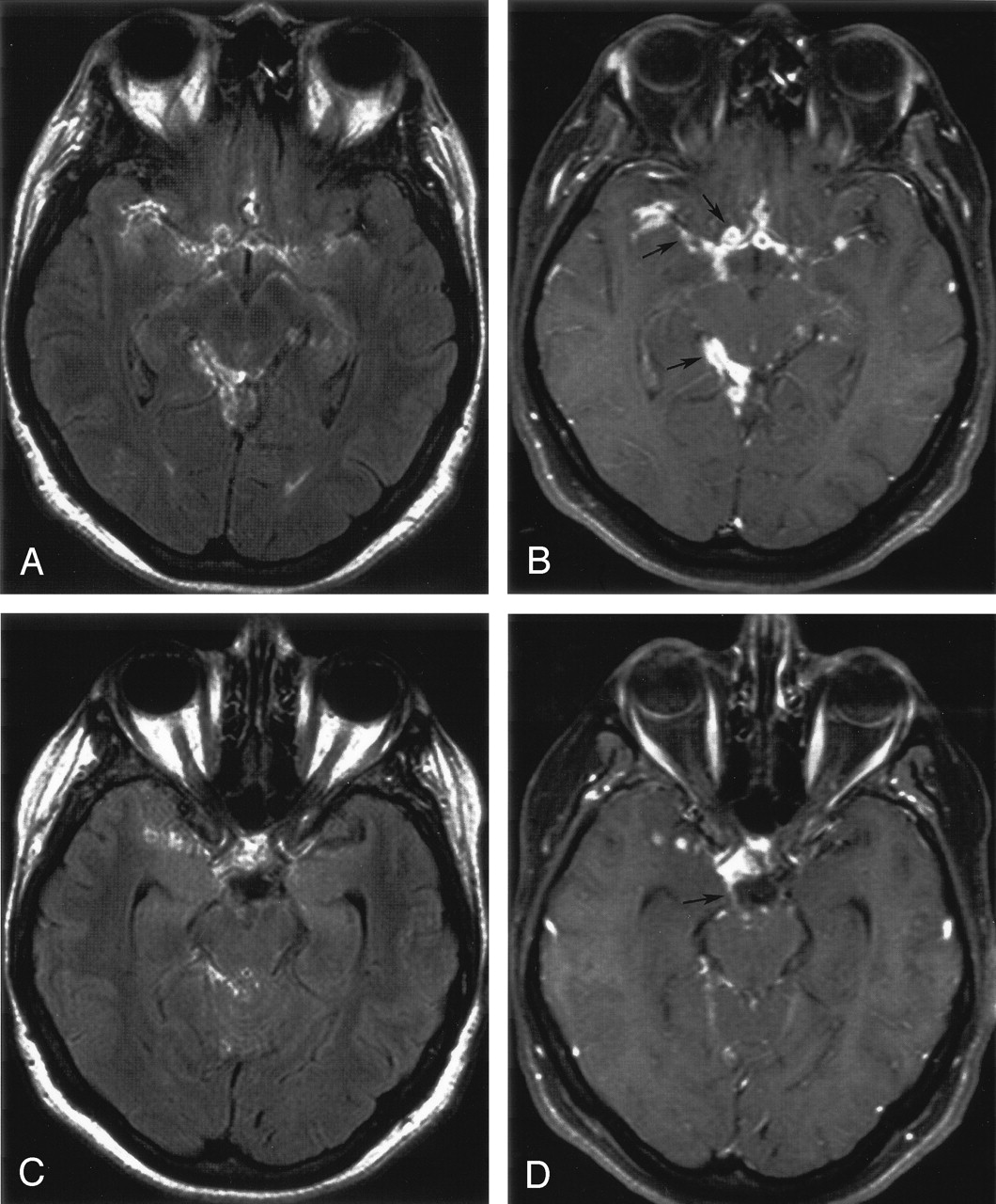
Tuberculous meningitis.
A, Contrast-enhanced FLAIR image shows mildly enhancing subarachnoid space lesions in the basilar cisterns, with extension into the sylvian fissures bilaterally, right ambient cistern, and quadrigeminal cistern.
B, Contrast-enhanced T1-weighted image with FS shows the enhancing lesions in the subarachnoid space more definitively (arrows). The contrast difference between the enhancing meninges and the adjacent brain is visually greater in the T1-weighted image.
C, Contrast-enhanced FLAIR image of the same patient shows enhancement in the region of the right third cranial nerve, but the nerve is not clearly seen.
D, The enhancement of the right third cranial nerve (arrow) is more distinct on the contrast-enhanced T1-weighted image with FS.

Transitional cell carcinoma of the urinary bladder with calvarial metastases and meningeal extension.
A, Contrast-enhanced FLAIR image shows localized mild pachymeningeal enhancement (arrows) adjacent to the focal destructive lesion of the right parietooccipital bone. The hyperintensity of adjacent white matter edema is clearer on contrast-enhanced FLAIR image. Note that the internal cerebral veins and cortical veins do not enhance on the FLAIR image. Similarly, the hypervascular pachymeninges (arrows) with increased blood pool does not enhance nearly as much as on the T1-weighted image in panel B.
B, The pachymeningeal enhancement is more apparent and appears thicker on a contrast-enhanced T1-weighted image with FS (arrow). The enhancing cortex is distinct from the underlying hypointense edema.
Longitudinal data were available in eight patients (Table 3). The interval between the initial MR image and follow-up imaging ranged from 6 days to 7 months. As a general rule, the aseptic and bacterial processes improved, but the meningeal involvement by tuberculosis and tumor progressed.
Longitudinal study of eight patients with meningeal abnormalities
Discussion
Infectious and neoplastic meningeal disease can produce serious complications, resulting in substantial morbidity and mortality. Early diagnosis is important so appropriate treatment can be instituted promptly to prevent major brain damage and permanent neurologic complications. Gadolinium-enhanced T1-weighted imaging has been the standard MR image used for evaluating meningeal disease. The competing signal intensity from CSF makes conventional T2-weighted images very insensitive for subarachnoid abnormalities. On the other hand, the FLAIR sequence effectively nullifies the signal intensity from CSF and provides heavy T2 weighting because of its very long echo time (7, 8). FLAIR imaging is known to be efficacious in the detection of subarachnoid hemorrhage (SAH), meningoencephalitis, and leptomeningeal metastases (9, 10). One study (9) went further in stating that contrast-enhanced T1-weighted imaging was far less sensitive than unenhanced FLAIR for leptomeningeal inflammatory and neoplastic disease. In the specific setting of intracranial leptomeningeal metastases, however, a subsequent larger study proved that contrast-enhanced T1-weighted imaging has higher sensitivity compared with unenhanced FLAIR (11).
One problem with postcontrast T1-weighted sequences is that enhancing cortical vessels can be confused with meningeal enhancement. Unlike T1-weighted images, postcontrast FLAIR images do not show contrast enhancement in vessels with slow-flowing blood. For this reason and based on their preliminary experience, Mathews et al (5) suggested that postcontrast FLAIR may be better for detecting superficial brain abnormalities, such as meningeal disease. In a companion in vitro study, they showed that the contrast between normal brain and lower concentrations of gadolinium was higher on FLAIR compare with T1-weighted images with or without magnetization transfer saturation. These observations stimulated a letter to the editor from Jackson and Hayman (12), who carried these analyses a step further by constructing two contrast models—one for gadolinium-enhanced CSF versus nonenhanced CSF and the other for gadolinium-enhanced CSF versus nonenhanced normal gray matter. Their models demonstrate that at concentrations of gadolinium below 0.7 mmol/L, the contrast is higher for fast FLAIR, and a concentration of 0.1–0.3 mmol/L results in the highest difference in contrast between fast FLAIR and T1-weighted images. In another in vitro phantom study, Mamourian et al (13) showed that gadolinium effects were visible on FLAIR images at concentrations four times lower than on T1-weighted images.
In support of the above data, contrast-enhanced FLAIR was reported to improve detection of leptomeningeal disease in a small group of pediatric patients (4). These investigators used both subjective and objective assessments of the presence and extent of leptomeningeal enhancement to reach their conclusions. Also, the T1-weighted images were always obtained after the FLAIR images. In another small series, Tsuchiya et al (3) concluded that contrast-enhanced FLAIR images can sometimes surpass contrast-enhanced T1-weighted images, but only five of the nine cases of leptomeningeal carcinomatosis were actually documented.
Contrary to the investigations and published reports discussed above, in a larger series by Singh et al (6), who are neuroradiologists, blinded to the results of cytology, reviewed 74 MR images of suspected leptomeningeal metastases. Results proved that fast FLAIR was less sensitive than standard T1-weighted spin-echo sequences for detecting contrast enhancement of neoplastic leptomeningeal disease.
Our results are in agreement with Singh et al (6) and suggest that contrast-enhanced T1-weighted MR imaging with FS is more sensitive than contrast-enhanced FLAIR for evaluation of meningeal lesions. Moreover, in a side-by-side comparison between the two sequences, contrast-enhanced T1-weighted images with FS made enhancing lesions more conspicuous and improved delineation of the location and extent of meningeal disease. The contrast between the enhancing lesions and nonenhancing tissues, such as brain and CSF, was subjectively greater on the T1-weighted images with FS (Figs 1–3). Also, pachymeningeal enhancement was more conspicuous on the T1-weighted images (Fig 3).
In our study, contrast-enhanced T1-weighted images with FS were rated better than or as effective as contrast-enhanced FLAIR in 25 of 33 studies. There was a clear preference for contrast-enhanced T1-weighted images with FS over contrast-enhanced FLAIR, which was significant (P < .01) based on an exact test of binomial proportions. In a few cases, the additive effects of T2 hyperintensity and enhancement made the abnormalities more apparent on the contrast-enhanced FLAIR images (Fig 4). On the FLAIR images, however, it was difficult to separate meningeal disease from hyperintense edema within the underlying cortex. Also, because precontrast FLAIR images were not available, it was not possible to determine how much of the FLAIR signal intensity was from preexisting T2 signal intensity and how much represented true enhancement. In several other cases, the parenchymal edema on the FLAIR images drew one’s attention to the area of meningeal abnormality, but it was more difficult to see the meningeal enhancement compared with the T1-weighted images.

Tuberculous meningitis.
A, Contrast-enhanced FLAIR image shows hyperintensity along the meninges and within several sulci of the left parietal lobe (arrowheads). A precontrast FLAIR image was not available to assess how much of the hyperintensity reflected T2 signal intensity and how much was true enhancement.
B, Contrast-enhanced T1-weighted image with FS reveals enhancement in the same area (arrows), but the enhancement is less intense and less extensive.
The experiments and conclusions of Jackson and Hayman (12) and Mamourian et al (13) were based on the assumption that the source of meningeal enhancement is leakage of contrast from pial vessels into the adjacent CSF. Because meningeal arteries do not have a blood-brain barrier, that assumption is likely valid in the case of normal meninges and acutely inflamed meninges. In the case of metastatic disease and chronic infections, however, the meninges are infiltrated by nodular tumor and thick inflammatory tissues that are very vascular and also have a component of interstitial space. In a busy clinical setting, the postcontrast images are generally obtained immediately after the contrast injection. As a result, probably a large component of the gadolinium is within vascular blood pool, and this component of enhancement is missed by FLAIR (Fig 2). The subarachnoid component of contrast would not be a factor unless delayed images were obtained. Thus, the proposed strength of FLAIR (lack of vascular enhancement) probably turns out to be a weakness.
Furthermore, as pointed out by Taoka et al (14), sulcal hyperintensity on FLAIR images can occur without apparent CSF abnormality. For example, as a result of mass effect and vascular disease, an increase in blood pool, a small amount of protein leakage, and “in-flow” enhancement of congested blood may contribute to sulcal hyperintensity on FLAIR images. These phenomena can lead to false-positive contrast-enhanced FLAIR images (Fig 1) unless the contrast images are carefully compared with the precontrast FLAIR images.
FLAIR images provide good visualization of the subarachnoid spaces within the cerebral fissures and sulci. On the other hand, images at the base of the brain and in the posterior fossa are often marred by flow artifacts, which can simulate meningeal enhancement on postcontrast images. For this reason, postcontrast FLAIR likely cannot be used as the primary imaging sequence for assessing meningeal disease. FLAIR can supplement, but cannot replace, T1-weighted sequences for postcontrast imaging.
Our study was not designed to evaluate the efficacy of FS in conjunction with gadolinium-enhanced T1-weighted imaging. That is part of another ongoing study at our institution. The addition of FS to contrast-enhanced T1-weighted imaging has been investigated previously and was shown to be helpful for the diagnosis of neoplastic and inflammatory diseases of the spine (15), as well as in head and neck lesions (16). FS improves visibility of subtle contrast enhancement by suppressing the high signal intensity of fat, increasing the dynamic range of gray-scale contrast, and eliminating the chemical shift artifact. The compressed gray scale would apply to images displayed on film, but when viewing on a workstation or PACS station adjustment of the window width and window level can compensate for the compressed gray scale. FS may improve visualization of meningeal enhancement in close proximity to fatty marrow within the calvaria and skull base.
There are several limitations to our study. Our subjects included only those with reported positive MR images for meningeal disease, so we cannot assess sensitivity or specificity of the imaging sequences. Our reviewers (J.R.H., J.F.H., S.G.I.) were not blinded to the presence or absence of disease, but rather were blinded to any original interpretations and which sequences were positive for disease. Our goal was to directly compare the two sequences to assess which sequence visually showed superior contrast enhancement and better depiction of the meningeal abnormality. Our cohort of 35 cases with positive MR images is comparable to the other major studies comparing FLAIR and T1-weighted sequences.
In our series, the contrast-enhanced T1-weighted images with FS were randomly obtained either before or after the contrast-enhanced FLAIR. The effect of delayed enhancement is not clear but may be related to the status of vascularity of the individual lesions that can affect the accumulation of the contrast medium at various scanning times. We did not study the specific mechanisms of enhancement in our cases, but it is likely because of a combination of vascular enhancement, interstitial accumulation of contrast medium, and leakage of contrast into the subarachnoid space. Because the meninges do not have a blood-brain barrier, the order or timing of the MR images after the contrast injection is probably not a significant factor, and analysis of our data revealed no statistically significant “order effect” regarding order of acquisition of the T1-weighted and FLAIR sequences.
Recently, Kikuchi et al (17) presented computer-simulated brain maps showing that image contrast between white matter and gray matter is greater on T1-weighted FLAIR compared with T1-weighted spin-echo sequences. The utility of the T1-weighted FLAIR sequence remains to be tested in a clinical setting.
Conclusion
In our limited case series, contrast-enhanced T1-weighted MR imaging with FS was superior to contrast-enhanced FLAIR imaging in most cases for depicting abnormal meningeal enhancement and subarachnoid space lesions in patients with intracranial meningeal disease. We propose that the FLAIR sequence fails to capture the signal intensity from the slow-flowing blood pool within the very vascular meningeal inflammatory and neoplastic lesions.
References
- Received February 5, 2004.
- Accepted after revision April 23, 2004.
- Copyright © American Society of Neuroradiology
In this issue


Jump to section
Related Articles
Cited By...
- Diagnostic Accuracy of MRI for Detection of Meningitis in Infants
- Potentially Reversible and Recognizable Acute Encephalopathic Syndromes: Disease Categorization and MRI Appearances
- Comparison of the Added Value of Contrast-Enhanced 3D Fluid-Attenuated Inversion Recovery and Magnetization-Prepared Rapid Acquisition of Gradient Echo Sequences in Relation to Conventional Postcontrast T1-Weighted Images for the Evaluation of Leptomeningeal Diseases at 3T