Advertisement

Main menu

  • Home
  • Content
    • Current Issue
    • Accepted Manuscripts
    • Article Preview
    • Past Issue Archive
    • Video Articles
    • AJNR Case Collection
    • Case of the Week Archive
    • Case of the Month Archive
    • Classic Case Archive
  • Special Collections
    • AJNR Awards
    • ASNR Foundation Special Collection
    • Most Impactful AJNR Articles
    • Photon-Counting CT
    • Spinal CSF Leak Articles (Jan 2020-June 2024)
  • Multimedia
    • AJNR Podcasts
    • AJNR SCANtastic
    • Trainee Corner
    • MRI Safety Corner
    • Imaging Protocols
  • For Authors
    • Submit a Manuscript
    • Submit a Video Article
    • Submit an eLetter to the Editor/Response
    • Manuscript Submission Guidelines
    • Statistical Tips
    • Fast Publishing of Accepted Manuscripts
    • Graphical Abstract Preparation
    • Imaging Protocol Submission
    • Author Policies
  • About Us
    • About AJNR
    • Editorial Board
    • Editorial Board Alumni
  • More
    • Become a Reviewer/Academy of Reviewers
    • Subscribers
    • Permissions
    • Alerts
    • Feedback
    • Advertisers
    • ASNR Home

User menu

  • Alerts
  • Log in

Search

  • Advanced search
American Journal of Neuroradiology

American Journal of Neuroradiology

ASHNR American Society of Functional Neuroradiology ASHNR American Society of Pediatric Neuroradiology ASSR
  • Alerts
  • Log in

Advanced Search

  • Home
  • Content
    • Current Issue
    • Accepted Manuscripts
    • Article Preview
    • Past Issue Archive
    • Video Articles
    • AJNR Case Collection
    • Case of the Week Archive
    • Case of the Month Archive
    • Classic Case Archive
  • Special Collections
    • AJNR Awards
    • ASNR Foundation Special Collection
    • Most Impactful AJNR Articles
    • Photon-Counting CT
    • Spinal CSF Leak Articles (Jan 2020-June 2024)
  • Multimedia
    • AJNR Podcasts
    • AJNR SCANtastic
    • Trainee Corner
    • MRI Safety Corner
    • Imaging Protocols
  • For Authors
    • Submit a Manuscript
    • Submit a Video Article
    • Submit an eLetter to the Editor/Response
    • Manuscript Submission Guidelines
    • Statistical Tips
    • Fast Publishing of Accepted Manuscripts
    • Graphical Abstract Preparation
    • Imaging Protocol Submission
    • Author Policies
  • About Us
    • About AJNR
    • Editorial Board
    • Editorial Board Alumni
  • More
    • Become a Reviewer/Academy of Reviewers
    • Subscribers
    • Permissions
    • Alerts
    • Feedback
    • Advertisers
    • ASNR Home
  • Follow AJNR on Twitter
  • Visit AJNR on Facebook
  • Follow AJNR on Instagram
  • Join AJNR on LinkedIn
  • RSS Feeds

AJNR is seeking candidates for the AJNR Podcast Editor. Read the position description.

Research ArticleBRAIN

Vascular Occlusion Sites Determine Differences in Lesion Growth from Early Apparent Diffusion Coefficient Lesion to Final Infarct

Jens Fiehler, Karina Knudsen, Götz Thomalla, Einar Goebell, Michael Rosenkranz, Cornelius Weiller, Joachim Röther, Hermann Zeumer and Thomas Kucinski
American Journal of Neuroradiology May 2005, 26 (5) 1056-1061;
Jens Fiehler
  • Find this author on Google Scholar
  • Find this author on PubMed
  • Search for this author on this site
Karina Knudsen
  • Find this author on Google Scholar
  • Find this author on PubMed
  • Search for this author on this site
Götz Thomalla
  • Find this author on Google Scholar
  • Find this author on PubMed
  • Search for this author on this site
Einar Goebell
  • Find this author on Google Scholar
  • Find this author on PubMed
  • Search for this author on this site
Michael Rosenkranz
  • Find this author on Google Scholar
  • Find this author on PubMed
  • Search for this author on this site
Cornelius Weiller
  • Find this author on Google Scholar
  • Find this author on PubMed
  • Search for this author on this site
Joachim Röther
  • Find this author on Google Scholar
  • Find this author on PubMed
  • Search for this author on this site
Hermann Zeumer
  • Find this author on Google Scholar
  • Find this author on PubMed
  • Search for this author on this site
Thomas Kucinski
  • Find this author on Google Scholar
  • Find this author on PubMed
  • Search for this author on this site
  • Article
  • Figures & Data
  • Info & Metrics
  • Responses
  • References
  • PDF
Loading

Article Figures & Data

Figures

  • Tables
  • Fig 1.
    • Download figure
    • Open in new tab
    • Download powerpoint
    Fig 1.

    Types of occlusion were defined on MRA, as follows: a + c was an occlusion of the ICA at the neck accompanied by an MCA embolism, or ICA/MCA occlusion; b, occlusion of the intracranial bifurcation of the ICA, or carotid T occlusion, which included one of 17 cases with an occlusion of the proximal anterior and middle cerebral artery; c, occlusion of the MCA trunk, which included nine of 48 occlusions of the bifurcation or trifurcation lateral to the medial lenticulostriate arteries; and d, occlusion of a single or multiple MCA branch occlusion with free trifurcation, which included three of 36 cases with an additional occlusion of the peripheral anterior cerebral artery.
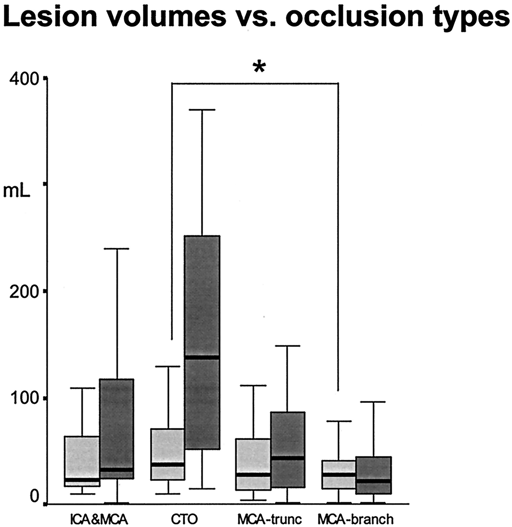
  • Fig 2.
    • Download figure
    • Open in new tab
    • Download powerpoint
    Fig 2.

    Boxplots of lesion volumes for ADCman <6 hours after stroke onset and lesion volume on days 5–8 (on T2-weighted or CT images) for each type of occlusion. Asterisk = significant differences in ADCman in multiple pairwise comparisons between types. Infarct volumes were significantly different (P < .05) for all types except for ICA/MCA versus MCA trunk occlusions (MCA-trunc). CTO = carotid-T occlusion.

  • Fig 3.
    • Download figure
    • Open in new tab
    • Download powerpoint
    Fig 3.

    Reperfusion assessed on day 1 after stroke onset for each type of occlusion in 65 patients treated with intravenous thrombolysis. CTO = carotid-T occlusion.

  • Fig 4.
    • Download figure
    • Open in new tab
    • Download powerpoint
    Fig 4.

    T2-weighted (b = 0 s/mm2, top) and ADC (bottom) images obtained in patients with proximal vascular occlusions show considerable lesion growth without recanalization. Left box, Images obtained in a 71-year-old man with an ICA/MCA occlusion treated with intravenous thrombolysis. Right box, Images in a 67-year-old woman with a carotid-T occlusion treated with craniotomy.

  • Fig 5.
    • Download figure
    • Open in new tab
    • Download powerpoint
    Fig 5.

    Smallest (top row) and largest (bottom row) ADC lesions <6 hours after stroke onset are depicted for each type of occlusion (arrow). Section showing the maximal extent of the lesion was chosen in each case. CTO = carotid-T occlusion.

Tables

  • Figures
  • Patient data

    OcclusionAge (y)Time of MR Imaging (h)Lesion Volume (cm3)NIHSS ScoreBarthel Index at 90 d
    ADCmanADC<550Infarct
    ICA/MCA (n = 19)61 (31–77)3.2 (1.1–5.1)23 (9–158)10 (0–87)32 (1–385)14 (8–22)100 (0–100)
    Carotid T (n = 17)69 (27–83)3.2 (1.5–5.5)37 (10–187)11 (1–67)138 (14–370)16 (9–24)43 (0–100)
    MCA trunk (n = 48)64 (35–90)2.8 (1.5–5.7)27 (4–156)11 (1–90)44 (1–355)15 (8–23)100 (0–100)
    MCA Branch (n = 36)63 (34–82)3.0 (1.0–5.0)27 (1–78)8 (0–41)21 (1–96)9 (3–20)100 (0–100)
    • Note.—Data are the median (range).

PreviousNext
Back to top

In this issue

American Journal of Neuroradiology: 26 (5)
American Journal of Neuroradiology
Vol. 26, Issue 5
1 May 2005
  • Table of Contents
  • Index by author
Advertisement
Print
Download PDF
Email Article

Thank you for your interest in spreading the word on American Journal of Neuroradiology.

NOTE: We only request your email address so that the person you are recommending the page to knows that you wanted them to see it, and that it is not junk mail. We do not capture any email address.

Enter multiple addresses on separate lines or separate them with commas.
Vascular Occlusion Sites Determine Differences in Lesion Growth from Early Apparent Diffusion Coefficient Lesion to Final Infarct
(Your Name) has sent you a message from American Journal of Neuroradiology
(Your Name) thought you would like to see the American Journal of Neuroradiology web site.
CAPTCHA
This question is for testing whether or not you are a human visitor and to prevent automated spam submissions.
Cite this article
Jens Fiehler, Karina Knudsen, Götz Thomalla, Einar Goebell, Michael Rosenkranz, Cornelius Weiller, Joachim Röther, Hermann Zeumer, Thomas Kucinski
Vascular Occlusion Sites Determine Differences in Lesion Growth from Early Apparent Diffusion Coefficient Lesion to Final Infarct
American Journal of Neuroradiology May 2005, 26 (5) 1056-1061;

Citation Manager Formats

  • BibTeX
  • Bookends
  • EasyBib
  • EndNote (tagged)
  • EndNote 8 (xml)
  • Medlars
  • Mendeley
  • Papers
  • RefWorks Tagged
  • Ref Manager
  • RIS
  • Zotero
0 Responses
Respond to this article
Share
Bookmark this article
Vascular Occlusion Sites Determine Differences in Lesion Growth from Early Apparent Diffusion Coefficient Lesion to Final Infarct
Jens Fiehler, Karina Knudsen, Götz Thomalla, Einar Goebell, Michael Rosenkranz, Cornelius Weiller, Joachim Röther, Hermann Zeumer, Thomas Kucinski
American Journal of Neuroradiology May 2005, 26 (5) 1056-1061;
del.icio.us logo Twitter logo Facebook logo Mendeley logo
  • Tweet Widget
  • Facebook Like
  • Google Plus One

Jump to section

  • Article
    • Abstract
    • Methods
    • Results
    • Occlusion Type and Initial Lesion Volume
    • Follow-Up of Lesion Volumes and Outcomes
    • Recanalization and Reperfusion
    • Discussion
    • Conclusion
    • Footnotes
    • References
  • Figures & Data
  • Info & Metrics
  • Responses
  • References
  • PDF

Related Articles

  • No related articles found.
  • PubMed
  • Google Scholar

Cited By...

  • Stroke Lesion Volumes and Outcome Are Not Different in Hemispheric Stroke Side Treated With Intravenous Thrombolysis Based on Magnetic Resonance Imaging Criteria
  • Potential for the Use of the Solitaire Stent for Recanalization of Middle Cerebral Artery Occlusion without a Susceptibility Vessel Sign
  • Comparison of 10 TTP and Tmax Estimation Techniques for MR Perfusion-Diffusion Mismatch Quantification in Acute Stroke
  • Hyperintense Vessels on Acute Stroke Fluid-Attenuated Inversion Recovery Imaging: Associations With Clinical and Other MRI Findings
  • Hyperacute stent placement in acute cervical internal carotid artery occlusions: the potential role of magnetic resonance imaging
  • Does Diffusion-Weighted Imaging Represent the Ischemic Core? An Evidence-Based Systematic Review
  • MRI-Based Selection for Intra-Arterial Stroke Therapy: Value of Pretreatment Diffusion-Weighted Imaging Lesion Volume in Selecting Patients With Acute Stroke Who Will Benefit From Early Recanalization
  • Predictors for malignant middle cerebral artery infarctions: A postmortem analysis
  • Crossref
  • Google Scholar

This article has not yet been cited by articles in journals that are participating in Crossref Cited-by Linking.

More in this TOC Section

  • Usefulness of Quantitative Susceptibility Mapping for the Diagnosis of Parkinson Disease
  • White Matter Alterations in the Brains of Patients with Active, Remitted, and Cured Cushing Syndrome: A DTI Study
  • Qualitative and Quantitative Analysis of MR Imaging Findings in Patients with Middle Cerebral Artery Stroke Implanted with Mesenchymal Stem Cells
Show more BRAIN

Similar Articles

Advertisement

Indexed Content

  • Current Issue
  • Accepted Manuscripts
  • Article Preview
  • Past Issues
  • Editorials
  • Editor's Choice
  • Fellows' Journal Club
  • Letters to the Editor
  • Video Articles

Cases

  • Case Collection
  • Archive - Case of the Week
  • Archive - Case of the Month
  • Archive - Classic Case

Special Collections

  • AJNR Awards
  • ASNR Foundation Special Collection
  • Most Impactful AJNR Articles
  • Photon-Counting CT
  • Spinal CSF Leak Articles (Jan 2020-June 2024)

More from AJNR

  • Trainee Corner
  • Imaging Protocols
  • MRI Safety Corner

Multimedia

  • AJNR Podcasts
  • AJNR Scantastics

Resources

  • Turnaround Time
  • Submit a Manuscript
  • Submit a Video Article
  • Submit an eLetter to the Editor/Response
  • Manuscript Submission Guidelines
  • Statistical Tips
  • Fast Publishing of Accepted Manuscripts
  • Graphical Abstract Preparation
  • Imaging Protocol Submission
  • Evidence-Based Medicine Level Guide
  • Publishing Checklists
  • Author Policies
  • Become a Reviewer/Academy of Reviewers
  • News and Updates

About Us

  • About AJNR
  • Editorial Board
  • Editorial Board Alumni
  • Alerts
  • Permissions
  • Not an AJNR Subscriber? Join Now
  • Advertise with Us
  • Librarian Resources
  • Feedback
  • Terms and Conditions
  • AJNR Editorial Board Alumni

American Society of Neuroradiology

  • Not an ASNR Member? Join Now

© 2025 by the American Society of Neuroradiology All rights, including for text and data mining, AI training, and similar technologies, are reserved.
Print ISSN: 0195-6108 Online ISSN: 1936-959X

Powered by HighWire