Advertisement

Main menu

  • Home
  • Content
    • Current Issue
    • Accepted Manuscripts
    • Article Preview
    • Past Issue Archive
    • Video Articles
    • AJNR Case Collection
    • Case of the Week Archive
    • Case of the Month Archive
    • Classic Case Archive
  • Special Collections
    • AJNR Awards
    • ASNR Foundation Special Collection
    • Most Impactful AJNR Articles
    • Photon-Counting CT
    • Spinal CSF Leak Articles (Jan 2020-June 2024)
  • Multimedia
    • AJNR Podcasts
    • AJNR SCANtastic
    • Trainee Corner
    • MRI Safety Corner
    • Imaging Protocols
  • For Authors
    • Submit a Manuscript
    • Submit a Video Article
    • Submit an eLetter to the Editor/Response
    • Manuscript Submission Guidelines
    • Statistical Tips
    • Fast Publishing of Accepted Manuscripts
    • Graphical Abstract Preparation
    • Imaging Protocol Submission
    • Author Policies
  • About Us
    • About AJNR
    • Editorial Board
    • Editorial Board Alumni
  • More
    • Become a Reviewer/Academy of Reviewers
    • Subscribers
    • Permissions
    • Alerts
    • Feedback
    • Advertisers
    • ASNR Home

User menu

  • Alerts
  • Log in

Search

  • Advanced search
American Journal of Neuroradiology

American Journal of Neuroradiology

ASHNR American Society of Functional Neuroradiology ASHNR American Society of Pediatric Neuroradiology ASSR
  • Alerts
  • Log in

Advanced Search

  • Home
  • Content
    • Current Issue
    • Accepted Manuscripts
    • Article Preview
    • Past Issue Archive
    • Video Articles
    • AJNR Case Collection
    • Case of the Week Archive
    • Case of the Month Archive
    • Classic Case Archive
  • Special Collections
    • AJNR Awards
    • ASNR Foundation Special Collection
    • Most Impactful AJNR Articles
    • Photon-Counting CT
    • Spinal CSF Leak Articles (Jan 2020-June 2024)
  • Multimedia
    • AJNR Podcasts
    • AJNR SCANtastic
    • Trainee Corner
    • MRI Safety Corner
    • Imaging Protocols
  • For Authors
    • Submit a Manuscript
    • Submit a Video Article
    • Submit an eLetter to the Editor/Response
    • Manuscript Submission Guidelines
    • Statistical Tips
    • Fast Publishing of Accepted Manuscripts
    • Graphical Abstract Preparation
    • Imaging Protocol Submission
    • Author Policies
  • About Us
    • About AJNR
    • Editorial Board
    • Editorial Board Alumni
  • More
    • Become a Reviewer/Academy of Reviewers
    • Subscribers
    • Permissions
    • Alerts
    • Feedback
    • Advertisers
    • ASNR Home
  • Follow AJNR on Twitter
  • Visit AJNR on Facebook
  • Follow AJNR on Instagram
  • Join AJNR on LinkedIn
  • RSS Feeds

AJNR is seeking candidates for the AJNR Podcast Editor. Read the position description.

Research ArticleBRAIN

Regional Cerebral Blood Flow After Acetazolamide Challenge in Patients with Dural Arteriovenous Fistula: Simple Way to Evaluate Intracranial Venous Hypertension

Jun Deguchi, Makoto Yamada, Hitoshi Kobata and Toshihiko Kuroiwa
American Journal of Neuroradiology May 2005, 26 (5) 1101-1106;
Jun Deguchi
  • Find this author on Google Scholar
  • Find this author on PubMed
  • Search for this author on this site
Makoto Yamada
  • Find this author on Google Scholar
  • Find this author on PubMed
  • Search for this author on this site
Hitoshi Kobata
  • Find this author on Google Scholar
  • Find this author on PubMed
  • Search for this author on this site
Toshihiko Kuroiwa
  • Find this author on Google Scholar
  • Find this author on PubMed
  • Search for this author on this site
  • Article
  • Figures & Data
  • Info & Metrics
  • Responses
  • References
  • PDF
Loading

Article Figures & Data

Figures

  • Tables
  • Fig 1.
    • Download figure
    • Open in new tab
    • Download powerpoint
    Fig 1.

    Case 21. Right lateral internal carotid angiograms show a Cognard type IV lesion in an asymptomatic patient.

    A, Preoperative image shows an anterior fossa DAVF with cortical venous reflux.

    B, Postoperative image shows disappearance of the DAVF and venous reflux.
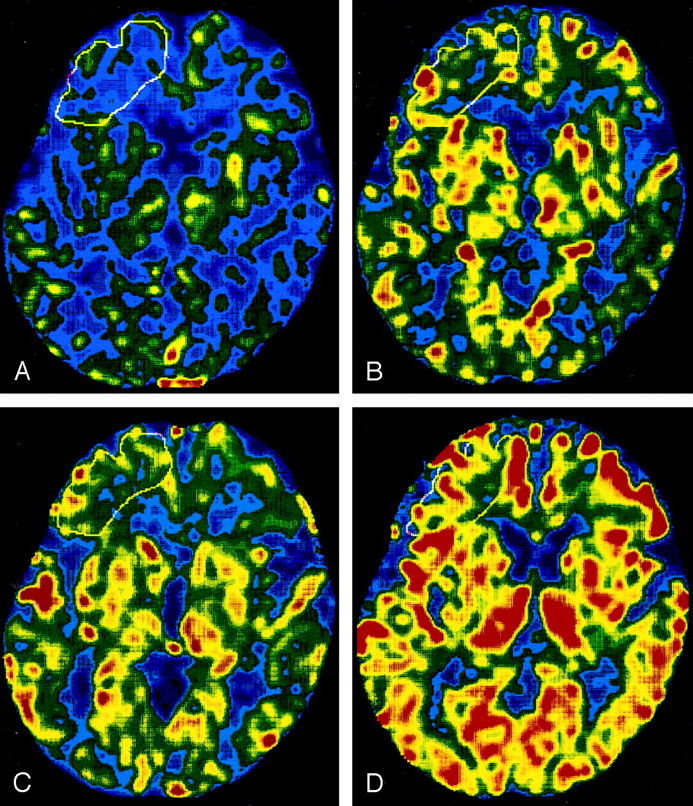
  • Fig 2.
    • Download figure
    • Open in new tab
    • Download powerpoint
    Fig 2.

    Case 21. rCBF maps for a Cognard type IV lesion in an asymptomatic patient.

    A and B, Before surgery, resting rCBF in the right frontal region (white outline) is low (A), and the response of rCBF to the acetazolamide challenge is limited (B).

    C and D, At 6 months after surgery, resting rCBF (C) and the response of rCBF (D) to the acetazolamide challenge is increased.

  • Fig 3.
    • Download figure
    • Open in new tab
    • Download powerpoint
    Fig 3.

    Differences in rCBF between groups according to Cognard angiographic classification.

    A, Resting rCBF significantly differs between type I and types III and IV.

    B, After acetazolamide challenge, rCBF significantly differs, even between type I and type II.

    C, Increase in rCBF due to acetazolamide (ΔF, or acetazolamide value minus resting value) is significantly different between type I and the other types.

Tables

  • Figures
    • View popup
    TABLE 1:

    Clinical summary of patients

    Patient/Age (y)/SexCognard ClassificationAffected SinusSymptomTreatment*
    DescriptionDue to Venous Hypertension?
    1/75/MIR sigmoidTinnitusNoTAE
    2/54/MISuperior sagittal sinusBulging scalp vesselsNoTAE
    3/78/FISuperior sagittal sinusAneurysm-like STA dilatationNoNone
    4/54/FIL cavernousNoneNANone
    5/58/MIR cavernousChemosis, diplopiaNoNone
    6/73/MIL cavernousDiplopiaNoNone
    7/75/FIL cavernousChemosisNoTVE
    8/62/MIIaL sigmoidTinnitusNoTAE, TVE
    9/75/MIIaR transverse sigmoidNoneNANone
    10/33/MIIbL transverse sigmoidHeadache, tinnitusYesTAE, TVE
    11/66/FIIbL cavernousDiplopia, disorientationYesTVE
    12/85/FIIbL cavernousChemosis, diplopiaNoTAE
    13/33/FIIa+bR transverse sigmoidHeadache, visual disturbanceYesTAE
    14/54/FIIa+bL transverse sigmoidDementiaYesTAE,TVE
    15/55/MIIIL transverse sigmoidHemorrhage, vertigo, convulsionYesTAE, TVE
    16/63/MIIIR transverse sigmoidHemorrhageYesTAE
    17/67/MIIIR transverse confluenceL lower quadrant homonymous anopsiaYesTAE, TVE
    18/72/FIIIL transverse confluenceDementiaYesTVE
    19/78/FIIIL transverseConvulsion, disturbed consciousnessYesTAE
    20/67/MIVL anterior fossaConvulsionYesSurgery
    21/74/MIVR anterior fossaNoneNASurgery
    • Note.—NA = Not applicable, STA = superficial temporal artery, TAE = transarterial embolization, TAV = transvenous embolization.

    • * All patients improved except for patients 3 and 12, whose condition was unchanged.

    • View popup
    TABLE 2:

    rCBF in patients with and those without symptoms caused by venous hypertension

    MeasureSymptomatic GroupAsymptomatic GroupP Value*
    Age (y)58.8 ± 15.369.4 ± 10.5.1118
    rCBF (mL/min/100g)
        Resting23.3 ± 5.133.2 ± 7.8.0092
        Acetazolamide27.1 ± 5.147.6 ± 10.9.0003
    ΔF†3.9 ± 2.714.4 ± 6.2.0002
    • * Mann-Whitney U test.

    • † Acetazolamide rCBF − resting rCBF.

    • View popup
    TABLE 3:

    Regional rCBF before and after treatment

    rCBF (ml/min/100g)Before Treatment6 Months After TreatmentP Value*
    Symptomatic patients (n = 10)
        Resting23.3 ± 5.126.8 ± 7.3.1849
        Acetazolamide27.1 ± 5.138.0 ± 12.4.0166
        ΔF†3.9 ± 2.711.2 ± 7.0.0367
    Asymptomatic patients (n = 4)
        Resting28.8 ± 8.230.1 ± 6.4>.05
        Acetazolamide38.4 ± 7.647.4 ± 8.6.0679
        ΔF†9.7 ± 2.617.2 ± 6.9.0679
    • * Wilcoxon signed rank test.

    • † Acetazolamide rCBF − resting rCBF.

PreviousNext
Back to top

In this issue

American Journal of Neuroradiology: 26 (5)
American Journal of Neuroradiology
Vol. 26, Issue 5
1 May 2005
  • Table of Contents
  • Index by author
Advertisement
Print
Download PDF
Email Article

Thank you for your interest in spreading the word on American Journal of Neuroradiology.

NOTE: We only request your email address so that the person you are recommending the page to knows that you wanted them to see it, and that it is not junk mail. We do not capture any email address.

Enter multiple addresses on separate lines or separate them with commas.
Regional Cerebral Blood Flow After Acetazolamide Challenge in Patients with Dural Arteriovenous Fistula: Simple Way to Evaluate Intracranial Venous Hypertension
(Your Name) has sent you a message from American Journal of Neuroradiology
(Your Name) thought you would like to see the American Journal of Neuroradiology web site.
CAPTCHA
This question is for testing whether or not you are a human visitor and to prevent automated spam submissions.
Cite this article
Jun Deguchi, Makoto Yamada, Hitoshi Kobata, Toshihiko Kuroiwa
Regional Cerebral Blood Flow After Acetazolamide Challenge in Patients with Dural Arteriovenous Fistula: Simple Way to Evaluate Intracranial Venous Hypertension
American Journal of Neuroradiology May 2005, 26 (5) 1101-1106;

Citation Manager Formats

  • BibTeX
  • Bookends
  • EasyBib
  • EndNote (tagged)
  • EndNote 8 (xml)
  • Medlars
  • Mendeley
  • Papers
  • RefWorks Tagged
  • Ref Manager
  • RIS
  • Zotero
0 Responses
Respond to this article
Share
Bookmark this article
Regional Cerebral Blood Flow After Acetazolamide Challenge in Patients with Dural Arteriovenous Fistula: Simple Way to Evaluate Intracranial Venous Hypertension
Jun Deguchi, Makoto Yamada, Hitoshi Kobata, Toshihiko Kuroiwa
American Journal of Neuroradiology May 2005, 26 (5) 1101-1106;
del.icio.us logo Twitter logo Facebook logo Mendeley logo
  • Tweet Widget
  • Facebook Like
  • Google Plus One

Jump to section

  • Article
    • Abstract
    • Methods
    • Results
    • Discussion
    • Conclusion
    • References
  • Figures & Data
  • Info & Metrics
  • Responses
  • References
  • PDF

Related Articles

  • No related articles found.
  • PubMed
  • Google Scholar

Cited By...

  • No citing articles found.
  • Crossref
  • Google Scholar

This article has not yet been cited by articles in journals that are participating in Crossref Cited-by Linking.

More in this TOC Section

  • Usefulness of Quantitative Susceptibility Mapping for the Diagnosis of Parkinson Disease
  • White Matter Alterations in the Brains of Patients with Active, Remitted, and Cured Cushing Syndrome: A DTI Study
  • Qualitative and Quantitative Analysis of MR Imaging Findings in Patients with Middle Cerebral Artery Stroke Implanted with Mesenchymal Stem Cells
Show more BRAIN

Similar Articles

Advertisement

Indexed Content

  • Current Issue
  • Accepted Manuscripts
  • Article Preview
  • Past Issues
  • Editorials
  • Editor's Choice
  • Fellows' Journal Club
  • Letters to the Editor
  • Video Articles

Cases

  • Case Collection
  • Archive - Case of the Week
  • Archive - Case of the Month
  • Archive - Classic Case

Special Collections

  • AJNR Awards
  • ASNR Foundation Special Collection
  • Most Impactful AJNR Articles
  • Photon-Counting CT
  • Spinal CSF Leak Articles (Jan 2020-June 2024)

More from AJNR

  • Trainee Corner
  • Imaging Protocols
  • MRI Safety Corner

Multimedia

  • AJNR Podcasts
  • AJNR Scantastics

Resources

  • Turnaround Time
  • Submit a Manuscript
  • Submit a Video Article
  • Submit an eLetter to the Editor/Response
  • Manuscript Submission Guidelines
  • Statistical Tips
  • Fast Publishing of Accepted Manuscripts
  • Graphical Abstract Preparation
  • Imaging Protocol Submission
  • Evidence-Based Medicine Level Guide
  • Publishing Checklists
  • Author Policies
  • Become a Reviewer/Academy of Reviewers
  • News and Updates

About Us

  • About AJNR
  • Editorial Board
  • Editorial Board Alumni
  • Alerts
  • Permissions
  • Not an AJNR Subscriber? Join Now
  • Advertise with Us
  • Librarian Resources
  • Feedback
  • Terms and Conditions
  • AJNR Editorial Board Alumni

American Society of Neuroradiology

  • Not an ASNR Member? Join Now

© 2025 by the American Society of Neuroradiology All rights, including for text and data mining, AI training, and similar technologies, are reserved.
Print ISSN: 0195-6108 Online ISSN: 1936-959X

Powered by HighWire