Advertisement

Main menu

  • Home
  • Content
    • Current Issue
    • Accepted Manuscripts
    • Article Preview
    • Past Issue Archive
    • Video Articles
    • AJNR Case Collection
    • Case of the Week Archive
    • Case of the Month Archive
    • Classic Case Archive
  • Special Collections
    • AJNR Awards
    • ASNR Foundation Special Collection
    • Most Impactful AJNR Articles
    • Photon-Counting CT
    • Spinal CSF Leak Articles (Jan 2020-June 2024)
  • Multimedia
    • AJNR Podcasts
    • AJNR SCANtastic
    • Trainee Corner
    • MRI Safety Corner
    • Imaging Protocols
  • For Authors
    • Submit a Manuscript
    • Submit a Video Article
    • Submit an eLetter to the Editor/Response
    • Manuscript Submission Guidelines
    • Statistical Tips
    • Fast Publishing of Accepted Manuscripts
    • Graphical Abstract Preparation
    • Imaging Protocol Submission
    • Author Policies
  • About Us
    • About AJNR
    • Editorial Board
    • Editorial Board Alumni
  • More
    • Become a Reviewer/Academy of Reviewers
    • Subscribers
    • Permissions
    • Alerts
    • Feedback
    • Advertisers
    • ASNR Home

User menu

  • Alerts
  • Log in

Search

  • Advanced search
American Journal of Neuroradiology

American Journal of Neuroradiology

ASHNR American Society of Functional Neuroradiology ASHNR American Society of Pediatric Neuroradiology ASSR
  • Alerts
  • Log in

Advanced Search

  • Home
  • Content
    • Current Issue
    • Accepted Manuscripts
    • Article Preview
    • Past Issue Archive
    • Video Articles
    • AJNR Case Collection
    • Case of the Week Archive
    • Case of the Month Archive
    • Classic Case Archive
  • Special Collections
    • AJNR Awards
    • ASNR Foundation Special Collection
    • Most Impactful AJNR Articles
    • Photon-Counting CT
    • Spinal CSF Leak Articles (Jan 2020-June 2024)
  • Multimedia
    • AJNR Podcasts
    • AJNR SCANtastic
    • Trainee Corner
    • MRI Safety Corner
    • Imaging Protocols
  • For Authors
    • Submit a Manuscript
    • Submit a Video Article
    • Submit an eLetter to the Editor/Response
    • Manuscript Submission Guidelines
    • Statistical Tips
    • Fast Publishing of Accepted Manuscripts
    • Graphical Abstract Preparation
    • Imaging Protocol Submission
    • Author Policies
  • About Us
    • About AJNR
    • Editorial Board
    • Editorial Board Alumni
  • More
    • Become a Reviewer/Academy of Reviewers
    • Subscribers
    • Permissions
    • Alerts
    • Feedback
    • Advertisers
    • ASNR Home
  • Follow AJNR on Twitter
  • Visit AJNR on Facebook
  • Follow AJNR on Instagram
  • Join AJNR on LinkedIn
  • RSS Feeds

AJNR is seeking candidates for the AJNR Podcast Editor. Read the position description.

OtherBRAIN

Active Hemorrhage into a Postresection Cavity Detected by Neuro-CT Angiography

Matthew T. Walker, Anoop Wattamwar, David Mellman and Jae Mo
American Journal of Neuroradiology May 2005, 26 (5) 1163-1165;
Matthew T. Walker
  • Find this author on Google Scholar
  • Find this author on PubMed
  • Search for this author on this site
Anoop Wattamwar
  • Find this author on Google Scholar
  • Find this author on PubMed
  • Search for this author on this site
David Mellman
  • Find this author on Google Scholar
  • Find this author on PubMed
  • Search for this author on this site
Jae Mo
  • Find this author on Google Scholar
  • Find this author on PubMed
  • Search for this author on this site
  • Article
  • Figures & Data
  • Info & Metrics
  • Responses
  • References
  • PDF
Loading

Abstract

Summary: We describe a case demonstrating active extravasation of contrast material into a hematoma resection cavity during CT angiography (CTA) that necessitated emergent reexploration, decompression, and hemostatic control. Our case highlights the value of neuro-CTA in the immediate postoperative setting and describes another scenario where CTA has added value. Prompt recognition of contrast extravasation is critical to the diagnosis and ultimately affects the quality of patient care.

CT angiography (CTA) is a fast, minimally invasive, and inexpensive neurovascular imaging technique that is gaining in popularity because of its availability and spatial resolution. As a relatively new technique, its technical strengths (spatial resolution) and limitations (temporal resolution) are being assessed in a variety of clinical scenarios, including aneurysms, vascular malformations, steno-occlusive disease, vasospasm, and trauma (1–8). Active extravasation, which indicates active bleeding, has been described during neuro-CTA examinations including ruptured aneurysms, subdural hematomas, and spontanteous parenchymal hematomas and is generally regarded as a medical emergency. Recognition and accurate interpretation of active extravasation can help localize areas of active bleeding.

Case Description

A 22-year-old man was thrown from a motorcycle and suffered severe head and facial trauma. The patient’s Glasgow coma score at the scene was 3 and improved to 6 in the emergency department. A noncontrast CT (NCCT) examination demonstrated a large left frontal and basal ganglionic hematoma with subfalcine herniation and brain stem compression (Fig 1A). An emergency craniotomy was performed to decompress the hematoma and achieve hemostasis. After reattaching the bone flap, the patient deteriorated clinically. The bone flap was removed and emergent NCCT and neuro-CTA were performed to assess the resection cavity. The NCCT showed reaccumulation of the hematoma (Fig 1B). The source data set from the CTA demonstrated multiple foci of high attenuation fluid (contrast) within the reaccumulated hematoma, including anteroinferiorly where the lenticulostriates reside (Fig 2A and B). The contrast foci were nonanatomic and did not conform to a known vascular structure. In addition, the contrast layered dependently in a fluid-fluid pattern. The patient was returned to the operating room for reexploration. The hematoma was again evacuated and multiple small active bleeders were cauterized. Two actively bleeding lenticulostriates were observed anteroinferiorly: one was clipped, and the other was cauterized. These lenticulostriates corresponded to one of the areas of active extravasation. The patient survived the injury and was transferred to a rehabilitation facility.

Fig 1.
  • Download figure
  • Open in new tab
  • Download powerpoint
Fig 1.

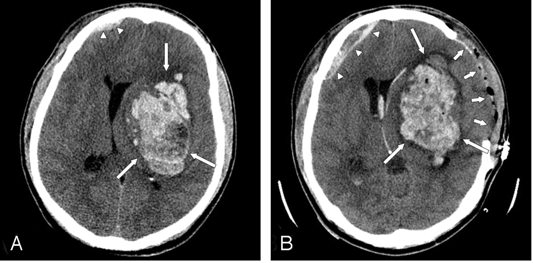
A, Axial CT scan at the level of the 3rd ventricle on admission showed a large left intrparenchymal hematoma (arrows) with vasogenic edema subflacine herniation. A small right frontal subdural hematoma is noted (arrowheads).

B, Axial CT scan at the level of the 3rd ventricle immediately after surgical decompression and subsequent clinical decline showed a left frontal craniectomy (short arrows), reaccumulation of the hematoma (long arrows), persistent subflacine herniation, and a slightly larger right frontal subdural (arrowheads).

Fig 2.
  • Download figure
  • Open in new tab
  • Download powerpoint
Fig 2.

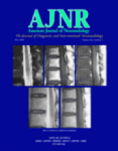
A, Source axial CTA image at the level of the bodies of the lateral ventricles showed a layering fluid collection in the ventral aspect of the hematoma including a dependent hyperattenuated component dorsally (long arrow) and a nondependent hypoattenuated component ventrally (short arrow). The hyperattenuated component represented contrast whereas the non-dependent component likely represents plasma.

B, Sagittal multiplanar reformatted image of the CTA source data set showed multiple areas of “nonanatomic puddling” of contrast (circles) within the hematoma indicating active extravastion during the CTA examination.

Discussion

Traumatic intracranial lesions can be broadly characterized as primary or secondary lesions, depending upon their temporal relationship with the initial injury (9). Primary lesions are a direct result of the traumatic force and can be characterized as neuronal injuries, hemorrhages, vascular injuries, or pia-arachnoid injuries. Primary hemorrhages occur in multiple spaces including subdural, epidural, subarachnoid, intraventricular, and intraparenchymal. In the blunt trauma patient, intraparenchymal hematomas arise due to sudden acceleration-deceleration and rotational forces, which then result in the disruption of arteries and/or veins such as the lenticulostriates and are frequently identified in the frontotemporal white matter or deep gray nuclei (9). Dissections or tears of small vessels such as the lenticulostriates are rare and difficult to detect because of current spatial resolution limitations and practice patterns: traumatic intracranial parenchymal hemorrhages do not routinely undergo vascular imaging. Although direct visualization of a vascular injury to a lenticulostriate is beyond the spatial and temporal resolution of current noninvasive techniques (CTA, MR angiography), the secondary sign of “contrast extravasation” can be identified.

Prompt recognition of a vascular injury and active hemorrhage is crucial for optimal patient care. Detection of active extravasation has been widely reported on conventional angiography in the setting of acute nontraumatic intracranial hemorrhage (10, 11). There have been several case reports of active extravasation detected during neuro-CTA including aneurysms, subdural hemorrhage, and nontraumatic intracranial hemorrhage (12–15).

Gosselin and Vieco (12) described two cases of active hemorrhage from ruptured aneurysms demonstrated on CT angiography. In those cases, irregularity of the wall of the aneurysm, nonanatomic collection of contrast, and contrast opacification of the preexisting hematoma on the delayed scan were seen. The term “hematoma opacification sign” was used to describe opacification of a previously nonopacified hematoma adjacent to an aneurysm. They also stressed the nonanatomic appearance of such a collection as critical in the accurate assessment of this finding. The outcome was fatal in both cases. Nakada et al (13) described a ruptured anterior communicating artery aneurysm that reruptured during the initial diagnostic CTA with obvious contrast extravasation around the anterior communicating artery. The rerupture was fatal.

Kinoshita et al (14) described contrast extravasation into a subdural hematoma during a CTA and accurately located the bleeding cortical artery.

Murai et al (15) evaluated the usefulness of CTA to detect active extravasation in the setting of acute nontraumatic spontaneous intracranial hemorrhage. In 31 consecutive patients, five cases of active extravasation were detected, three of which showed hematoma enlargement. Of the 26 cases without extravasation, the hematomas remained stable on follow-up imaging. They concluded that 3D-CTA was valuable in the detection of persistent hemorrhage in the subset of patients studied.

In the current case, initial decompression was performed, followed by reaccumulation of the hematoma. CTA demonstrated multiple areas of contrast “puddling,” which represented active bleeding and prompted emergent reexploration. Some of the puddling was deep in the cavity in the expected location of the lenticulostriate vessels. The CTA identified areas of active bleeding and helped guide the neurosurgeon to areas of active bleeding and eventual hemostatic control.

As CT technology advances—including higher section profiles, longer detector configurations and increased computing power—the temporal and spatial resolution of CTA will improve. Although desirable in most situations, improved temporal resolution can be a source of error for the detection of active extravasation when the leak is slow instead of brisk. In this circumstance, extravasation may only be detected on a delayed CT scan after a neuro-CTA (B Dillon, personal communication). If an active hemorrhage is suspected, adding a delayed CT of the brain after neuro-CTA may help reduce false-negative examinations and improve specificity.

Conclusion

Identifying active extravasation of contrast during a neuro-CTA is critical for timely management and optimal patient care. The sine qua non of active extravasation into a hematoma is “nonanatomic puddling of contrast” and can be recognized on the source CTA data set. A delayed CT scan after neuro-CTA may help identify slower hemorrhages and improve specificity.

References

  1. ↵
    Chappell ET, Moure FC, Good MC. Comparison of computed tomographic angiography with digital subtraction angiography in the diagnosis of cerebral aneurysms: a meta-analysis. Neurosurgery 2003;52:624–631
    CrossRefPubMed
  2. Jayaraman MV, Mayo-Smith WW, Tung GA, et al. Detection of intracranial aneurysms: multi-detector row CT angiography compared with DSA. Radiology 2004;230:510–518
    PubMed
  3. Sasiadek M, Hendrich B, Turek T, et al. Our own experience with CT angiography in early diagnosis of cerebral vascular malformations. Neurol Neurochir Pol 2000;34(6 suppl):48–55
  4. Tanaka H, Numaguchi Y, Konno S, et al. Initial experience with helical CT and 3D reconstruction in therapeutic planning of cerebral AVMs: comparison with 3D time-of-flight MRA and digital subtraction angiography. J Comput Assist Tomogr 1997;21:811–817
    CrossRefPubMed
  5. Skutta B, Furst G, Eilers J, et al. Intracranial stenoocclusive disease: double-detector helical CT angiography versus digital subtraction angiography. AJNR Am J Neuroradiol 1999;20:791–799
    Abstract/FREE Full Text
  6. Anderson GB, Ashforth R, Steinke DE, Findlay JM. CT angiography for the detection of cerebral vasospasm in patients with acute subarachnoid hemorrhage. AJNR Am J Neuroradiol 2000;21:1011–1015
    Abstract/FREE Full Text
  7. Otawara Y, Ogasawara K, Ogawa A, et al. Evaluation of vasospasm after subarachnoid hemorrhage by use of multislice computed tomographic angiography. Neurosurgery 2002;51:939–943
    CrossRefPubMed
  8. ↵
    Hollingworth W, Nathens AB, Kanne JP, et al. The diagnostic accuracy of computed tomography angiography for traumatic or atherosclerotic lesions of the carotid and vertebral arteries: a systematic review. Eur J Radiol 2003;48:88–102
    CrossRefPubMed
  9. ↵
    Gentry LR. Head trauma. In: Atlas SW, ed. Magnetic resonance imaging of the brain and spine. 3rd ed. Philadelphia: Lippincott, Williams & Wilkins;2002 :1059–1098
  10. ↵
    Komiyama M, Yasui T, Tamura K, et al. Simultaneous bleeding from multiple lenticulostriate arteries in hypertensive intracerebral haemorrhage. Neuroradiology 1995;37:129–130
    PubMed
  11. ↵
    Mizukami M, Araki G, Mihara H, et al. Arteriographically visualized extravasation in hypertensive intracerebral hemorrhage: report of seven cases. Stroke 1972;3:527–537
    Abstract/FREE Full Text
  12. ↵
    Gosselin MV, Vieco PT. Active hemorrhage of intracranial aneurysm: diagnosis by CT angiography. J Comput Assist Tomogr 1997;21:22–24
    CrossRefPubMed
  13. ↵
    Nakada M, Akaike S, Futami K. Rupture of an aneurysm during three-dimensional computerized tomography. J Neurosurg 2000;93:900
    PubMed
  14. ↵
    Kinoshita Y, Machi T, Satoh Y, et al. Extravation of contrast media in acute cerebral hematoma during three-dimensional CT angiography: a case report. No Shinkei Geka 1999;27:157–161
    PubMed
  15. ↵
    Murai Y, Takaki R, Ikeda Y, et al. Three-dimensional computerized tomography angiography in patients with hyperacute intracerebral hemorrhage. J Neurosurg 1999;91:424–431
    PubMed
  • Received July 28, 2004.
  • Accepted after revision August 6, 2004.
  • Copyright © American Society of Neuroradiology
PreviousNext
Back to top

In this issue

American Journal of Neuroradiology: 26 (5)
American Journal of Neuroradiology
Vol. 26, Issue 5
1 May 2005
  • Table of Contents
  • Index by author
Advertisement
Print
Download PDF
Email Article

Thank you for your interest in spreading the word on American Journal of Neuroradiology.

NOTE: We only request your email address so that the person you are recommending the page to knows that you wanted them to see it, and that it is not junk mail. We do not capture any email address.

Enter multiple addresses on separate lines or separate them with commas.
Active Hemorrhage into a Postresection Cavity Detected by Neuro-CT Angiography
(Your Name) has sent you a message from American Journal of Neuroradiology
(Your Name) thought you would like to see the American Journal of Neuroradiology web site.
CAPTCHA
This question is for testing whether or not you are a human visitor and to prevent automated spam submissions.
Cite this article
Matthew T. Walker, Anoop Wattamwar, David Mellman, Jae Mo
Active Hemorrhage into a Postresection Cavity Detected by Neuro-CT Angiography
American Journal of Neuroradiology May 2005, 26 (5) 1163-1165;

Citation Manager Formats

  • BibTeX
  • Bookends
  • EasyBib
  • EndNote (tagged)
  • EndNote 8 (xml)
  • Medlars
  • Mendeley
  • Papers
  • RefWorks Tagged
  • Ref Manager
  • RIS
  • Zotero
0 Responses
Respond to this article
Share
Bookmark this article
Active Hemorrhage into a Postresection Cavity Detected by Neuro-CT Angiography
Matthew T. Walker, Anoop Wattamwar, David Mellman, Jae Mo
American Journal of Neuroradiology May 2005, 26 (5) 1163-1165;
del.icio.us logo Twitter logo Facebook logo Mendeley logo
  • Tweet Widget
  • Facebook Like
  • Google Plus One

Jump to section

  • Article
    • Abstract
    • Case Description
    • Discussion
    • Conclusion
    • References
  • Figures & Data
  • Info & Metrics
  • Responses
  • References
  • PDF

Related Articles

  • No related articles found.
  • PubMed
  • Google Scholar

Cited By...

  • Contrast extravasation on CT angiography predicts hematoma expansion in intracerebral hemorrhage
  • Crossref
  • Google Scholar

This article has not yet been cited by articles in journals that are participating in Crossref Cited-by Linking.

More in this TOC Section

  • Optimal MRI Sequence for Identifying Occlusion Location in Acute Stroke: Which Value of Time-Resolved Contrast-Enhanced MRA?
  • Evaluating the Effects of White Matter Multiple Sclerosis Lesions on the Volume Estimation of 6 Brain Tissue Segmentation Methods
  • Quiet PROPELLER MRI Techniques Match the Quality of Conventional PROPELLER Brain Imaging Techniques
Show more BRAIN

Similar Articles

Advertisement

Indexed Content

  • Current Issue
  • Accepted Manuscripts
  • Article Preview
  • Past Issues
  • Editorials
  • Editor's Choice
  • Fellows' Journal Club
  • Letters to the Editor
  • Video Articles

Cases

  • Case Collection
  • Archive - Case of the Week
  • Archive - Case of the Month
  • Archive - Classic Case

Special Collections

  • AJNR Awards
  • ASNR Foundation Special Collection
  • Most Impactful AJNR Articles
  • Photon-Counting CT
  • Spinal CSF Leak Articles (Jan 2020-June 2024)

More from AJNR

  • Trainee Corner
  • Imaging Protocols
  • MRI Safety Corner

Multimedia

  • AJNR Podcasts
  • AJNR Scantastics

Resources

  • Turnaround Time
  • Submit a Manuscript
  • Submit a Video Article
  • Submit an eLetter to the Editor/Response
  • Manuscript Submission Guidelines
  • Statistical Tips
  • Fast Publishing of Accepted Manuscripts
  • Graphical Abstract Preparation
  • Imaging Protocol Submission
  • Evidence-Based Medicine Level Guide
  • Publishing Checklists
  • Author Policies
  • Become a Reviewer/Academy of Reviewers
  • News and Updates

About Us

  • About AJNR
  • Editorial Board
  • Editorial Board Alumni
  • Alerts
  • Permissions
  • Not an AJNR Subscriber? Join Now
  • Advertise with Us
  • Librarian Resources
  • Feedback
  • Terms and Conditions
  • AJNR Editorial Board Alumni

American Society of Neuroradiology

  • Not an ASNR Member? Join Now

© 2025 by the American Society of Neuroradiology All rights, including for text and data mining, AI training, and similar technologies, are reserved.
Print ISSN: 0195-6108 Online ISSN: 1936-959X

Powered by HighWire