
Article Figures & Data
Figures
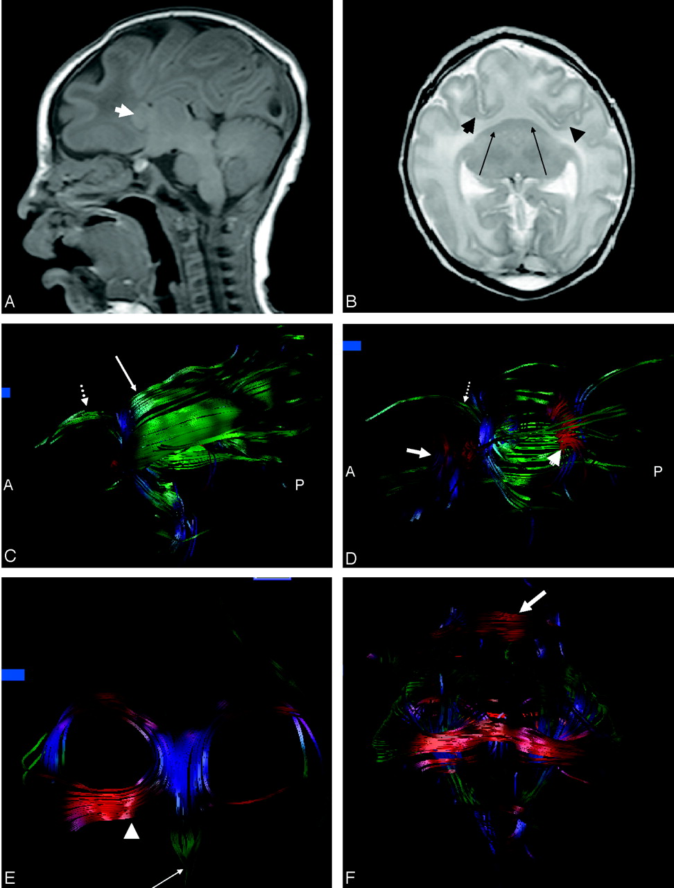
- Fig 1.
A, Sagittal T1-weighted images show preservation of the infratentorial contents. There is no third ventricle or definite callosal fibers. Arrow indicates fused caudates.
B, Axial T2-weighted image shows marked frontal lobe hypoplasia resulting in anterior and medial displacement of the sylvian fissures (arrowheads). The fused crescent-shaped caudates are indicated by the arrows. Note the fused thalami.
C, On the sagittal image derived from fiber tracking, there is no demarcation between the inferior and superior longitudinal fasciculus. Note corticospinal tracts (dotted arrow) arching anteriorly toward the expected location of the motor cortex. Some fibers (long arrow) of the posterior limbs of the internal capsules are seen arching dorsally cranial to the frontooccipital fasciculus; these are presumed to be projection fibers to the sensory cortex on the ventrolateral portion of the holosphere.
D, On the sagittal view, some of the fibers have been removed from the frontooccipital fasciculus to allow visualization of the splenium (arrowhead). The dotted arrow indicates corticospinal tracts, and the long arrow indicates fused caudates.
E, On the coronal image derived from fiber tracking, the thickened dysplastic fornices are foreshortened in an anteroposterior direction. The rostral extension of the fornices divide into horizontally oriented asymmetric paramedian structures similar to the posterior projections of the anterior commissure (arrowhead) and a single median structure similar to the precommissural fornix (arrow) but thicker and longer than the usual appearance of the precommissural fornix in a neonate.
F, The fused caudates have been removed from this coronal image to allow visualization of the midline fibers connecting the frontooccipital fasciculi. The fornices can be seen dorsal to the connecting fibers. A band of transversely oriented white matter is seen over the frontal convexity (arrow).
In this issue


Jump to section
Related Articles
Cited By...
- No citing articles found.