Advertisement

Main menu

  • Home
  • Content
    • Current Issue
    • Accepted Manuscripts
    • Article Preview
    • Past Issue Archive
    • Video Articles
    • AJNR Case Collection
    • Case of the Week Archive
    • Case of the Month Archive
    • Classic Case Archive
  • Special Collections
    • AJNR Awards
    • ASNR Foundation Special Collection
    • Most Impactful AJNR Articles
    • Photon-Counting CT
    • Spinal CSF Leak Articles (Jan 2020-June 2024)
  • Multimedia
    • AJNR Podcasts
    • AJNR SCANtastic
    • Trainee Corner
    • MRI Safety Corner
    • Imaging Protocols
  • For Authors
    • Submit a Manuscript
    • Submit a Video Article
    • Submit an eLetter to the Editor/Response
    • Manuscript Submission Guidelines
    • Statistical Tips
    • Fast Publishing of Accepted Manuscripts
    • Graphical Abstract Preparation
    • Imaging Protocol Submission
    • Author Policies
  • About Us
    • About AJNR
    • Editorial Board
    • Editorial Board Alumni
  • More
    • Become a Reviewer/Academy of Reviewers
    • Subscribers
    • Permissions
    • Alerts
    • Feedback
    • Advertisers
    • ASNR Home

User menu

  • Alerts
  • Log in

Search

  • Advanced search
American Journal of Neuroradiology

American Journal of Neuroradiology

ASHNR American Society of Functional Neuroradiology ASHNR American Society of Pediatric Neuroradiology ASSR
  • Alerts
  • Log in

Advanced Search

  • Home
  • Content
    • Current Issue
    • Accepted Manuscripts
    • Article Preview
    • Past Issue Archive
    • Video Articles
    • AJNR Case Collection
    • Case of the Week Archive
    • Case of the Month Archive
    • Classic Case Archive
  • Special Collections
    • AJNR Awards
    • ASNR Foundation Special Collection
    • Most Impactful AJNR Articles
    • Photon-Counting CT
    • Spinal CSF Leak Articles (Jan 2020-June 2024)
  • Multimedia
    • AJNR Podcasts
    • AJNR SCANtastic
    • Trainee Corner
    • MRI Safety Corner
    • Imaging Protocols
  • For Authors
    • Submit a Manuscript
    • Submit a Video Article
    • Submit an eLetter to the Editor/Response
    • Manuscript Submission Guidelines
    • Statistical Tips
    • Fast Publishing of Accepted Manuscripts
    • Graphical Abstract Preparation
    • Imaging Protocol Submission
    • Author Policies
  • About Us
    • About AJNR
    • Editorial Board
    • Editorial Board Alumni
  • More
    • Become a Reviewer/Academy of Reviewers
    • Subscribers
    • Permissions
    • Alerts
    • Feedback
    • Advertisers
    • ASNR Home
  • Follow AJNR on Twitter
  • Visit AJNR on Facebook
  • Follow AJNR on Instagram
  • Join AJNR on LinkedIn
  • RSS Feeds

AJNR is seeking candidates for the AJNR Podcast Editor. Read the position description.

Research ArticleHEAD AND NECK

A Potential Pitfall of MR Imaging for Assessing Mandibular Invasion of Squamous Cell Carcinoma in the Oral Cavity

A. Imaizumi, N. Yoshino, I. Yamada, K. Nagumo, T. Amagasa, K. Omura, N. Okada and T. Kurabayashi
American Journal of Neuroradiology January 2006, 27 (1) 114-122;
A. Imaizumi
  • Find this author on Google Scholar
  • Find this author on PubMed
  • Search for this author on this site
N. Yoshino
  • Find this author on Google Scholar
  • Find this author on PubMed
  • Search for this author on this site
I. Yamada
  • Find this author on Google Scholar
  • Find this author on PubMed
  • Search for this author on this site
K. Nagumo
  • Find this author on Google Scholar
  • Find this author on PubMed
  • Search for this author on this site
T. Amagasa
  • Find this author on Google Scholar
  • Find this author on PubMed
  • Search for this author on this site
K. Omura
  • Find this author on Google Scholar
  • Find this author on PubMed
  • Search for this author on this site
N. Okada
  • Find this author on Google Scholar
  • Find this author on PubMed
  • Search for this author on this site
T. Kurabayashi
  • Find this author on Google Scholar
  • Find this author on PubMed
  • Search for this author on this site
  • Article
  • Figures & Data
  • Info & Metrics
  • Responses
  • References
  • PDF
Loading

Article Figures & Data

Figures

  • Tables
  • Fig 1.
    • Download figure
    • Open in new tab
    • Download powerpoint
    Fig 1.

    A 67-year-old man with left floor-of-mouth carcinoma, showing a true-positive result for cortical invasion with both MR imaging and CT.

    A, Axial T1-weighted MR image (560/14).

    B, Axial bone algorithm CT image.

    Both MR and CT image reveal destruction of the cortex (arrows) adjacent to the tumor mass. These findings were histopathologically confirmed.
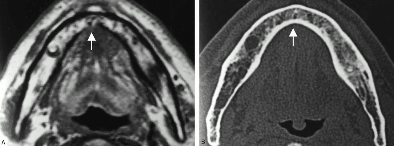
  • Fig 2.
    • Download figure
    • Open in new tab
    • Download powerpoint
    Fig 2.

    A 48-year-old woman with left floor-of-mouth carcinoma showing a true-negative result for cortical invasion with both MR imaging and CT.

    A, Axial T1-weighted MR image (560/14).

    B, Axial bone algorithm CT image.

    Both MR and CT image reveal intact lingual cortex (arrows) adjacent to the tumor mass. Histopathologic examination after marginal mandibulectomy confirmed no tumor invasion into the mandible.

  • Fig 3.
    • Download figure
    • Open in new tab
    • Download powerpoint
    Fig 3.

    A 70-year-old man with anterior floor-of-mouth carcinoma showing a false-positive result with MR imaging and true-negative result with CT for cortical invasion.

    A, Axial T1-weighted MR image (700/14).

    B, Axial bone algorithm CT image.

    The lingual cortex (arrow) is suspected to be involved by the tumor mass on T1-weighted MR image, whereas it is intact on CT. Histopathologic examination after marginal mandibulectomy confirmed no tumor invasion into the mandible. Chemical shift artifacts induced by bone marrow fat are considered to account for the false-positive result with MR imaging. That is, the black line of the cortex adjacent to the tumor mass is thought to be obscured by spatial misplacement of bone marrow fat.

  • Fig 4.
    • Download figure
    • Open in new tab
    • Download powerpoint
    Fig 4.

    A 46-year-old man with anterior floor-of mouth carcinoma showing a false-positive result with MR imaging and true-negative result with CT for cortical invasion.

    A, Axial T1-weighted MR image (560/14).

    B, Axial bone algorithm CT image.

    The lingual cortex (arrow) is suspected to be involved by the tumor mass on T1-weighted MR image, whereas it is intact on CT. Histopathologic examination after marginal mandibulectomy confirmed no tumor invasion into the mandible. As in Fig 3, misevaluation with MR imaging is considered to be due to chemical shift artifacts.
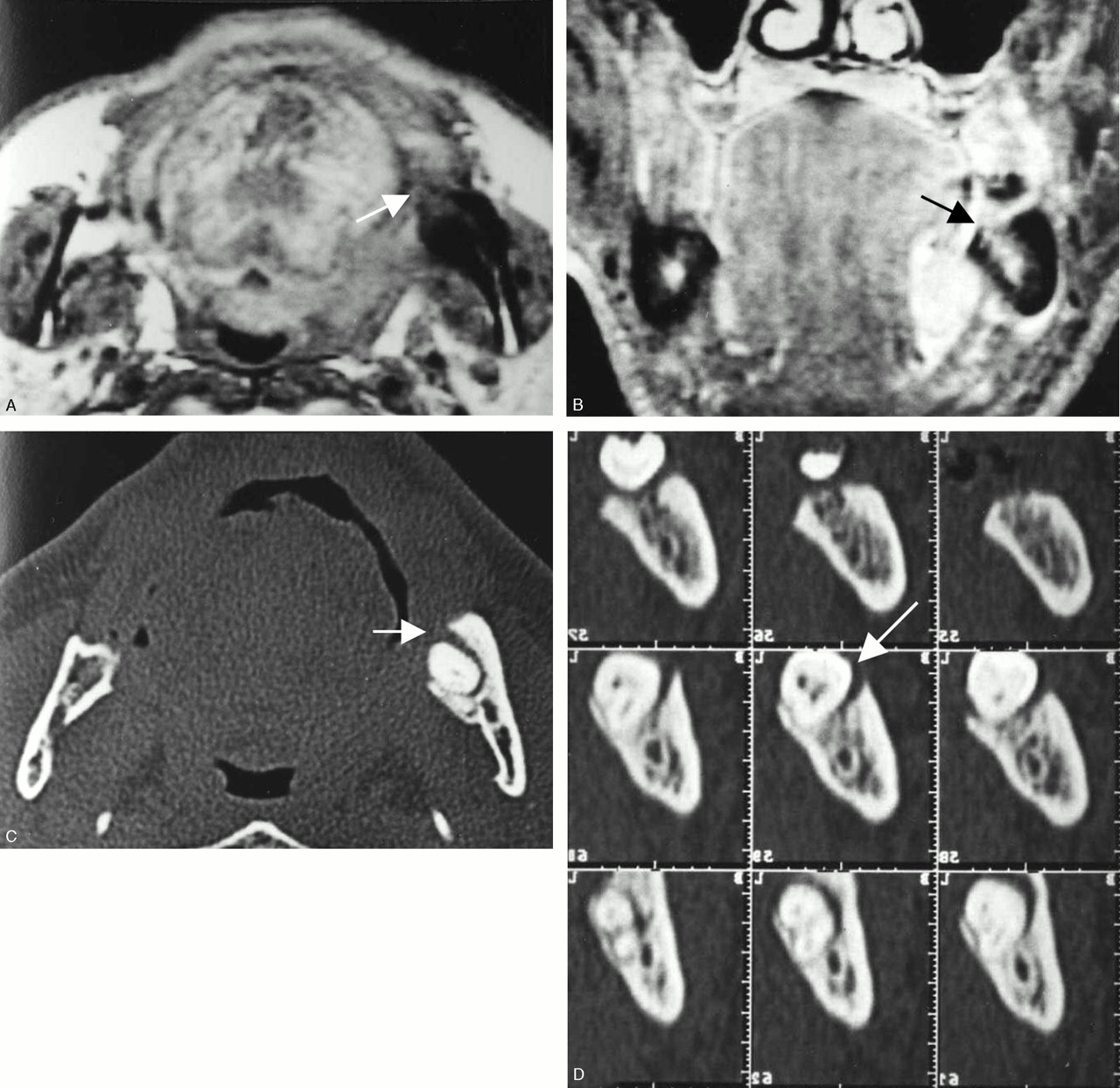
  • Fig 5.
    • Download figure
    • Open in new tab
    • Download powerpoint
    Fig 5.

    A 62-year-old man with left floor-of mouth carcinoma showing a false-positive result with MR imaging and true-negative result with CT for cortical invasion.

    A, Axial T1-weighted image (560/14).

    B, Contrast-enhanced coronal T1-weighted image (700/14).

    C, Axial bone algorithm CT image.

    D, Dental CT reformatted images.

    MR images reveal diffuse abnormal signal intensity of the bone marrow in the left molar region (A and B, arrow), strongly suggestive of tumor invasion. On the other hand, CT reveals bone absorption with relatively smooth margins around the tooth root (C and D, arrow), suggestive of periodontal disease. Histopathogic examination after marginal mandibulectomy confirmed no tumor invasion into the mandible.

  • Fig 6.
    • Download figure
    • Open in new tab
    • Download powerpoint
    Fig 6.
    • Download figure
    • Open in new tab
    • Download powerpoint
    Fig 6.

    A 69-year-old man with left lower gingival carcinoma showing a false-positive result with MR imaging and true-negative result with CT for the involvement of inferior alveolar canal.

    A, Axial T1-weighted image (560/14).

    B, Contrast-enhanced coronal T1-weighted image (520/14).

    C, Axial bone algorithm CT image.

    D, Dental CT reformatted images.

    E, Photomicrograph of the surgical specimen (Hematoxylin and eosin stain; original magnification, ×5.3)

    MR images reveal abnormal signal intensity of bone marrow in the left molar region reaching the inferior alveolar canal (B, arrow), suggestive of inferior alveolar canal involvement. However, no involvement of the canal (D, arrow) is suspected on CT images. The photomicrograph of the surgical specimen reveals tumor invasion into the bone marrow with small focal alveolar bone absorption. Reactive fibrous change spreads in the bone marrow and reaches the inferior alveolar canal (E, arrow), which accounts for the overestimating of tumor extent with MR imaging.

  • Fig 7.
    • Download figure
    • Open in new tab
    • Download powerpoint
    Fig 7.
    • Download figure
    • Open in new tab
    • Download powerpoint
    Fig 7.

    MR images of a normal volunteer obtained by different imaging methods

    A, T1-weighted image (560/14) with ordinary setting of phase- and frequency-encoding directions.

    B, T1-weighted image (560/14) in which phase- and frequency-encoding directions are swapped.

    C, T1-weighted image (560/14) with fat saturation. ν = frequency-encoding direction

    Despite the absence of tumor mass, the lingual cortex of the mandible is obscured in A (arrowhead). However, it is intact when phase- and frequency-directions are swapped (B) or fat signal intensity is suppressed (C). These findings confirm that the cortical defect in A is attributed to chemical shift artifact of bone marrow fat. Spatial misplacement of the fat occurs along the y axis direction in A and along the x axis direction in B.

Tables

  • Figures
    • View popup
    Table 1:

    Diagnostic criteria of MR imaging and CT in evaluating tumor invasion into the mandible

    Mandibular cortical invasion
        MRI, CT: Defect of the cortical bone adjacent to the tumor mass
    Bone marrow involvement
        MRI: Abnormal signal intensity of bone marrow* contiguous to the cortical defect
        CT: Trabecular disruption contiguous to the cortical defect
    Inferior alveolar canal involvement
        MRI, CT: Bone marrow involvement reaching the inferior alveolar canal
    • * Hypointense on T1-weighted and hyperintense on fat-suppressed T2-weighted image, with enhancement after contrast administration.

    • View popup
    Table 2:

    Diagnostic accuracy of MR imaging and CT in evaluating bone invasion

    TPTNFPFNSensitivitySpecificityPPVNPVAccuracy
    No. of Cases%
    Mandibular cortical invasion
        MR imaging24141219654*679374
        CT25233010088*8910094
    Bone marrow involvement
        MR imaging2421519681839588
        CT252330100888910094
    Inferior alveolar canal involvement
        MR imaging53214010070†2610073
        CT5442010096†7110096
    • Note:—TP indicates true-positive; TN, true-negative; FP, false-positive; FN, false-negative; PPV, positive predictive value; NPV, negative predictive value.

    • * P = .004;

    • † P = .002 (McNemar test).

PreviousNext
Back to top

In this issue

American Journal of Neuroradiology: 27 (1)
American Journal of Neuroradiology
Vol. 27, Issue 1
January, 2006
  • Table of Contents
  • Index by author
Advertisement
Print
Download PDF
Email Article

Thank you for your interest in spreading the word on American Journal of Neuroradiology.

NOTE: We only request your email address so that the person you are recommending the page to knows that you wanted them to see it, and that it is not junk mail. We do not capture any email address.

Enter multiple addresses on separate lines or separate them with commas.
A Potential Pitfall of MR Imaging for Assessing Mandibular Invasion of Squamous Cell Carcinoma in the Oral Cavity
(Your Name) has sent you a message from American Journal of Neuroradiology
(Your Name) thought you would like to see the American Journal of Neuroradiology web site.
CAPTCHA
This question is for testing whether or not you are a human visitor and to prevent automated spam submissions.
Cite this article
A. Imaizumi, N. Yoshino, I. Yamada, K. Nagumo, T. Amagasa, K. Omura, N. Okada, T. Kurabayashi
A Potential Pitfall of MR Imaging for Assessing Mandibular Invasion of Squamous Cell Carcinoma in the Oral Cavity
American Journal of Neuroradiology Jan 2006, 27 (1) 114-122;

Citation Manager Formats

  • BibTeX
  • Bookends
  • EasyBib
  • EndNote (tagged)
  • EndNote 8 (xml)
  • Medlars
  • Mendeley
  • Papers
  • RefWorks Tagged
  • Ref Manager
  • RIS
  • Zotero
0 Responses
Respond to this article
Share
Bookmark this article
A Potential Pitfall of MR Imaging for Assessing Mandibular Invasion of Squamous Cell Carcinoma in the Oral Cavity
A. Imaizumi, N. Yoshino, I. Yamada, K. Nagumo, T. Amagasa, K. Omura, N. Okada, T. Kurabayashi
American Journal of Neuroradiology Jan 2006, 27 (1) 114-122;
del.icio.us logo Twitter logo Facebook logo Mendeley logo
  • Tweet Widget
  • Facebook Like
  • Google Plus One

Jump to section

  • Article
    • Abstract
    • Methods
    • Results
    • Discussion
    • Conclusion
    • References
  • Figures & Data
  • Info & Metrics
  • Responses
  • References
  • PDF

Related Articles

  • No related articles found.
  • PubMed
  • Google Scholar

Cited By...

  • Anterior mandible canal communications: a potential portal of entry for tumour spread
  • Crossref
  • Google Scholar

This article has not yet been cited by articles in journals that are participating in Crossref Cited-by Linking.

More in this TOC Section

  • Correlation of Apparent Diffusion Coefficient at 3T with Prognostic Parameters of Retinoblastoma
  • Parathyroid Lesions: Characterization with Dual-Phase Arterial and Venous Enhanced CT of the Neck
  • MR Evaluation of Sinonasal Angiomatous Polyp
Show more HEAD AND NECK

Similar Articles

Advertisement

Indexed Content

  • Current Issue
  • Accepted Manuscripts
  • Article Preview
  • Past Issues
  • Editorials
  • Editor's Choice
  • Fellows' Journal Club
  • Letters to the Editor
  • Video Articles

Cases

  • Case Collection
  • Archive - Case of the Week
  • Archive - Case of the Month
  • Archive - Classic Case

Special Collections

  • AJNR Awards
  • ASNR Foundation Special Collection
  • Most Impactful AJNR Articles
  • Photon-Counting CT
  • Spinal CSF Leak Articles (Jan 2020-June 2024)

More from AJNR

  • Trainee Corner
  • Imaging Protocols
  • MRI Safety Corner

Multimedia

  • AJNR Podcasts
  • AJNR Scantastics

Resources

  • Turnaround Time
  • Submit a Manuscript
  • Submit a Video Article
  • Submit an eLetter to the Editor/Response
  • Manuscript Submission Guidelines
  • Statistical Tips
  • Fast Publishing of Accepted Manuscripts
  • Graphical Abstract Preparation
  • Imaging Protocol Submission
  • Evidence-Based Medicine Level Guide
  • Publishing Checklists
  • Author Policies
  • Become a Reviewer/Academy of Reviewers
  • News and Updates

About Us

  • About AJNR
  • Editorial Board
  • Editorial Board Alumni
  • Alerts
  • Permissions
  • Not an AJNR Subscriber? Join Now
  • Advertise with Us
  • Librarian Resources
  • Feedback
  • Terms and Conditions
  • AJNR Editorial Board Alumni

American Society of Neuroradiology

  • Not an ASNR Member? Join Now

© 2025 by the American Society of Neuroradiology All rights, including for text and data mining, AI training, and similar technologies, are reserved.
Print ISSN: 0195-6108 Online ISSN: 1936-959X

Powered by HighWire