
Article Figures & Data
Figures
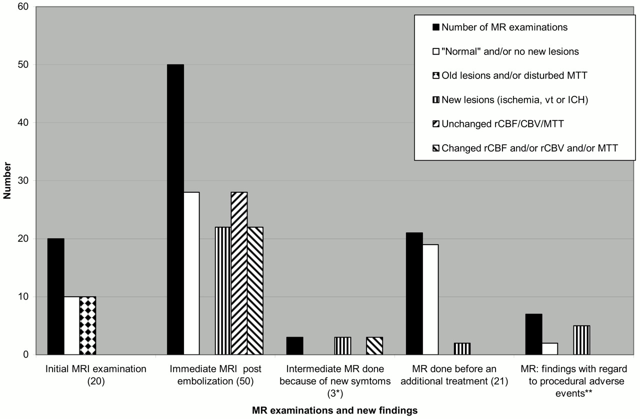
- Fig 1.
MR imaging examinations during the study. MTT indicates mean transit time; rCBF/CBV, regional cerebral blood flow/volume; Vt, venous thrombosis; ICH, intracerebral hemorrhage. *All 3 examinations in one patient. **Included in the 22 postprocedural MRI with new MR lesions. The 10 follow-up examinations are not included in this figure.
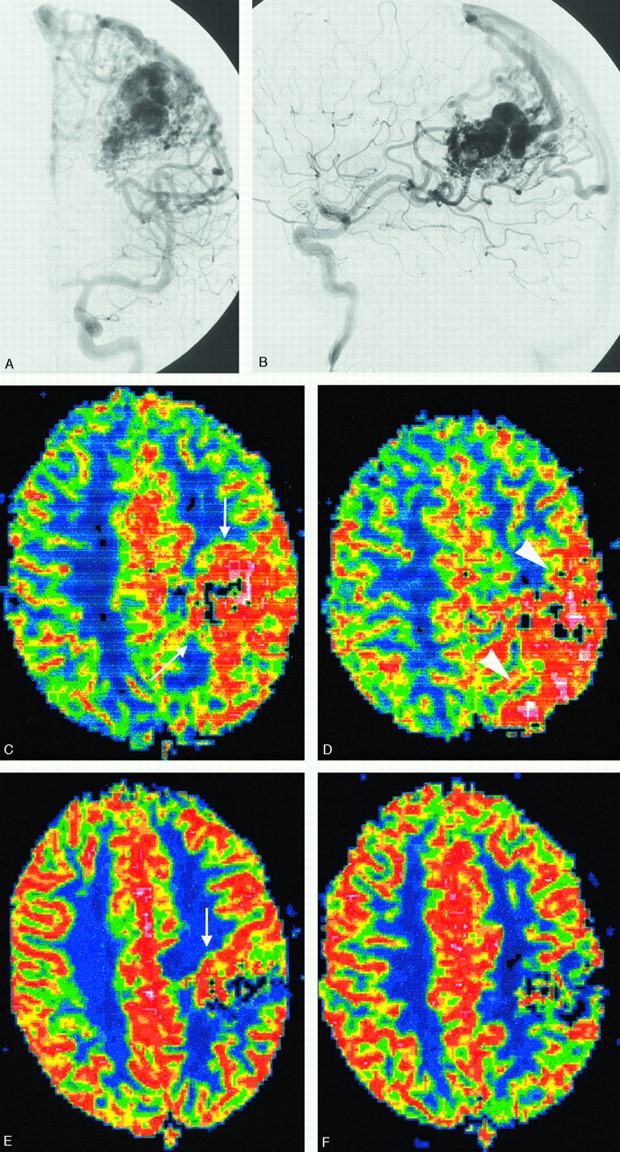
- Fig 2.
Parieto-occipital AVM before and after treatment.
A and B, Left internal carotid artery (ICA) angiograms in frontal and lateral projections demonstrating a parieto-occipital high-flow AVM with multiple feeders and cortical veins draining to the superior sagittal sinus (pre-embolization).
C and D, Axial rCBFs illustrating the increased perfusion in the AVM nidus (arrow) as well as in the widespread distribution of the cortical draining veins before treatment.
E and F, Almost corresponding rCBFs made after 2 uneventful sessions of embolization. The images show a decrease in AVM nidus size and rCBF in the area of the previously enlarged and draining veins. The patient was then sent for subsequent radiation therapy.
- Fig 3.
Patient with a small temporal AVM.
A and B, Frontal projections of internal carotid angiogram done before and after an uneventful embolization. One of 2 draining veins is patent after the treatment.
C and D, Illustrate the cast of glue on unsubtracted DSA and on coronal MR (2D FLASH, performed 26 hours after treatment). The susceptibility artifacts caused by the glue are well illustrated but an associated hematoma could not be definitely defined.
E–G, Axial MR (T2-weighted, DWI b 1000, and ADC maps) performed after treatment show a vasogenic edema in the surroundings of the AVM nidus (arrowhead).
H, Axial CT, performed 2 days later in conjunction with sudden onset of minor symptoms, shows a small hematoma in the area of previous edema. The AVM showed a complete and spontaneous occlusion at follow-up.
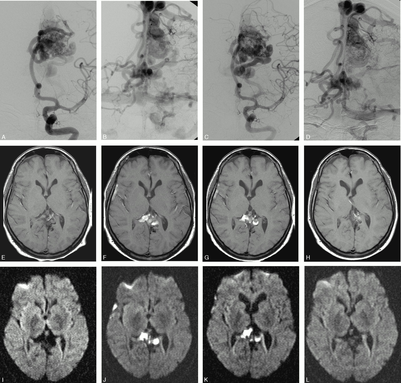
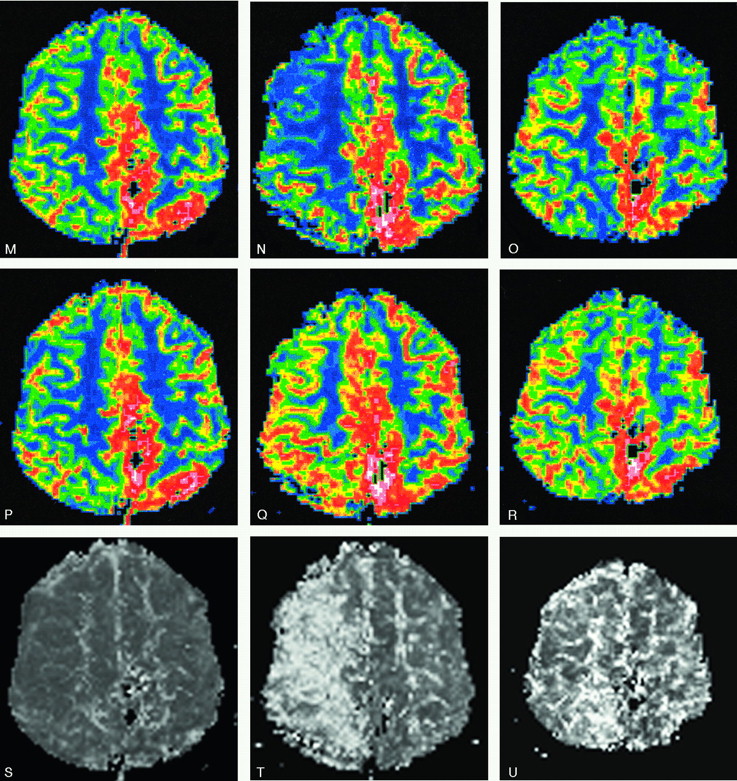
- Fig 4.
Parietal high-flow AVM with deep and bilateral cortical drainage.
A–D, Frontal left internal carotid angiogram, early and late phase, before and after the second embolization. Patent venous drainage is seen after the treatment.
E–L, Axial MR (E–H, T1-weighted, and I–L, DWI [b 1000]) immediately posttreatment, at 23 days, 35 days after treatment, and before a third treatment, 3 months later. The images illustrate the development of venous thrombosis 3 weeks after treatment in the deep and contralateral cortical veins with a complete resolution at follow-up.
M–U, rCBF (M–O) and rCBV (P–R) with the corresponding MTT (S–U): unchanged PI images after treatment (left column) are followed by a dramatic increase in MTT (severe drop in rCBF with a mild rCBV increase) 3 weeks later (middle column). A slow but almost total normalization of PI pattern was seen 9 days later (ie, after hypervolemic hemodilution) (right column). The patient was left with no symptoms.
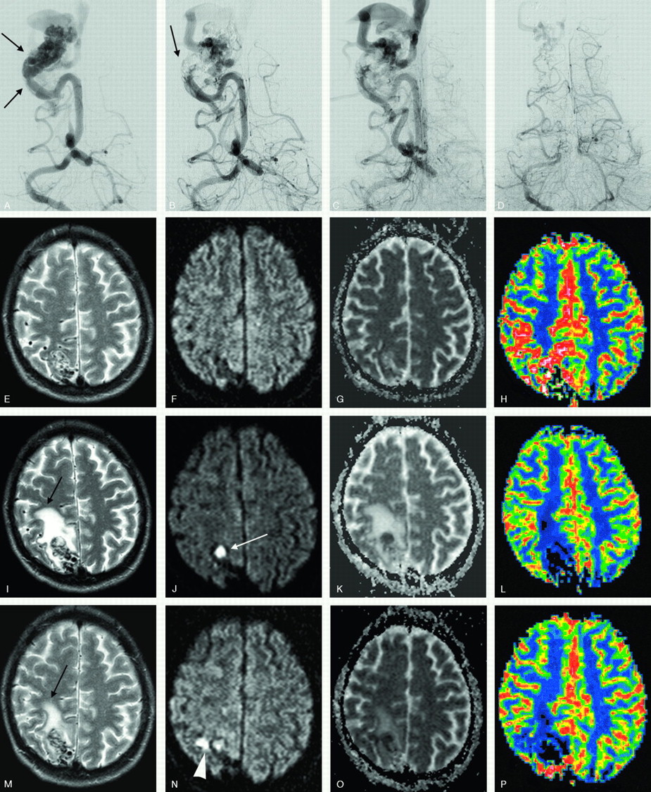
- Fig 5.
Occipital AVM before and after treatment.
A, Frontal right vertebral angiogram demonstrates a small, high-flow, parieto-occipital AVM with an enlarged feeder supplying 2 intranidal fistulas (arrows) before embolization.
B and C, Same projection after partial embolization. Glue can be seen at the site of the fistulas, and the venous drainage remained patent.
D, DSA, performed 6 weeks later (same projection) in conjunction with and before an additional embolization. A spontaneous occlusion of the feeder supplying the fistula is noted with a partial reduction of the nidus. The vein is still patent but reduced in size.
E–P, Axial T2-weighted, DWI, ADC maps, and rCBF done after the first embolization (E–H), before (I–L), and after (M–P) the second treatment. A vasogenic edema (black arrow) evolved in between the treatments, because of a spontaneous thrombosis of the feeder, nidus plus draining vein (white arrow), and decreased immediately after the second embolization. A small, perinidal ischemic lesion is seen after the last treatment (arrowhead). The patient experienced minor and transient headaches in between the embolizations.
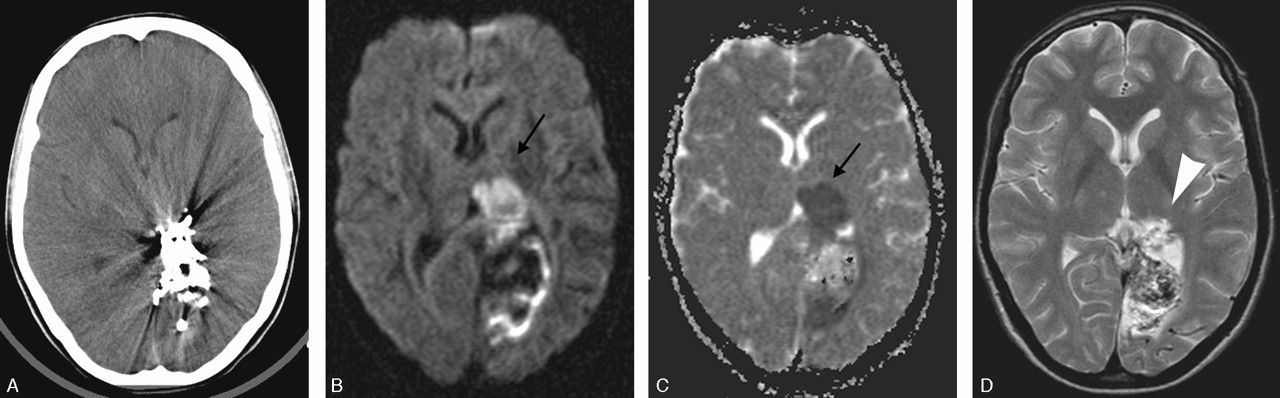
- Fig 6.
Patient with an occipital AVM. Procedure-related technical events occurred during treatment (reflux of glue and a glued catheter).
A, Axial CT performed the day after treatment illustrates the severe artifacts caused by the glue, but no obvious ischemic lesion is noted.
B and C, Corresponding sections on postprocedural MR-DWI/ADC demonstrate a rather large ischemic lesion (arrows) within the left thalamic nucleus (not seen on the CT image).
D, Axial T2-weighted MR image at follow-up shows a small and permanent infarct in the posterior portion of the left thalamus (arrowhead).


Tables
Patient/AVM Clinical Presentation Location (Lobes) Extension (Parenchymal) Size (ml) Venous Drainage High Flow, Fistula Spetzler-Martin Grade Bleeding 10 Frontal 3 Cortical 5 <6 6 Cortical (c) 14 9 I 4 Temporal 4 WM 5 6–10 2 Single 4 II 5 ICH 6 Parietal 4 Mixed (C+WM) 9 >10–20 8 Multiple 10 III 6 IVH 1 Occipital 2 Intraventr. 1 >20 5 Deep (D) 2 IV 6 EDH 1 Mixed* 5 Dural 1 Mixed (C+D) 5 V 0 SAH 2 Cerebellar 2 Epilepsy 6 Dural 1 (venous ectasia 9/21 (±f.stenos)) Incidental finding 5 21 21 21/21 21 21 9/21 21 Note:—
Mixed* indicates extending into more than one lobe; WM, white matter; Mixed (c+wm), located cortical and in the white matter (deep location); Intraventr, intraventricular location; Mixed (c+d), mixed cortical and deep venous drainage; F.stenos, functional stenosis; Spetzler-Martin grades I–V (according to size, location, and venous drainage).
Size (ml) No. of Procedures Occlusion Rate and Patients <6 6–10 11–20 >20 Endovascular proc./AVM 1 6 1 3 10 2 1 3 1 10 4 2 2 16 6 1 6 8 1 8 No. of patients 6 2 8 5 21/50 Occlusion rate %/AVM 100 6 1 1 8 85–99 1 4 2 7 70–84 2 3 5 55–69 1 1 <55 No. of patients 6 2 8 5 21/21 Note:—Proc. indicates procedure; AVM, arteriovenous malformation.
Type of Event Patients No. of Events Comment Extravasation 3 4* During contrast injection (3) During catheterization (1) Adverse migration of glue (including reflux) 3 (1**) 3 During embolizations Emboli 1 1 Transient Glued catheter 1** 1 Intentionally left behind Total 7 9 Note:—
4* indicates 2/4 extravasations took place in the same patient but in 2 separate procedures;
1** , one patient had both glue migration and a glued catheter.
- Table 4:
New lesions on MRI/DWI posttreatment and in between treatments (type of lesion, number, size, and location)
Posttreatment Between post- and next pretreatment MRI Number Location Size Number Number Location Total lesions Ischemic (i) 23 (6 - ai) 4 C <4 7 1 PN 24 1 SC 4–10 7 3 BGL 11–20 5 9 PN >20 4 1 WM 5 C-bell Venous 8 8 PN <4 5 13* 9 C 21 Thrombosis/clots (vt) 4–10 3 3 deep.v 1 PN Uncertain 4 4 PN <4 3 4 (i or vt) 4–10 1 ICH 4 1 PN <10 1 2 6 1 C 10–20 2 1 SC >20 1 1 BGL Vasogenic edema 4 (→2 ICH) (<10 2) 1** PN/SC 5 (10–20 2) Total 43 17 (4 patients) 60/60 Note:—ICH indicates intracerebral hematoma; ai, lesions related to catheterization during the diagnostic part of the procedure; C, cortical; SC, subcortical; BGL, basal ganglia; PN, perinidal (close to the nidus); WM, white matter; C-bell, cerebellar hemisphere; Deep, deep veins.
* Progressive venous thrombosis >2 weeks after treatment.
** Developed vasogenic edema because of combined thrombosis of a feeding artery, the nidus, and vein (represented as 1/13 vt above).
Signs and Symptoms 0–5 Days after Each Embolization New Symptoms Occurring after Discharge/between Embolizations At Last Evaluation*** Outcome (Related to Embolization) Transient Reduced Unchanged Worse Hemiparesis 2 (1*) 1** 2 1 1 mRS 2 (**) Paresthesis 1 1* 2 Dysphasia and/or dysarthria/dyscalcyli 2 (1*) 2 Hemianopsia 2 (1*) 1 2 1 1 mRS 2 Dipoplia 2 (1*) 2 Seventh nerve palsy 1 (1*) 1* 2 Allodynia 1 1 1 mRS 1 Miscellaneous 2 2 4 Headache 1* (ev) 1 Dyscoordination 1 Confusion 1* Total 12 (7 patients) 7 (4 patients) 16 3 3 Note:—
1* indicates that one patient in the category had combined symptoms and is included in the other subgroups (hemiparesis plus N VII palsy in one, hemianopsia plus diplopia plus confusion in a second, dysphasia and headache in a third, and parestesis plus N VII palsy in a fourth patient);
** , patient with ICH 2 months after second embolization (uncertain if related to treatment);
*** , evaluation within 12 months after complete occlusion (8 patients) or before radiotherapy (13 patients); mRS, modified Rankin scale; 1, minor symptoms; 2, some restriction in lifestyle.
In this issue


Jump to section
Related Articles
Cited By...
- Selection of Patients for Treatment of Brain Arteriovenous Malformations by the Transvenous Approach: Relationship with Venous Anatomy and Risk of Hemorrhagic Complications
- Complications of Endovascular Treatments for Brain Arteriovenous Malformations: A Nationwide Surveillance
- Neurological outcomes and cure rates of embolization of brain arteriovenous malformations with n-butyl cyanoacrylate or Onyx: a meta-analysis
- Hemorrhagic Complications after Endovascular Treatment of Cerebral Arteriovenous Malformations
- A Translational Paradigm for the Preclinical Evaluation of the Stroke Neuroprotectant Tat-NR2B9c in Gyrencephalic Nonhuman Primates
- Microsurgical retrieval of an endovascular microcatheter trapped during Onyx embolization of a cerebral arteriovenous malformation
- Brain Arteriovenous Malformation Treatment Using a Combination of Onyx and a New Detachable Tip Microcatheter, SONIC: Short-Term Results
- Territorial and Microvascular Perfusion Impairment in Brain Arteriovenous Malformations