Advertisement

Main menu

  • Home
  • Content
    • Current Issue
    • Accepted Manuscripts
    • Article Preview
    • Past Issue Archive
    • Video Articles
    • AJNR Case Collection
    • Case of the Week Archive
    • Case of the Month Archive
    • Classic Case Archive
  • Special Collections
    • AJNR Awards
    • ASNR Foundation Special Collection
    • Most Impactful AJNR Articles
    • Photon-Counting CT
    • Spinal CSF Leak Articles (Jan 2020-June 2024)
  • Multimedia
    • AJNR Podcasts
    • AJNR SCANtastic
    • Trainee Corner
    • MRI Safety Corner
    • Imaging Protocols
  • For Authors
    • Submit a Manuscript
    • Submit a Video Article
    • Submit an eLetter to the Editor/Response
    • Manuscript Submission Guidelines
    • Statistical Tips
    • Fast Publishing of Accepted Manuscripts
    • Graphical Abstract Preparation
    • Imaging Protocol Submission
    • Author Policies
  • About Us
    • About AJNR
    • Editorial Board
    • Editorial Board Alumni
  • More
    • Become a Reviewer/Academy of Reviewers
    • Subscribers
    • Permissions
    • Alerts
    • Feedback
    • Advertisers
    • ASNR Home

User menu

  • Alerts
  • Log in

Search

  • Advanced search
American Journal of Neuroradiology

American Journal of Neuroradiology

ASHNR American Society of Functional Neuroradiology ASHNR American Society of Pediatric Neuroradiology ASSR
  • Alerts
  • Log in

Advanced Search

  • Home
  • Content
    • Current Issue
    • Accepted Manuscripts
    • Article Preview
    • Past Issue Archive
    • Video Articles
    • AJNR Case Collection
    • Case of the Week Archive
    • Case of the Month Archive
    • Classic Case Archive
  • Special Collections
    • AJNR Awards
    • ASNR Foundation Special Collection
    • Most Impactful AJNR Articles
    • Photon-Counting CT
    • Spinal CSF Leak Articles (Jan 2020-June 2024)
  • Multimedia
    • AJNR Podcasts
    • AJNR SCANtastic
    • Trainee Corner
    • MRI Safety Corner
    • Imaging Protocols
  • For Authors
    • Submit a Manuscript
    • Submit a Video Article
    • Submit an eLetter to the Editor/Response
    • Manuscript Submission Guidelines
    • Statistical Tips
    • Fast Publishing of Accepted Manuscripts
    • Graphical Abstract Preparation
    • Imaging Protocol Submission
    • Author Policies
  • About Us
    • About AJNR
    • Editorial Board
    • Editorial Board Alumni
  • More
    • Become a Reviewer/Academy of Reviewers
    • Subscribers
    • Permissions
    • Alerts
    • Feedback
    • Advertisers
    • ASNR Home
  • Follow AJNR on Twitter
  • Visit AJNR on Facebook
  • Follow AJNR on Instagram
  • Join AJNR on LinkedIn
  • RSS Feeds

AJNR is seeking candidates for the AJNR Podcast Editor. Read the position description.

Research ArticleBRAIN

Bone-Subtraction CT Angiography for the Evaluation of Intracranial Aneurysms

B.F. Tomandl, T. Hammen, E. Klotz, H. Ditt, B. Stemper and M. Lell
American Journal of Neuroradiology January 2006, 27 (1) 55-59;
B.F. Tomandl
  • Find this author on Google Scholar
  • Find this author on PubMed
  • Search for this author on this site
T. Hammen
  • Find this author on Google Scholar
  • Find this author on PubMed
  • Search for this author on this site
E. Klotz
  • Find this author on Google Scholar
  • Find this author on PubMed
  • Search for this author on this site
H. Ditt
  • Find this author on Google Scholar
  • Find this author on PubMed
  • Search for this author on this site
B. Stemper
  • Find this author on Google Scholar
  • Find this author on PubMed
  • Search for this author on this site
M. Lell
  • Find this author on Google Scholar
  • Find this author on PubMed
  • Search for this author on this site
  • Article
  • Figures & Data
  • Info & Metrics
  • Responses
  • References
  • PDF
Loading

Article Figures & Data

Figures

  • Fig 1.
    • Download figure
    • Open in new tab
    • Download powerpoint
    Fig 1.

    Illustration of the bone-removal process.

    A, Sagittal multiplanar reformation of the data after bone removal shows the contrast-enhanced vascular structures (arrows) as well as brain tissue.

    B, Sagittal maximum intensity projection image (15-mm thickness) demonstrates the intraosseous parts of the internal carotid artery (short arrow) as well as the origin of the ophthalmic artery (long arrow).
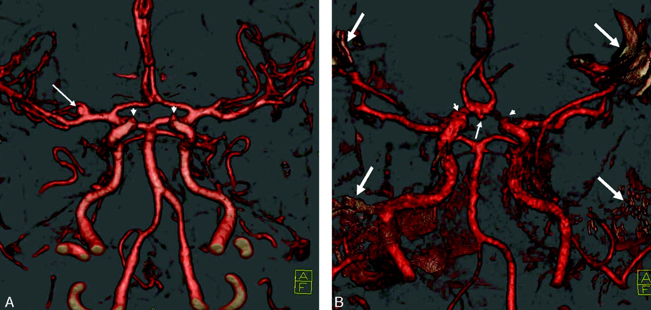
  • Fig 2.
    • Download figure
    • Open in new tab
    • Download powerpoint
    Fig 2.

    Different qualities of bone removal, anterior-inferior view. Note the visibility of the origin of the ophthalmic artery in both images (arrowheads).

    A, “Excellent” bone removal without artifacts. There is a small aneurysm of the right middle cerebral artery (arrow).

    B, “Moderate” result with remnants of bone (arrows) because of movement of the patient during one of the scans. The arteries within the skull base are still visible. There is a small aneurysm of the anterior communicating artery (arrow).
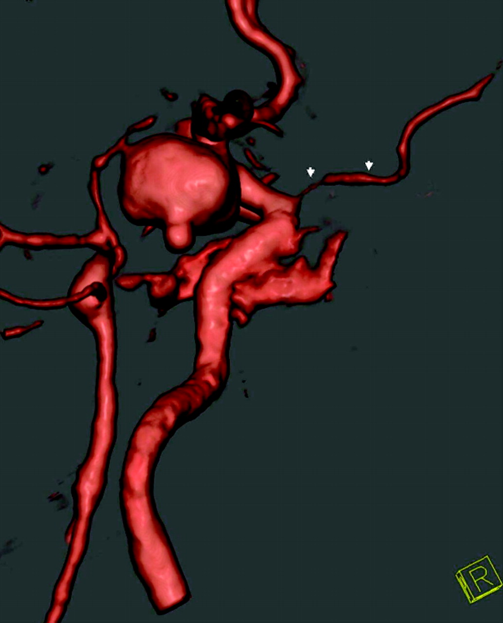
  • Fig 3.
    • Download figure
    • Open in new tab
    • Download powerpoint
    Fig 3.

    Optimal visualization of the ophthalmic artery (arrowheads) of the right ICA in this patient with a large aneurysm of the ICA from a lateral view.

  • Fig 4.
    • Download figure
    • Open in new tab
    • Download powerpoint
    Fig 4.

    Prominent cavernous sinus (arrowheads).

    A, 3D volume-rendered image with high opacity and shading does not allow evaluation of the intracavernous part of the left ICA completely on this medial view.

    B, Transparent (low-opacity) volume-rendered image allows for a good evaluation of the intracavernous portion of the ICA (arrow).

  • Fig 5.
    • Download figure
    • Open in new tab
    • Download powerpoint
    Fig 5.

    Aneurysm of the left infraclinoidal ICA, left-superior view.

    A, On standard CT angiography the aneurysm is hardly visible (arrow) due to obscuration by the anterior clinoid process.

    B, Bone-subtraction CT angiography (BSCTA) clearly shows the complete shape of the aneurysm (arrow).

  • Fig 6.
    • Download figure
    • Open in new tab
    • Download powerpoint
    Fig 6.

    Comparison of digital subtraction angiography (DSA), 3D DSA and BSCTA. Patient with a large aneurysm of the right ICA (arrows), right anterior oblique view.

    A, DSA.

    B, 3D DSA, direct volume rendering.

    C, BSCTA. The cavernous sinus does not disturb the interpretation of the image (arrowheads).

  • Fig 7.
    • Download figure
    • Open in new tab
    • Download powerpoint
    Fig 7.

    Use of BSCTA for therapy planning of an intracavernous aneurysm of the left ICA (arrows).

    A, BSCTA, lateral view of the left ICA.

    B, DSA, lateral view of the left ICA.

    C, Transparent (low-opacity) volume rendering allows visualization of the borders of the aneurysm’s broad neck (arrowheads) from an anterior view, which is useful for therapy planning. This aneurysm was finally treated by means of stent-protected coiling.

PreviousNext
Back to top

In this issue

American Journal of Neuroradiology: 27 (1)
American Journal of Neuroradiology
Vol. 27, Issue 1
January, 2006
  • Table of Contents
  • Index by author
Advertisement
Print
Download PDF
Email Article

Thank you for your interest in spreading the word on American Journal of Neuroradiology.

NOTE: We only request your email address so that the person you are recommending the page to knows that you wanted them to see it, and that it is not junk mail. We do not capture any email address.

Enter multiple addresses on separate lines or separate them with commas.
Bone-Subtraction CT Angiography for the Evaluation of Intracranial Aneurysms
(Your Name) has sent you a message from American Journal of Neuroradiology
(Your Name) thought you would like to see the American Journal of Neuroradiology web site.
CAPTCHA
This question is for testing whether or not you are a human visitor and to prevent automated spam submissions.
Cite this article
B.F. Tomandl, T. Hammen, E. Klotz, H. Ditt, B. Stemper, M. Lell
Bone-Subtraction CT Angiography for the Evaluation of Intracranial Aneurysms
American Journal of Neuroradiology Jan 2006, 27 (1) 55-59;

Citation Manager Formats

  • BibTeX
  • Bookends
  • EasyBib
  • EndNote (tagged)
  • EndNote 8 (xml)
  • Medlars
  • Mendeley
  • Papers
  • RefWorks Tagged
  • Ref Manager
  • RIS
  • Zotero
0 Responses
Respond to this article
Share
Bookmark this article
Bone-Subtraction CT Angiography for the Evaluation of Intracranial Aneurysms
B.F. Tomandl, T. Hammen, E. Klotz, H. Ditt, B. Stemper, M. Lell
American Journal of Neuroradiology Jan 2006, 27 (1) 55-59;
del.icio.us logo Twitter logo Facebook logo Mendeley logo
  • Tweet Widget
  • Facebook Like
  • Google Plus One

Jump to section

  • Article
    • Abstract
    • Materials and Methods
    • Results
    • Discussion
    • Conclusion
    • Footnotes
    • References
  • Figures & Data
  • Info & Metrics
  • Responses
  • References
  • PDF

Related Articles

  • No related articles found.
  • PubMed
  • Google Scholar

Cited By...

  • Surveillance of Unruptured Intracranial Saccular Aneurysms Using Noncontrast 3D-Black-Blood MRI: Comparison of 3D-TOF and Contrast-Enhanced MRA with 3D-DSA
  • Three-dimensional image fusion of CTA and angiography for real-time guidance during neurointerventional procedures
  • Diagnostic Impact of Bone-Subtraction CT Angiography for Patients with Acute Subarachnoid Hemorrhage
  • Bone-Subtracted Spinal CT Angiography Using Nonrigid Registration for Better Visualization of Arterial Feeders in Spinal Arteriovenous Fistulas
  • Improved Arterial Visualization in Cerebral CT Perfusion-Derived Arteriograms Compared with Standard CT Angiography: A Visual Assessment Study
  • Crossref
  • Google Scholar

This article has not yet been cited by articles in journals that are participating in Crossref Cited-by Linking.

More in this TOC Section

  • Usefulness of Quantitative Susceptibility Mapping for the Diagnosis of Parkinson Disease
  • White Matter Alterations in the Brains of Patients with Active, Remitted, and Cured Cushing Syndrome: A DTI Study
  • Qualitative and Quantitative Analysis of MR Imaging Findings in Patients with Middle Cerebral Artery Stroke Implanted with Mesenchymal Stem Cells
Show more BRAIN

Similar Articles

Advertisement

Indexed Content

  • Current Issue
  • Accepted Manuscripts
  • Article Preview
  • Past Issues
  • Editorials
  • Editor's Choice
  • Fellows' Journal Club
  • Letters to the Editor
  • Video Articles

Cases

  • Case Collection
  • Archive - Case of the Week
  • Archive - Case of the Month
  • Archive - Classic Case

Special Collections

  • AJNR Awards
  • ASNR Foundation Special Collection
  • Most Impactful AJNR Articles
  • Photon-Counting CT
  • Spinal CSF Leak Articles (Jan 2020-June 2024)

More from AJNR

  • Trainee Corner
  • Imaging Protocols
  • MRI Safety Corner

Multimedia

  • AJNR Podcasts
  • AJNR Scantastics

Resources

  • Turnaround Time
  • Submit a Manuscript
  • Submit a Video Article
  • Submit an eLetter to the Editor/Response
  • Manuscript Submission Guidelines
  • Statistical Tips
  • Fast Publishing of Accepted Manuscripts
  • Graphical Abstract Preparation
  • Imaging Protocol Submission
  • Evidence-Based Medicine Level Guide
  • Publishing Checklists
  • Author Policies
  • Become a Reviewer/Academy of Reviewers
  • News and Updates

About Us

  • About AJNR
  • Editorial Board
  • Editorial Board Alumni
  • Alerts
  • Permissions
  • Not an AJNR Subscriber? Join Now
  • Advertise with Us
  • Librarian Resources
  • Feedback
  • Terms and Conditions
  • AJNR Editorial Board Alumni

American Society of Neuroradiology

  • Not an ASNR Member? Join Now

© 2025 by the American Society of Neuroradiology All rights, including for text and data mining, AI training, and similar technologies, are reserved.
Print ISSN: 0195-6108 Online ISSN: 1936-959X

Powered by HighWire