
I read with interest the recent case report by Burtis et al regarding findings of MR imaging in craniospinal hypotension.1 The authors described an enlarged intrathecal spinal vein connecting with engorged “epidural” venous plexus through the intervertebral vein. In addition, the engorged venous system in the intraspinal extrathecal space, prominent in the ventral aspect, has been interpreted to be the “epidural” vein in the previous reports of this disease.2–4 The veins posterior to the theca, the posterior internal vertebral venous plexus, are exactly situated in the epidural space proper, though the veins anterior to the theca, the anterior internal vertebral venous plexus (AIVVP), are not in the space. The AIVVP consists of 2 longitudinal venous spaces between 2 thin layers of the posterior longitudinal ligament, the superficial (dorsal) and deep (ventral) layers5–7 (Fig 1). The walls of the AIVVP have all 3 layers of a vein: intima, media, and adventitia. Each AIVVP locates in the lateral part of the spinal canal symmetrically, and joins at the midline by retrocorporeal anastomoses. Although existing MR imaging cannot depict the layer relationships, anatomically correct expression of the venous system is desirable.
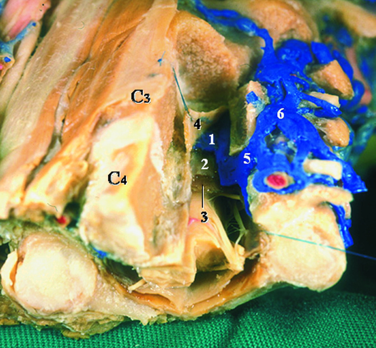
Anterocaudal view of a cadaver specimen after left vertebrectomy shows layer relationships between the AIVVP and surrounding structures. The AIVVP (1) is formed by 2 layers of the posterior longitudinal ligament, the superficial layer (2) just anterior to the dural theca, (3) and deep layer (4). The intervertebral vein (5) and vertebral vein (6) are also shown.
References
Reply:
After thorough review of Dr. Shimizu’s image references and written response to our case report on intracranial hypotension, we are in complete concurrence with his assertion that the anterior internal vertebral venous plexus (AIVVP) is not situated within the epidural space proper. Rather, the AIVVP is within the layers of the posterior longitudinal ligament.
We are grateful for his comprehensive review of the pertinent anatomy in this case and for his letter regarding the error.
- Copyright © American Society of Neuroradiology
In this issue


Jump to section
Related Articles
Cited By...
- No citing articles found.