Fig 2.

Fig 2.
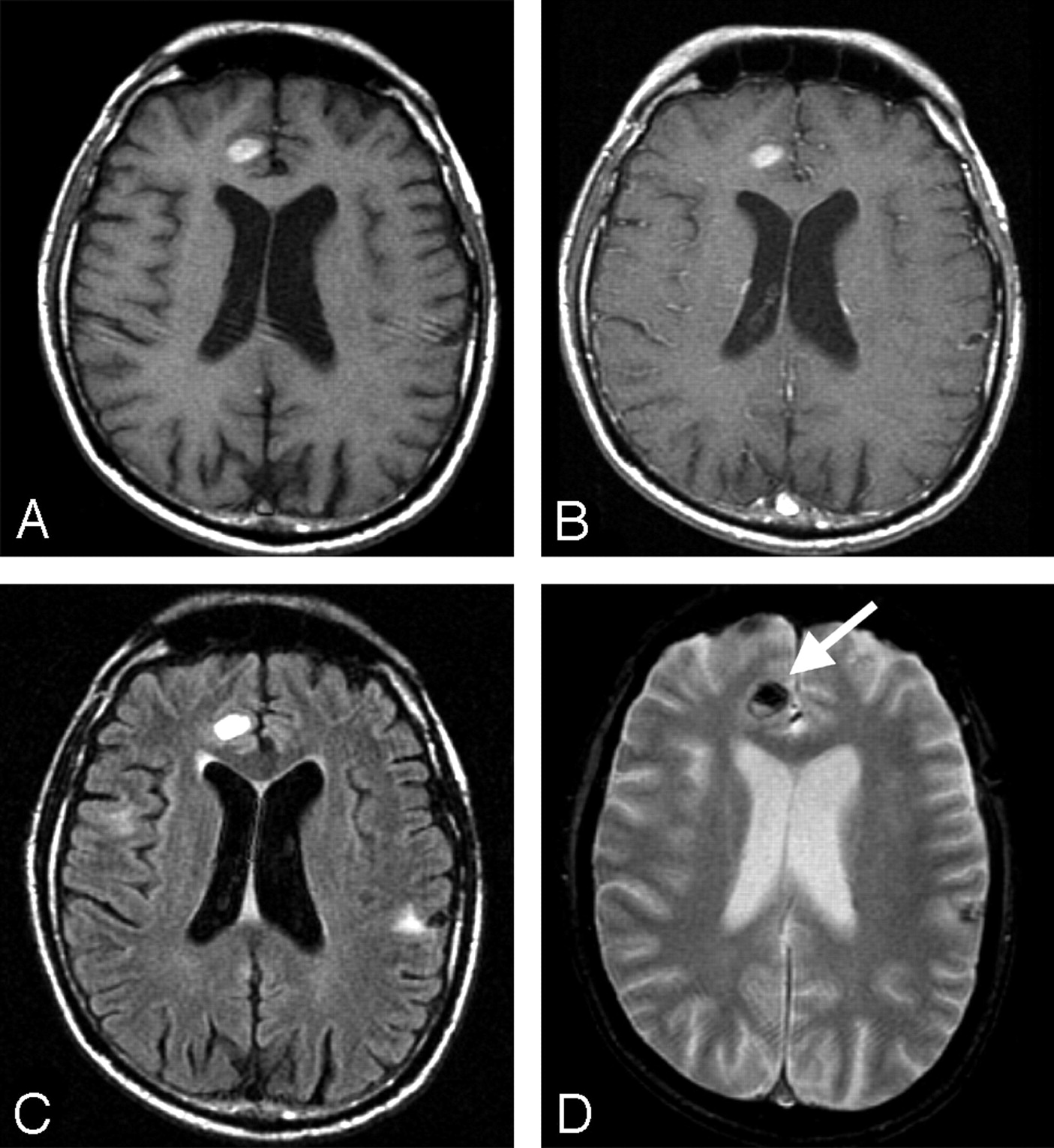
Combined susceptibility effect and T1-weighted hyperintensity were seen in only one quarter of lesions, but this combination was 16 times more likely with melanoma than with lung cancer metastases. Intrinsic T1-weighted hyperintensity (A), minimal enhancement (B), minimal surrounding edema on a FLAIR image (C), and susceptibility effect (D) are shown in metastatic melanoma in the right frontal lobe.


Related Articles
Cited By...
- Malignant melanoma with central nervous system involvement in a dog treated with surgery, radiotherapy and chemotherapy
- Metastatic melanoma of unknown primary in the temporalis muscle
- 123I-BZA2 as a Melanin-Targeted Radiotracer for the Identification of Melanoma Metastases: Results and Perspectives of a Multicenter Phase III Clinical Trial