
Article Figures & Data
Figures
- Fig 1.
Near-occlusion ICA stenosis with distal ICA collapse, threadlike “string sign.” CTA axial source image (A) near the skull base and coronal MPR (B) showing collapse of the right distal ICA (thin arrow) in comparison to normal caliber left distal ICA (arrowhead). These arteries are continuous from the proximal carotid bulb and are headed toward the carotid canal. Both features should be identified on MPRs with reference to the axial source images to distinguish collapsed ICAs from other vessels, especially the ascending pharyngeal artery. C, 3D-rendered (left) and oblique sagittal MPR (right) of the right carotid arteries showing the severe carotid bulb stenosis, appearing amputated on reformatted images. There is collapse of the right distal ICA (thin arrow), similar in size to the ascending pharyngeal artery on the oblique sagittal MPR (thick solid arrow). The identification of both these vessels was confirmed on the axial source images and other MPRs by identifying their origins and anatomic continuations.
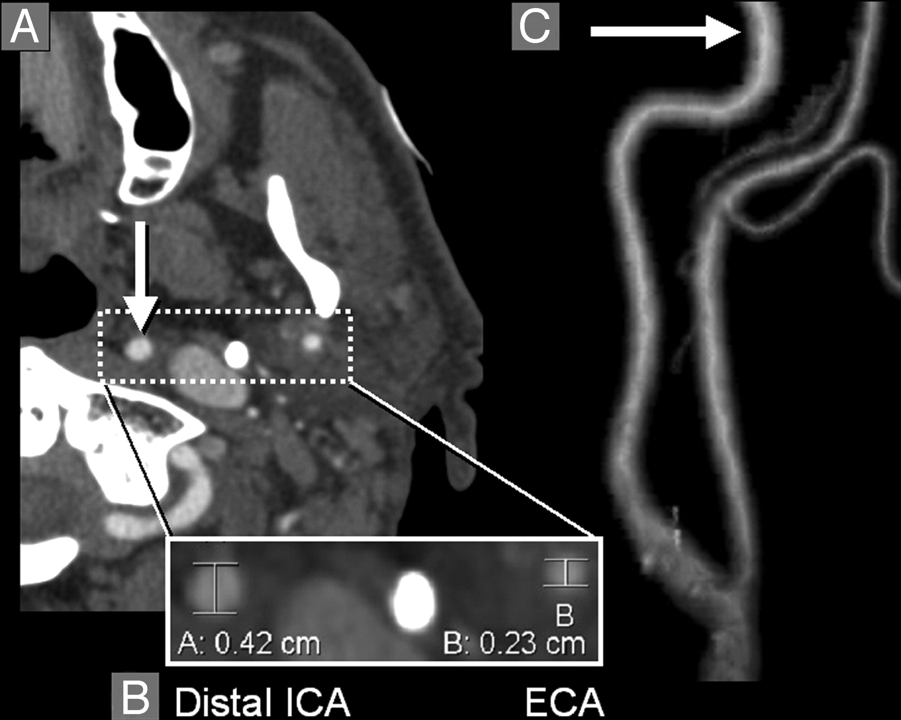
- Fig 2.
Normal distal ICA. A, Axial CTA image at the level of the left distal ICA (arrow) and distal ECA (both enclosed by dashed-line box). Densely calcified styloid process is between the ICA and the ECA. B, Magnification of the distal ICA and distal ECA with diameter measurements; the distal ICA diameter (A, 0.42 cm) is substantially larger than the distal ECA diameter (B, 0.23 cm). Densely calcified styloid process is between the ICA and the ECA. C, 3D-rendered image of the left distal ICA and distal ECA, showing their normal relationship (arrow, distal ICA).
- Fig 3.
Decreased diameter of distal ICA lumen associated with near-occlusion stenosis. A, Axial CTA image at the level of the left distal ICA (arrow) and distal ECA (both enclosed by dashed-line box). B, Magnification of the distal ICA and distal ECA with diameter measurements; in this case, the distal ICA diameter (B, 0.26 cm) is smaller than the distal ECA diameter (A, 0.32 cm). C, 3D-rendered image of the left distal ICA and distal ECA, showing the decreased distal ICA (arrow, distal ICA), secondary to near-occlusion ICA stenosis in this case. Distal ICA reduction that is near equal in diameter to distal ECA is substantially decreased from expected normal as shown in Fig 2). The maximum left ICA bulb stenosis measured 0.07 cm, shown on 3D-rendered image as an amputated segment. Calcification surrounds segments of the left carotid bifurcation.
- Fig 4.
ROC curves depicting the performance of each model in identifying near-occlusion stenosis. The model is more accurate the further the curve lies above the reference line (greatest area under the ROC curve). A, Ratio of the distal ICA lumen to the contralateral distal ICA lumen (dICA:dICA*, solid line), the most accurate model to identify near-occlusion stenosis. B, Distal ICA lumen (dICA, dotted line) is the second most accurate model. C, Ratio of the distal ICA lumen to that of the ipsilateral ECA (ICA: ECA, dot-dashed line), is the third most accurate model. D, The ICA stenosis model (ICA, dashed line) is the least accurate to identify near-occlusion stenosis.


Tables
- Table 1:
Validity statistics of near occlusion single-variable test models (in order of decreasing accuracy)
Sensitivity Specificity PPV NPV dlCA:dlCA* (≤0.87), n = 216 97.3 89.4 65.5 99.4 dlCA (≤3.5 mm), n = 240 95.2 89.9 66.7 98.9 dlCA:ECA (≤1.27), n = 231 90.5 87.3 61.3 97.6 ICA (≤1.3 mm), n = 179 90.2 84.1 62.7 96.7 Note: —dlCA:dlCA* indicates ratio of the distal internal carotid artery (ICA) diameter to that of the contralateral distal ICA; dlCA, diameter of the distal ICA; dlCA:ECA, ratio of the distal ICA diameter to the distal external carotid artery diameter; ICA, narrowest diameter of the ICA bulb stenosis. All diameters are expressed in millimeters. PPV indicates positive predictive value; NPV, negative predictive value. Values demonstrate the ability of each model to identify near occlusion stenosis, based on threshold values for each model. The threshold values were based on the model’s receiver operating characteristic curve and assigned to maximize sensitivity and specificity for each test model. Positive and negative predictive values were calculated for all data. The threshold values (in parentheses) are reported under the model title, along with the population size (n).
- Table 2:
Validity statistics for the paired permutations of near occlusion single-variable test models (in order of decreasing accuracy)
Sensitivity Specificity PPV NPV dlCA + dlCA:dlCA* (n = 235) 91.9 96.0 81.0 98.4 dlCA:dlCA* + dlCA:ECA (n = 234) 89.2 95.4 78.6 97.9 dlCA + dlCA:ECA (n = 239) 88.1 95.4 80.4 97.4 ICA + dlCA (n = 231) 87.8 96.8 85.7 97.4 ICA + dlCA:dlCA* (n = 225) 86.8 96.3 82.5 97.3 ICA + dICA:ECA (n = 229) 82.9 96.8 85.0 96.3 Note:—dlCA:dlCA*, ratio of the distal internal carotid artery (ICA) diameter to that of the contralateral distal ICA; dlCA, diameter of the distal ICA; dlCA:ECA, ratio of the distal ICA diameter to the distal external carotid artery diameter; ICA, narrowest diameter of the ICA bulb stenosis. All diameters are expressed in millimeters. PPV indicates positive predictive value; NPV, negative predictive value. Values were obtained from contingency tables created to evaluate paired permutations of the single-variable test models. Positive and negative predictive values were calculated for all data. Population size (n) is reported for each permutation.
In this issue


Jump to section
Related Articles
Cited By...
- Diagnosing Carotid Near-Occlusion with Phase-Contrast MRI
- Assessment of Apparent Internal Carotid Tandem Occlusion on High-Resolution Vessel Wall Imaging: Comparison with Digital Subtraction Angiography
- Carotid Near-Occlusion: A Comprehensive Review, Part 2--Prognosis and Treatment, Pathophysiology, Confusions, and Areas for Improvement
- Carotid Near-Occlusion: A Comprehensive Review, Part 1--Definition, Terminology, and Diagnosis
- Multilevel Assessment of Atherosclerotic Extent Using a 40-Section Multidetector Scanner after Transient Ischemic Attack or Ischemic Stroke
- Factors associated with early outcome in patients with large-vessel carotid strokes
- The Relation of Carotid Calcium Volume with Carotid Artery Stenosis in Symptomatic Patients
- Should Modeling Methodology Suppress Anatomic Excellence?
- Window Settings for the Study of Calcified Carotid Plaques with Multidetector CT Angiography
- Contrast-Enhanced MR Angiography Is Not More Accurate Than Unenhanced 2D Time-of-Flight MR Angiography for Determining >=70% Internal Carotid Artery Stenosis
- Response to Letter by Bladin et al
- Simplification of the Residual Lumen Geometry in Measuring Carotid Stenosis
- Carotid Stenosis Index Revisited With Direct CT Angiography Measurement of Carotid Arteries to Quantify Carotid Stenosis