
Article Figures & Data
Figures
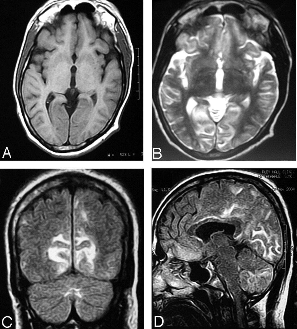
- Fig 1.
Prechelation MR imaging.
A–C, T1- and T2-weighted axial and FLAIR (respectively) coronal MR images show cortical gray matter and subcortical white matter lesion in the occipital lobe with edema and sulcal effacement.
D, FLAIR sagittal image shows involvement of subcortical white matter in the frontal regions, parieto-occipital lobe, body of the corpus callosum, and cerebellum.
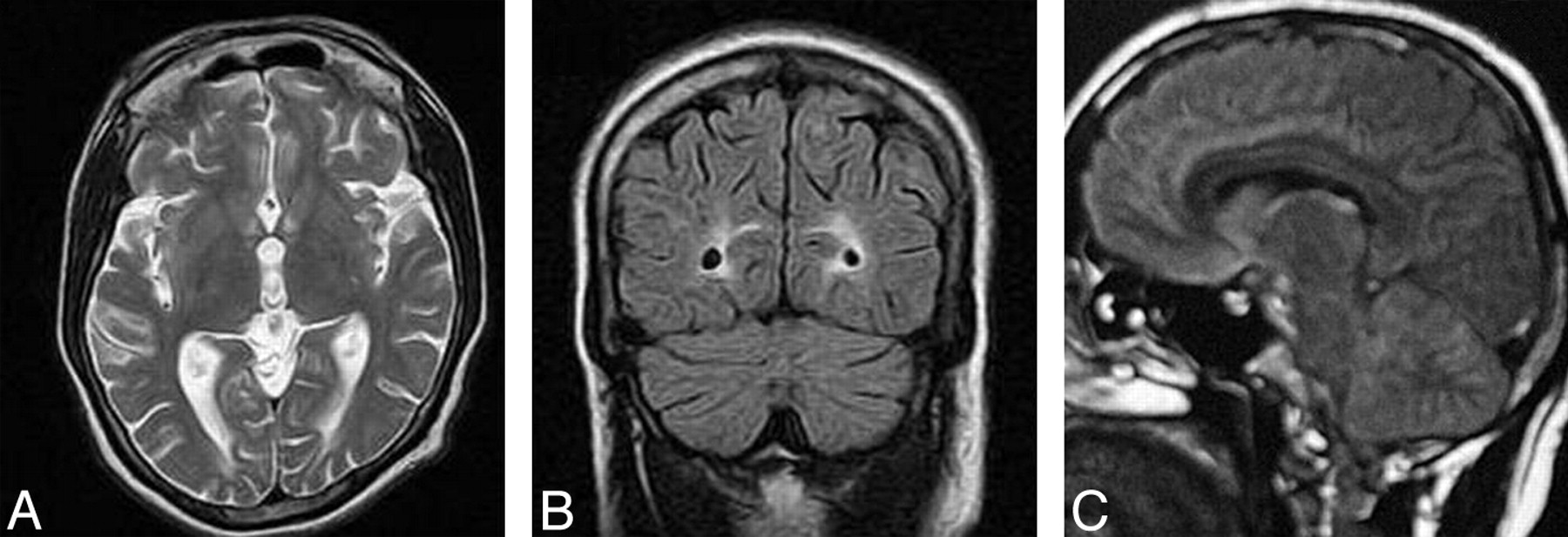
- Fig 2.
Postchelation MR imaging.
A and B, T2-weighted axial and FLAIR coronal images show near-total resolution. Few focal subcortical white matter lesions are still seen in the occipital lobe.
C, FLAIR sagittal images show near-total resolution.


