Advertisement

Main menu

  • Home
  • Content
    • Current Issue
    • Accepted Manuscripts
    • Article Preview
    • Past Issue Archive
    • Video Articles
    • AJNR Case Collection
    • Case of the Week Archive
    • Case of the Month Archive
    • Classic Case Archive
  • Special Collections
    • AJNR Awards
    • ASNR Foundation Special Collection
    • Most Impactful AJNR Articles
    • Photon-Counting CT
    • Spinal CSF Leak Articles (Jan 2020-June 2024)
  • Multimedia
    • AJNR Podcasts
    • AJNR SCANtastic
    • Trainee Corner
    • MRI Safety Corner
    • Imaging Protocols
  • For Authors
    • Submit a Manuscript
    • Submit a Video Article
    • Submit an eLetter to the Editor/Response
    • Manuscript Submission Guidelines
    • Statistical Tips
    • Fast Publishing of Accepted Manuscripts
    • Graphical Abstract Preparation
    • Imaging Protocol Submission
    • Author Policies
  • About Us
    • About AJNR
    • Editorial Board
    • Editorial Board Alumni
  • More
    • Become a Reviewer/Academy of Reviewers
    • Subscribers
    • Permissions
    • Alerts
    • Feedback
    • Advertisers
    • ASNR Home

User menu

  • Alerts
  • Log in

Search

  • Advanced search
American Journal of Neuroradiology

American Journal of Neuroradiology

ASHNR American Society of Functional Neuroradiology ASHNR American Society of Pediatric Neuroradiology ASSR
  • Alerts
  • Log in

Advanced Search

  • Home
  • Content
    • Current Issue
    • Accepted Manuscripts
    • Article Preview
    • Past Issue Archive
    • Video Articles
    • AJNR Case Collection
    • Case of the Week Archive
    • Case of the Month Archive
    • Classic Case Archive
  • Special Collections
    • AJNR Awards
    • ASNR Foundation Special Collection
    • Most Impactful AJNR Articles
    • Photon-Counting CT
    • Spinal CSF Leak Articles (Jan 2020-June 2024)
  • Multimedia
    • AJNR Podcasts
    • AJNR SCANtastic
    • Trainee Corner
    • MRI Safety Corner
    • Imaging Protocols
  • For Authors
    • Submit a Manuscript
    • Submit a Video Article
    • Submit an eLetter to the Editor/Response
    • Manuscript Submission Guidelines
    • Statistical Tips
    • Fast Publishing of Accepted Manuscripts
    • Graphical Abstract Preparation
    • Imaging Protocol Submission
    • Author Policies
  • About Us
    • About AJNR
    • Editorial Board
    • Editorial Board Alumni
  • More
    • Become a Reviewer/Academy of Reviewers
    • Subscribers
    • Permissions
    • Alerts
    • Feedback
    • Advertisers
    • ASNR Home
  • Follow AJNR on Twitter
  • Visit AJNR on Facebook
  • Follow AJNR on Instagram
  • Join AJNR on LinkedIn
  • RSS Feeds

AJNR is seeking candidates for the AJNR Podcast Editor. Read the position description.

Research ArticleHEAD AND NECK

Para-Cavernous Sinus Venous Structures: Anatomic Variations and Pathologic Conditions Evaluated on Fat-Suppressed 3D Fast Gradient-Echo MR Images

S. Tanoue, H. Kiyosue, M. Okahara, Y. Sagara, Y. Hori, J. Kashiwagi and H. Mori
American Journal of Neuroradiology May 2006, 27 (5) 1083-1089;
S. Tanoue
  • Find this author on Google Scholar
  • Find this author on PubMed
  • Search for this author on this site
H. Kiyosue
  • Find this author on Google Scholar
  • Find this author on PubMed
  • Search for this author on this site
M. Okahara
  • Find this author on Google Scholar
  • Find this author on PubMed
  • Search for this author on this site
Y. Sagara
  • Find this author on Google Scholar
  • Find this author on PubMed
  • Search for this author on this site
Y. Hori
  • Find this author on Google Scholar
  • Find this author on PubMed
  • Search for this author on this site
J. Kashiwagi
  • Find this author on Google Scholar
  • Find this author on PubMed
  • Search for this author on this site
H. Mori
  • Find this author on Google Scholar
  • Find this author on PubMed
  • Search for this author on this site
  • Article
  • Figures & Data
  • Info & Metrics
  • Responses
  • References
  • PDF
Loading

Article Figures & Data

Figures

  • Tables
  • Fig 1.
    • Download figure
    • Open in new tab
    • Download powerpoint
    Fig 1.

    Schematic drawings of variations of the SMCV. Type A, SMCV connects with the proximal SPS and flows into the frontal aspect of the cavernous sinus. Type B, The SMCV connects with the lateral aspect of the cavernous sinus independently. Type C, The SMCV turns downward and connects with the pterygoid plexus through the middle cranial fossa. Type D, The SMCV runs across the pyramidal ridge and connects with the superior petrosal sinus or transverse sinus via the tentorial sinus. SOV indicates the superior orbital vein; PP, pterygoid plexus; SuPS, superior petrosal sinus; SS, sigmoid sinus; and TS, transverse sinus.

  • Fig 2.
    • Download figure
    • Open in new tab
    • Download powerpoint
    Fig 2.

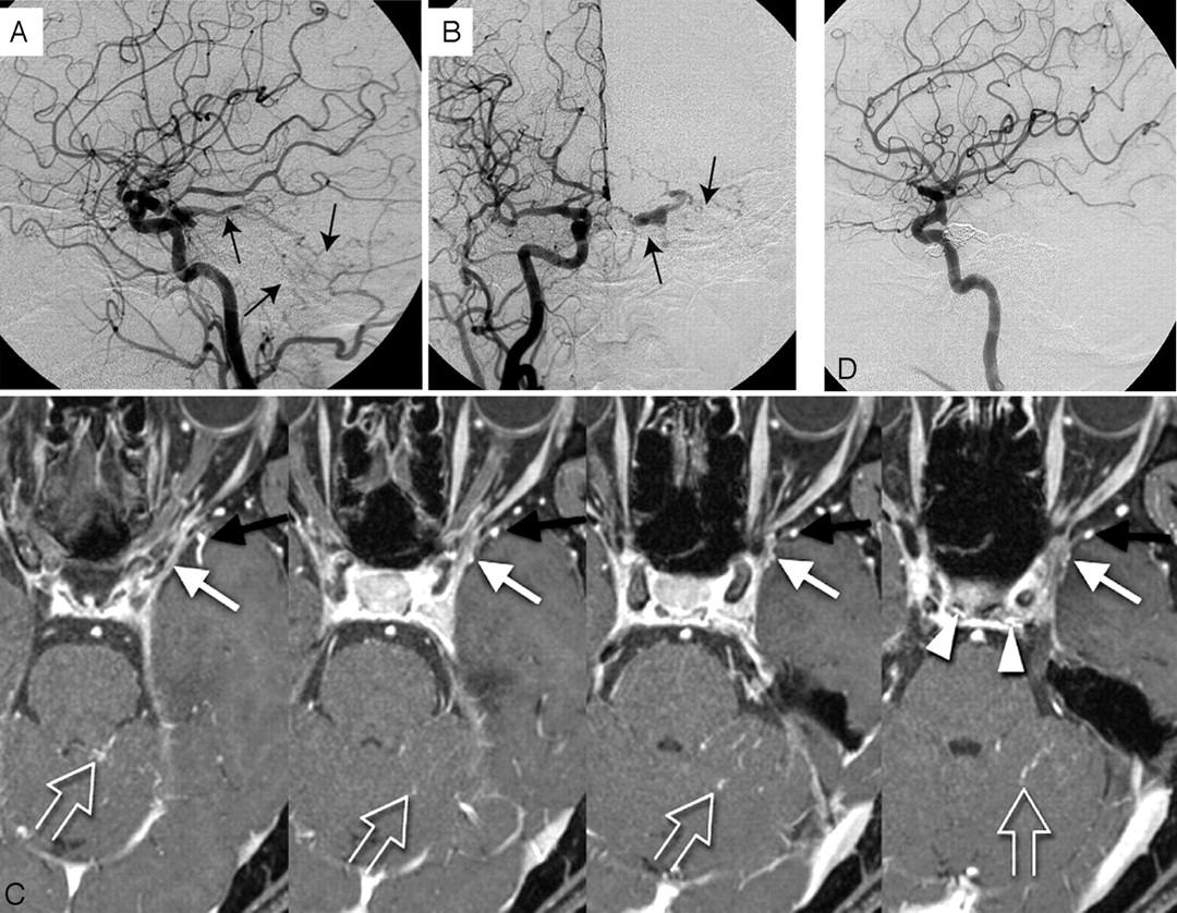
    Type A, A 69-year-old man with left maxillary cancer. The SMCV runs along the lesser sphenoid ridge (white arrows) to connect with the proximal segment of the SPS (black arrows) and connects with the anterior potion of cavernous sinus (arrowhead).

  • Fig 3.
    • Download figure
    • Open in new tab
    • Download powerpoint
    Fig 3.

    Type C, A 60-year-old woman with right acoustic neurinoma. The SMCV runs along the sphenoid ridge, turns downward, and connects with the pterygoid plexus via the foramen ovale (white arrow). Note the hypoplastic SPS connects with the cavernous sinus (black arrow). This pattern is categorized as type C.

  • Fig 4.
    • Download figure
    • Open in new tab
    • Download powerpoint
    Fig 4.

    Type D, A 58-year-old man with metastatic brain tumor from lung cancer. Under the temporal lobe, the SMCV runs across the pyramidal ridge posteriorly and connects with the transverse sinus via the tentorial sinus (white arrows).

  • Fig 5.
    • Download figure
    • Open in new tab
    • Download powerpoint
    Fig 5.

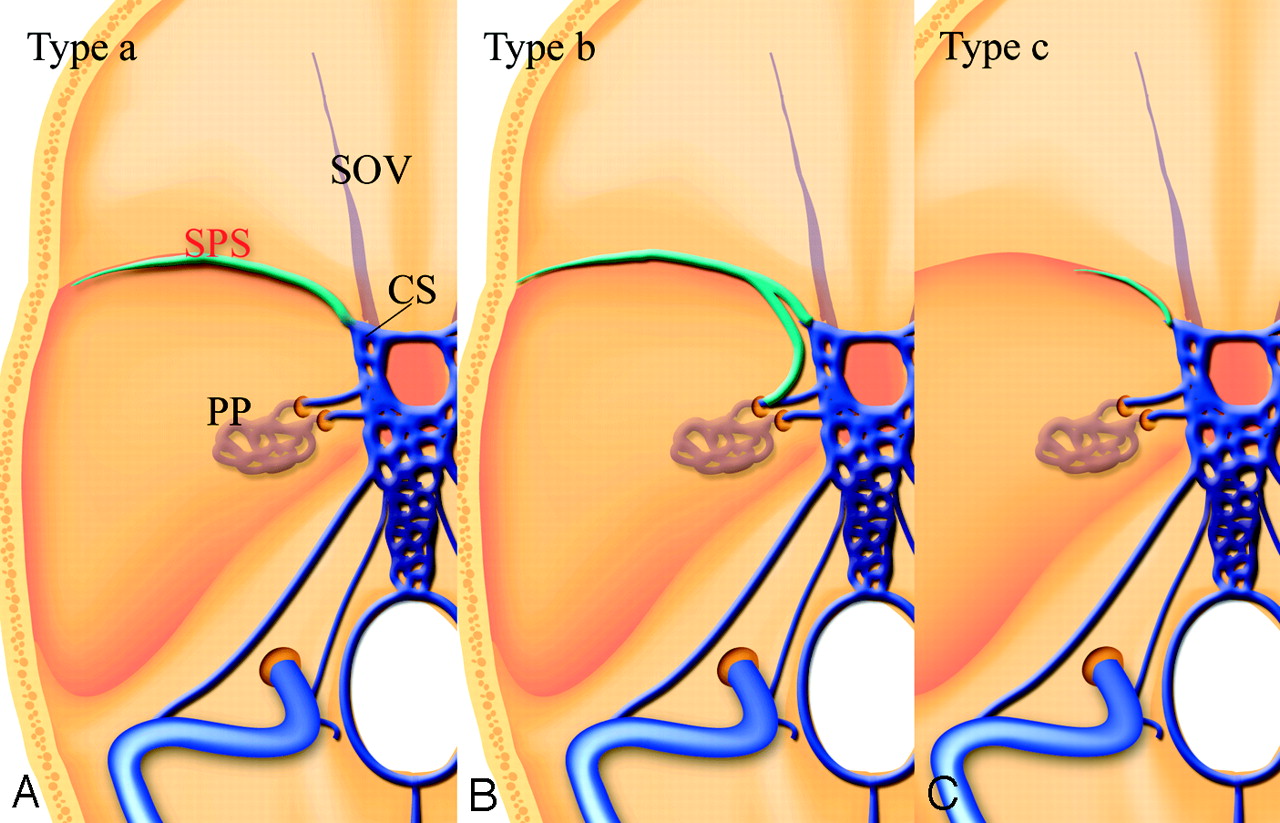
    Schematic drawings of variations of the SPS. Type a, The SPS runs along the lesser sphenoid wing and connects to the anterior aspect of the cavernous sinus. Type b, The SPS runs along the lesser sphenoid wing and connects with the foramen ovale plexus or the pterygoid plexus. Type c, the hypoplastic SPS connects with the cavernous sinus. CS indicates the cavernous sinus

  • Fig 6.
    • Download figure
    • Open in new tab
    • Download powerpoint
    Fig 6.

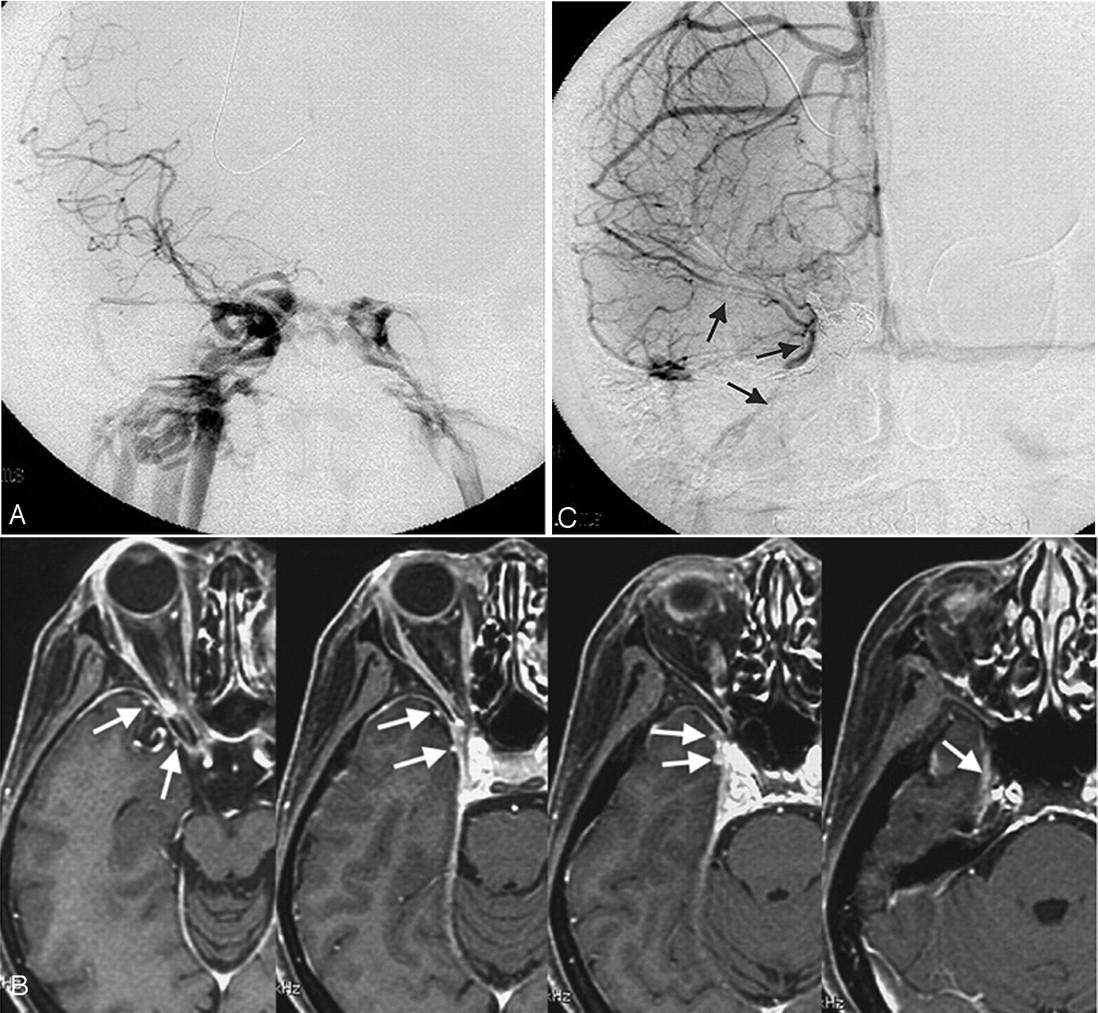
    Mixed type with type A and B. A 57-year-old woman with left cavernous dural arteriovenous fistula.

    A, Lateral view; B, frontal view. Right carotid arteriogram demonstrates a left cavernous dural arteriovenous fistula draining into the cerebellar cortical veins via the superior petrosal sinus (arrows). Retrograde opacification of the SMCV is not shown.

    C, On the contrast-enhanced gradient-echo MR image, a small venous structure connecting to lateral wall of the cavernous sinus, thought to be the SMCV, is observed (white arrow). The findings indicate the potential risk of cortical venous reflux to the medial temporal lobe. Another prominent SMCV flowed into the pterygoid plexus with no connection to the cavernous sinus (black arrow). Note the dilated inferior hypophyseal arteries joining the posteroinferior aspect of the cavernous sinus (arrowheads) and cortical venous reflux to the cerebellar veins (open arrows).

    D, This patient was treated by transvenous embolization, and the shunt disappeared completely.

  • Fig 7.
    • Download figure
    • Open in new tab
    • Download powerpoint
    Fig 7.

    Type C, A 43-year-old woman with a right direct carotid cavernous fistula.

    A, Frontal view of a right carotid arteriogram demonstrates a right carotid cavernous fistula. Retrograde opacification of the SMCV is not shown.

    B, On the contrast-enhanced gradient-echo MR image, 2 SMCVs are conjoined beside the cavernous sinus and flow through the middle carnial fossa (arrows). This patient was treated by transarterial and transvenous coil-packing of the right cavernous sinus.

    C, The carotid angiogram immediately after embolization demonstrates no visualization of the shunt. Note the right SMCV flowing beside the coil mass (arrows).

  • Fig 8.
    • Download figure
    • Open in new tab
    • Download powerpoint
    Fig 8.

    Schematic drawings of the developmental anatomy of cavernous and para-cavernous venous structures in the embryonic stage. SSS indicates superior sagittal sinus; PTS, primitive tentorial sinus; PS, prootic sinus; PMS, primitive maxillary sinus; and IPS, inferior petrosal sinus.

    A, Lateral view; B, axial view. In an 8-week embryo, cerebral venous structures develop from primitive dural plexuses surrounding primitive cerebral vesicles. The plexuses fuse to form venous sinuses and cortical veins. Two major primitive sinuses, the primitive tentorial sinus and the prootic sinus, contribute to the development of cavernous sinus and para-cavernous sinus veins. The primitive SMCV is connected with the transverse sinus via the primitive tentorial sinus.

    C, In a 12-week embryo, after several weeks, the primitive SMCV is elongated and develops anteromedially to form the SMCV as a development of the cerebral hemisphere. The prootic sinus contributes to form the superior ophthalmic vein, the cavernous sinus, and the foramen ovale venous plexus.

    D, Axial view; E, lateral view. In a developed embryo, the SMCV makes further anteromedial development in the prenatal stage; however, in many patients, there is no direct connection between the SMCV and the cavernous sinus. Secondary anastomosis after birth may form a connection between the SMCV and the cavernous sinus, and the connection to the primitive tentorial sinus subsequently degenerates.

Tables

  • Figures
  • Frequency of each variation of superficial middle cerebral vein and sphenoparietal sinus

    Superficial Middle Cerebral VeinSphenoparietal Sinus
    TypeNo. of Sites (%)TypeNo. of Sites (%)
    A29 (39)a52 (72)
    B22 (30)b3 (4)
    C8 (11)c12 (14)
    D6 (8)|⟶(B 4
    Mixed type2 (2)C 3
    D 5
PreviousNext
Back to top

In this issue

American Journal of Neuroradiology: 27 (5)
American Journal of Neuroradiology
Vol. 27, Issue 5
May 2006
  • Table of Contents
  • Index by author
Advertisement
Print
Download PDF
Email Article

Thank you for your interest in spreading the word on American Journal of Neuroradiology.

NOTE: We only request your email address so that the person you are recommending the page to knows that you wanted them to see it, and that it is not junk mail. We do not capture any email address.

Enter multiple addresses on separate lines or separate them with commas.
Para-Cavernous Sinus Venous Structures: Anatomic Variations and Pathologic Conditions Evaluated on Fat-Suppressed 3D Fast Gradient-Echo MR Images
(Your Name) has sent you a message from American Journal of Neuroradiology
(Your Name) thought you would like to see the American Journal of Neuroradiology web site.
CAPTCHA
This question is for testing whether or not you are a human visitor and to prevent automated spam submissions.
Cite this article
S. Tanoue, H. Kiyosue, M. Okahara, Y. Sagara, Y. Hori, J. Kashiwagi, H. Mori
Para-Cavernous Sinus Venous Structures: Anatomic Variations and Pathologic Conditions Evaluated on Fat-Suppressed 3D Fast Gradient-Echo MR Images
American Journal of Neuroradiology May 2006, 27 (5) 1083-1089;

Citation Manager Formats

  • BibTeX
  • Bookends
  • EasyBib
  • EndNote (tagged)
  • EndNote 8 (xml)
  • Medlars
  • Mendeley
  • Papers
  • RefWorks Tagged
  • Ref Manager
  • RIS
  • Zotero
0 Responses
Respond to this article
Share
Bookmark this article
Para-Cavernous Sinus Venous Structures: Anatomic Variations and Pathologic Conditions Evaluated on Fat-Suppressed 3D Fast Gradient-Echo MR Images
S. Tanoue, H. Kiyosue, M. Okahara, Y. Sagara, Y. Hori, J. Kashiwagi, H. Mori
American Journal of Neuroradiology May 2006, 27 (5) 1083-1089;
del.icio.us logo Twitter logo Facebook logo Mendeley logo
  • Tweet Widget
  • Facebook Like
  • Google Plus One

Jump to section

  • Article
    • Abstract
    • Materials and Methods
    • Results
    • Discussion
    • Conclusion
    • References
  • Figures & Data
  • Info & Metrics
  • Responses
  • References
  • PDF

Related Articles

  • No related articles found.
  • PubMed
  • Google Scholar

Cited By...

  • Petrobasal Vein: A Previously Unrecognized Vein Directly Connecting the Superior Petrosal Sinus with the Emissary Vein of the Foramen Ovale
  • Middle Cranial Fossa Sphenoidal Region Dural Arteriovenous Fistulas: Anatomic and Treatment Considerations
  • Venous structures at the craniocervical junction: anatomical variations evaluated by multidetector row CT
  • Crossref
  • Google Scholar

This article has not yet been cited by articles in journals that are participating in Crossref Cited-by Linking.

More in this TOC Section

  • Correlation of Apparent Diffusion Coefficient at 3T with Prognostic Parameters of Retinoblastoma
  • Parathyroid Lesions: Characterization with Dual-Phase Arterial and Venous Enhanced CT of the Neck
  • Efficacy of Diffusion-Weighted Imaging for the Differentiation between Lymphomas and Carcinomas of the Nasopharynx and Oropharynx: Correlations of Apparent Diffusion Coefficients and Histologic Features
Show more HEAD AND NECK

Similar Articles

Advertisement

Indexed Content

  • Current Issue
  • Accepted Manuscripts
  • Article Preview
  • Past Issues
  • Editorials
  • Editor's Choice
  • Fellows' Journal Club
  • Letters to the Editor
  • Video Articles

Cases

  • Case Collection
  • Archive - Case of the Week
  • Archive - Case of the Month
  • Archive - Classic Case

Special Collections

  • AJNR Awards
  • ASNR Foundation Special Collection
  • Most Impactful AJNR Articles
  • Photon-Counting CT
  • Spinal CSF Leak Articles (Jan 2020-June 2024)

More from AJNR

  • Trainee Corner
  • Imaging Protocols
  • MRI Safety Corner

Multimedia

  • AJNR Podcasts
  • AJNR Scantastics

Resources

  • Turnaround Time
  • Submit a Manuscript
  • Submit a Video Article
  • Submit an eLetter to the Editor/Response
  • Manuscript Submission Guidelines
  • Statistical Tips
  • Fast Publishing of Accepted Manuscripts
  • Graphical Abstract Preparation
  • Imaging Protocol Submission
  • Evidence-Based Medicine Level Guide
  • Publishing Checklists
  • Author Policies
  • Become a Reviewer/Academy of Reviewers
  • News and Updates

About Us

  • About AJNR
  • Editorial Board
  • Editorial Board Alumni
  • Alerts
  • Permissions
  • Not an AJNR Subscriber? Join Now
  • Advertise with Us
  • Librarian Resources
  • Feedback
  • Terms and Conditions
  • AJNR Editorial Board Alumni

American Society of Neuroradiology

  • Not an ASNR Member? Join Now

© 2025 by the American Society of Neuroradiology All rights, including for text and data mining, AI training, and similar technologies, are reserved.
Print ISSN: 0195-6108 Online ISSN: 1936-959X

Powered by HighWire