Advertisement

Main menu

  • Home
  • Content
    • Current Issue
    • Accepted Manuscripts
    • Article Preview
    • Past Issue Archive
    • Video Articles
    • AJNR Case Collection
    • Case of the Week Archive
    • Case of the Month Archive
    • Classic Case Archive
  • Special Collections
    • AJNR Awards
    • ASNR Foundation Special Collection
    • Most Impactful AJNR Articles
    • Photon-Counting CT
    • Spinal CSF Leak Articles (Jan 2020-June 2024)
  • Multimedia
    • AJNR Podcasts
    • AJNR SCANtastic
    • Trainee Corner
    • MRI Safety Corner
    • Imaging Protocols
  • For Authors
    • Submit a Manuscript
    • Submit a Video Article
    • Submit an eLetter to the Editor/Response
    • Manuscript Submission Guidelines
    • Statistical Tips
    • Fast Publishing of Accepted Manuscripts
    • Graphical Abstract Preparation
    • Imaging Protocol Submission
    • Author Policies
  • About Us
    • About AJNR
    • Editorial Board
    • Editorial Board Alumni
  • More
    • Become a Reviewer/Academy of Reviewers
    • Subscribers
    • Permissions
    • Alerts
    • Feedback
    • Advertisers
    • ASNR Home

User menu

  • Alerts
  • Log in

Search

  • Advanced search
American Journal of Neuroradiology

American Journal of Neuroradiology

ASHNR American Society of Functional Neuroradiology ASHNR American Society of Pediatric Neuroradiology ASSR
  • Alerts
  • Log in

Advanced Search

  • Home
  • Content
    • Current Issue
    • Accepted Manuscripts
    • Article Preview
    • Past Issue Archive
    • Video Articles
    • AJNR Case Collection
    • Case of the Week Archive
    • Case of the Month Archive
    • Classic Case Archive
  • Special Collections
    • AJNR Awards
    • ASNR Foundation Special Collection
    • Most Impactful AJNR Articles
    • Photon-Counting CT
    • Spinal CSF Leak Articles (Jan 2020-June 2024)
  • Multimedia
    • AJNR Podcasts
    • AJNR SCANtastic
    • Trainee Corner
    • MRI Safety Corner
    • Imaging Protocols
  • For Authors
    • Submit a Manuscript
    • Submit a Video Article
    • Submit an eLetter to the Editor/Response
    • Manuscript Submission Guidelines
    • Statistical Tips
    • Fast Publishing of Accepted Manuscripts
    • Graphical Abstract Preparation
    • Imaging Protocol Submission
    • Author Policies
  • About Us
    • About AJNR
    • Editorial Board
    • Editorial Board Alumni
  • More
    • Become a Reviewer/Academy of Reviewers
    • Subscribers
    • Permissions
    • Alerts
    • Feedback
    • Advertisers
    • ASNR Home
  • Follow AJNR on Twitter
  • Visit AJNR on Facebook
  • Follow AJNR on Instagram
  • Join AJNR on LinkedIn
  • RSS Feeds

AJNR is seeking candidates for the AJNR Podcast Editor. Read the position description.

Research ArticleINTERVENTIONAL

Embolization of Acutely Ruptured and Unruptured Wide-Necked Cerebral Aneurysms Using the Neuroform2 Stent without Pretreatment with Antiplatelets: A Single Center Experience

V. Katsaridis, C. Papagiannaki and C. Violaris
American Journal of Neuroradiology May 2006, 27 (5) 1123-1128;
V. Katsaridis
  • Find this author on Google Scholar
  • Find this author on PubMed
  • Search for this author on this site
C. Papagiannaki
  • Find this author on Google Scholar
  • Find this author on PubMed
  • Search for this author on this site
C. Violaris
  • Find this author on Google Scholar
  • Find this author on PubMed
  • Search for this author on this site
  • Article
  • Figures & Data
  • Info & Metrics
  • Responses
  • References
  • PDF
Loading

Article Figures & Data

Figures

  • Fig 1.
    • Download figure
    • Open in new tab
    • Download powerpoint
    Fig 1.

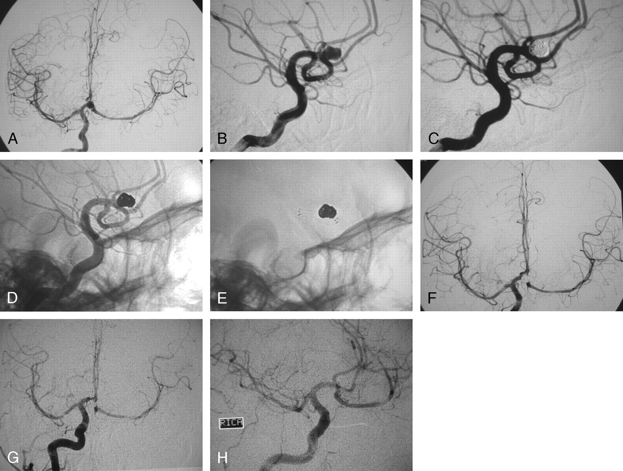
    A, Digital subtraction angiography (DSA), working projection, in a 69-year-old woman who presented with subarachnoid hemorrhage from a ruptured wide-necked left internal carotid artery (ICA) aneurysm at the origin of the posterior communicating artery (PcomA). A pseudoaneurysm is noted at the fundus of the ruptured aneurysm.

    B, DSA, working projection. The aneurysm is shown embolized after the deployment of a 4 × 15-mm Neuroform2 stent in the ICA. No contrast material in the aneurysm is shown, and the PcomA is shown patent.

    C, Unsubtracted angiography, working projection, showing the stent in the ICA and the coils in the aneurysm.

    D, Unsubtracted view, working projection, showing the markers of the stent and the coils.

    E, Follow-up DSA of the left ICA at 17 months, anteroposterior projection. The aneurysm is not visualized and all the branches are patent.

    F, Follow-up DSA the left ICA at 17 months, lateral projection. The aneurysm is not visualized and all the branches are patent.

  • Fig 2.
    • Download figure
    • Open in new tab
    • Download powerpoint
    Fig 2.

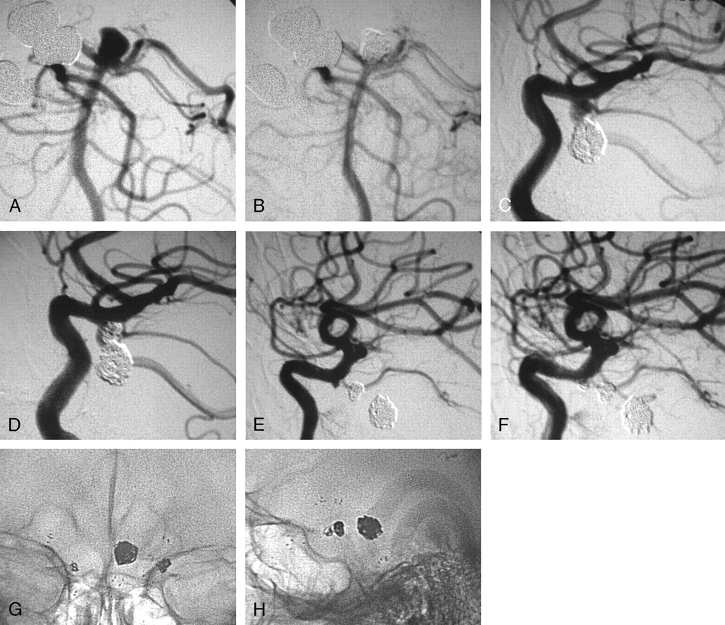
    A, Digital subtraction angiography (DSA), anteroposterior projection, of a 32-year-old woman with aplasia of the left internal carotid artery (ICA) harboring an incidental aneurysm of the anterior communicating artery (AcomA). The whole left hemisphere is supplied by the right ICA through the AcomA. Patency of the AcomA after the embolization was of the utmost importance.

    B, DSA, working projection for the embolization of the wide-necked AcomA aneurysm.

    C, DSA, working projection, showing the aneurysm embolized after the deployment of a 3 × 15-mm Neuroform2 stent in the A1 segment of the right anterior cerebral artery (ACA) extending to the AcomA and to the A2 segment of the left ACA, covering the neck of the aneurysm.

    D, Unsubtracted angiography, working projection, showing the stent in the AcomA complex and the coils in the aneurysm.

    E, Unsubtracted view, working projection, showing the markers of the stent and the coils.

    F, DSA, anteroposterior projection, after the embolization of the aneurysm showing the patency of the AcomA and the adequate supply of the left hemisphere.

    G, Follow-up DSA at 6 months, anteroposterior projection, showing the adequate supply of the left hemisphere from the right ICA.

    H, Follow-up DSA at 6 months, working projection, showing the persistent occlusion of the aneurysm and the patency of all the branches of the AcomA complex.

  • Fig 3.
    • Download figure
    • Open in new tab
    • Download powerpoint
    Fig 3.

    A 30-year-old man presented with subarachnoid hemorrhage, and angiography revealed he harbored 3 aneurysms: 1 at the basilar tip and 1 on each internal carotid artery (ICA) at the origin of both posterior communicating arteries. The wide-necked basilar tip aneurysm was considered the ruptured one and was treated first with coil embolization.

    A, Digital subtraction angiography (DSA), working projection, showing the wide-necked basilar tip aneurysm incorporating in the neck the proximal left posterior cerebral artery.

    B, DSA, working projection. The aneurysm is shown embolized after the deployment of a 3.5 × 15-mm Neuroform2 stent in the basilar artery extending into the left posterior cerebral artery.

    C, DSA, working projection showing a wide-necked left ICA aneurysm at the origin of the posterior communicating artery.

    D, DSA, working projection. The left ICA aneurysm is shown embolized with the use of a 4 × 15-mm Neuroform2 stent. The posterior communicating artery is shown patent despite the prolapse of a small coil loop in its lumen.

    E, DSA, working projection, showing a third small aneurysm on the right ICA by the origin of the posterior communicating artery which was not opacified by the ICA injection.

    F, DSA, working projection. The aneurysm is shown embolized after the deployment of another 4 × 15-mm Neuroform2 stent.

    G, Unsubtracted view, anteroposterior projection, showing the proximal and distal markers of the 3 stents and the 3 coil masses inside the 3 aneurysms.

    H, Unsubtracted view, lateral projection, showing the proximal and distal markers of the 3 stents and the 3 coil masses inside the 3 aneurysms. Follow-up DSA is not available for this patient because he was a resident of a different country and returned home in excellent clinical condition.

PreviousNext
Back to top

In this issue

American Journal of Neuroradiology: 27 (5)
American Journal of Neuroradiology
Vol. 27, Issue 5
May 2006
  • Table of Contents
  • Index by author
Advertisement
Print
Download PDF
Email Article

Thank you for your interest in spreading the word on American Journal of Neuroradiology.

NOTE: We only request your email address so that the person you are recommending the page to knows that you wanted them to see it, and that it is not junk mail. We do not capture any email address.

Enter multiple addresses on separate lines or separate them with commas.
Embolization of Acutely Ruptured and Unruptured Wide-Necked Cerebral Aneurysms Using the Neuroform2 Stent without Pretreatment with Antiplatelets: A Single Center Experience
(Your Name) has sent you a message from American Journal of Neuroradiology
(Your Name) thought you would like to see the American Journal of Neuroradiology web site.
CAPTCHA
This question is for testing whether or not you are a human visitor and to prevent automated spam submissions.
Cite this article
V. Katsaridis, C. Papagiannaki, C. Violaris
Embolization of Acutely Ruptured and Unruptured Wide-Necked Cerebral Aneurysms Using the Neuroform2 Stent without Pretreatment with Antiplatelets: A Single Center Experience
American Journal of Neuroradiology May 2006, 27 (5) 1123-1128;

Citation Manager Formats

  • BibTeX
  • Bookends
  • EasyBib
  • EndNote (tagged)
  • EndNote 8 (xml)
  • Medlars
  • Mendeley
  • Papers
  • RefWorks Tagged
  • Ref Manager
  • RIS
  • Zotero
0 Responses
Respond to this article
Share
Bookmark this article
Embolization of Acutely Ruptured and Unruptured Wide-Necked Cerebral Aneurysms Using the Neuroform2 Stent without Pretreatment with Antiplatelets: A Single Center Experience
V. Katsaridis, C. Papagiannaki, C. Violaris
American Journal of Neuroradiology May 2006, 27 (5) 1123-1128;
del.icio.us logo Twitter logo Facebook logo Mendeley logo
  • Tweet Widget
  • Facebook Like
  • Google Plus One

Jump to section

  • Article
    • Abstract
    • Patients and Techniques
    • Results
    • Discussion
    • Conclusion
    • References
  • Figures & Data
  • Info & Metrics
  • Responses
  • References
  • PDF

Related Articles

  • No related articles found.
  • PubMed
  • Google Scholar

Cited By...

  • Use of the pCONus as an adjunct to coil embolization of acutely ruptured aneurysms
  • Clinical and angiographic outcomes after stent-assisted coiling of cerebral aneurysms with Enterprise and Neuroform stents: a comparative analysis of the literature
  • Complications in Stent-Assisted Endovascular Therapy of Ruptured Intracranial Aneurysms and Relevance to Antiplatelet Administration: A Systematic Review
  • Silent embolism after stent-assisted coiling of cerebral aneurysms: diffusion-weighted MRI study of 75 cases
  • Stent usage in the treatment of intracranial aneurysms: past, present and future
  • Stent assisted coiling of the ruptured wide necked intracranial aneurysm
  • Immediate and Midterm Results following Treatment of Recently Ruptured Intracranial Aneurysms with the Pipeline Embolization Device
  • Y stenting using kissing stents for the treatment of bifurcation aneurysms
  • Stent-Supported Aneurysm Coiling: A Literature Survey of Treatment and Follow-Up
  • Stent-Assisted Coiling in Acutely Ruptured Intracranial Aneurysms: A Qualitative, Systematic Review of the Literature
  • Neuroform Stent-Assisted Coiling of Unruptured Intracranial Aneurysms: Short- and Midterm Results from a Single-Center Experience with 68 Patients
  • Endovascular Treatment of Wide-Neck Middle Cerebral Artery Aneurysms with Stents: A Review of 16 Cases
  • Acutely ruptured intracranial saccular aneurysms treated with stent assisted coiling: complications and outcomes in 42 consecutive patients
  • Crossref
  • Google Scholar

This article has not yet been cited by articles in journals that are participating in Crossref Cited-by Linking.

More in this TOC Section

  • SAVE vs. Solumbra Techniques for Thrombectomy
  • Contrast-Induced Encephalopathy after NeuroIR
  • CT Perfusion&Reperfusion in Acute Ischemic Stroke
Show more Interventional

Similar Articles

Advertisement

Indexed Content

  • Current Issue
  • Accepted Manuscripts
  • Article Preview
  • Past Issues
  • Editorials
  • Editor's Choice
  • Fellows' Journal Club
  • Letters to the Editor
  • Video Articles

Cases

  • Case Collection
  • Archive - Case of the Week
  • Archive - Case of the Month
  • Archive - Classic Case

Special Collections

  • AJNR Awards
  • ASNR Foundation Special Collection
  • Most Impactful AJNR Articles
  • Photon-Counting CT
  • Spinal CSF Leak Articles (Jan 2020-June 2024)

More from AJNR

  • Trainee Corner
  • Imaging Protocols
  • MRI Safety Corner

Multimedia

  • AJNR Podcasts
  • AJNR Scantastics

Resources

  • Turnaround Time
  • Submit a Manuscript
  • Submit a Video Article
  • Submit an eLetter to the Editor/Response
  • Manuscript Submission Guidelines
  • Statistical Tips
  • Fast Publishing of Accepted Manuscripts
  • Graphical Abstract Preparation
  • Imaging Protocol Submission
  • Evidence-Based Medicine Level Guide
  • Publishing Checklists
  • Author Policies
  • Become a Reviewer/Academy of Reviewers
  • News and Updates

About Us

  • About AJNR
  • Editorial Board
  • Editorial Board Alumni
  • Alerts
  • Permissions
  • Not an AJNR Subscriber? Join Now
  • Advertise with Us
  • Librarian Resources
  • Feedback
  • Terms and Conditions
  • AJNR Editorial Board Alumni

American Society of Neuroradiology

  • Not an ASNR Member? Join Now

© 2025 by the American Society of Neuroradiology All rights, including for text and data mining, AI training, and similar technologies, are reserved.
Print ISSN: 0195-6108 Online ISSN: 1936-959X

Powered by HighWire