Advertisement

Main menu

  • Home
  • Content
    • Current Issue
    • Accepted Manuscripts
    • Article Preview
    • Past Issue Archive
    • Video Articles
    • AJNR Case Collection
    • Case of the Week Archive
    • Case of the Month Archive
    • Classic Case Archive
  • Special Collections
    • AJNR Awards
    • ASNR Foundation Special Collection
    • Most Impactful AJNR Articles
    • Photon-Counting CT
    • Spinal CSF Leak Articles (Jan 2020-June 2024)
  • Multimedia
    • AJNR Podcasts
    • AJNR SCANtastic
    • Trainee Corner
    • MRI Safety Corner
    • Imaging Protocols
  • For Authors
    • Submit a Manuscript
    • Submit a Video Article
    • Submit an eLetter to the Editor/Response
    • Manuscript Submission Guidelines
    • Statistical Tips
    • Fast Publishing of Accepted Manuscripts
    • Graphical Abstract Preparation
    • Imaging Protocol Submission
    • Author Policies
  • About Us
    • About AJNR
    • Editorial Board
    • Editorial Board Alumni
  • More
    • Become a Reviewer/Academy of Reviewers
    • Subscribers
    • Permissions
    • Alerts
    • Feedback
    • Advertisers
    • ASNR Home

User menu

  • Alerts
  • Log in

Search

  • Advanced search
American Journal of Neuroradiology

American Journal of Neuroradiology

ASHNR American Society of Functional Neuroradiology ASHNR American Society of Pediatric Neuroradiology ASSR
  • Alerts
  • Log in

Advanced Search

  • Home
  • Content
    • Current Issue
    • Accepted Manuscripts
    • Article Preview
    • Past Issue Archive
    • Video Articles
    • AJNR Case Collection
    • Case of the Week Archive
    • Case of the Month Archive
    • Classic Case Archive
  • Special Collections
    • AJNR Awards
    • ASNR Foundation Special Collection
    • Most Impactful AJNR Articles
    • Photon-Counting CT
    • Spinal CSF Leak Articles (Jan 2020-June 2024)
  • Multimedia
    • AJNR Podcasts
    • AJNR SCANtastic
    • Trainee Corner
    • MRI Safety Corner
    • Imaging Protocols
  • For Authors
    • Submit a Manuscript
    • Submit a Video Article
    • Submit an eLetter to the Editor/Response
    • Manuscript Submission Guidelines
    • Statistical Tips
    • Fast Publishing of Accepted Manuscripts
    • Graphical Abstract Preparation
    • Imaging Protocol Submission
    • Author Policies
  • About Us
    • About AJNR
    • Editorial Board
    • Editorial Board Alumni
  • More
    • Become a Reviewer/Academy of Reviewers
    • Subscribers
    • Permissions
    • Alerts
    • Feedback
    • Advertisers
    • ASNR Home
  • Follow AJNR on Twitter
  • Visit AJNR on Facebook
  • Follow AJNR on Instagram
  • Join AJNR on LinkedIn
  • RSS Feeds

AJNR is seeking candidates for the AJNR Podcast Editor. Read the position description.

OtherREVIEW ARTICLE

Multiple Sclerosis: The Role of MR Imaging

Y. Ge
American Journal of Neuroradiology June 2006, 27 (6) 1165-1176;
Y. Ge
  • Find this author on Google Scholar
  • Find this author on PubMed
  • Search for this author on this site
  • Article
  • Figures & Data
  • Info & Metrics
  • Responses
  • References
  • PDF
Loading

Article Figures & Data

Figures

  • Tables
  • Fig 1.
    • Download figure
    • Open in new tab
    • Download powerpoint
    Fig 1.

    Axial T2-weighted images of a 43-year-old RRMS patient show prominent perivascular spaces (short arrows), which project radially and are aligned with lesions, following the course and configuration of deep venular structures. This may be associated with perivascular inflammation, which initiates the development of new lesions (long arrow). These prominent perivascular spaces might have implications for differentiating primary from secondary demyelinating lesions.
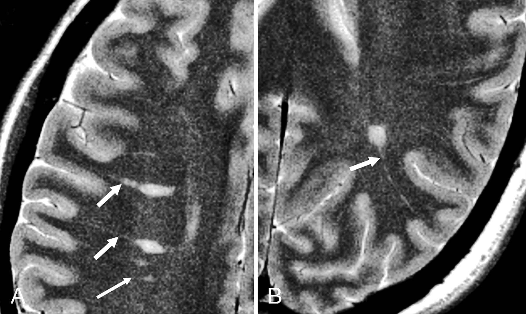
  • Fig 2.
    • Download figure
    • Open in new tab
    • Download powerpoint
    Fig 2.

    A 30-year-old female RRMS patient shown on T2WI (A), FLAIR (B), and contrast-enhanced T1WI. The lesions on FLAIR are usually prominent and several small lesions are depicted only on FLAIR (arrows). The lesion enhancement can be nodule (as shown in this case) or ringlike on T1-weighted imaging.

  • Fig 3.
    • Download figure
    • Open in new tab
    • Download powerpoint
    Fig 3.

    MS lesion (arrow) in corpus callosum on FLAIR imaging is failed to be picked up on T2-weighted imaging.

  • Fig 4.
    • Download figure
    • Open in new tab
    • Download powerpoint
    Fig 4.

    Averaged magnetization transfer ratio histograms from 3 groups (healthy control, RRMS, and SPMS) for global NAGM (A) and NAWM (B) tissues. Lower normalized peak height in SPMS population indicates relatively less residual normal brain tissue compared with that in RRMS patients.
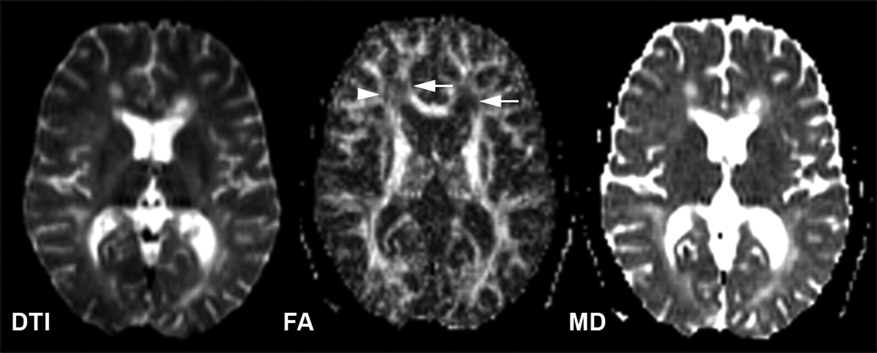
  • Fig 5.
    • Download figure
    • Open in new tab
    • Download powerpoint
    Fig 5.

    DTI (left, b = 0), FA (middle), and MD (right) maps of a 31-year-old female patient with RRMS. The decreased value of FA and increased value of MD for the lesions (arrows) are shown in their maps. Note that the decreased FA (arrowhead) in some white matter areas is probably due to fiber crossing.

  • Fig 6.
    • Download figure
    • Open in new tab
    • Download powerpoint
    Fig 6.

    Fiber tractography in a patient with MS (A) and a healthy volunteer (B). All the MS plaques (arrows) were marked and constructed in 3D. Note the reduced number of fibers when they traverse white matter lesions in the patient.

  • Fig 7.
    • Download figure
    • Open in new tab
    • Download powerpoint
    Fig 7.

    Axial T2-weighed (TE/TR = 90/2500 msec) image of a 26-year-old woman MS patient superimposed with the MR spectroscopy volume of interest. Spectra from 2 lesions (2 and 4) and 2 contralateral NAWM regions (1 and 3) are shown on common intensity and chemical shift (ppm) scales.

  • Fig 8.
    • Download figure
    • Open in new tab
    • Download powerpoint
    Fig 8.

    Axial gradient-echo imaging in a 29-year-old patient with MS (A) and a 34-year-old healthy volunteer (B). Greater hypointense signal intensities, which may be associated with excessive iron deposition, are seen in all ferruginated neurons in a patient compared with a healthy volunteer.

Tables

  • Figures
    • View popup
    Table 1:

    MR imaging characteristics in multiple sclerosis

    MethodsClassic Imaging Features
    Conventional MRIMultiple lesions: periventricular > peripheral
    Ovoid shape
    Dilated perivascular space
    Optic nerve, U fiber, and callosal involvement
    Generalized atrophy at relatively younger age
    Enhancing lesions (ring, rim, or solid)
    Gradually increased number of lesions
    MTRDecrease in lesions
    Decrease in NAWM—precede new lesion
    Lower in nonenhancing than in enhancing lesions
    Lower in ischemic than in demyelinating lesions
    Lowest in the core of ring enhancing lesion
    Lower in gray matter than in white matter
    DTIHigher MD and lower FA in lesions than in NAWM
    Higher MD and lower FA in NAWM than in normal white matter
    Not reliable to differentiate enhancing and nonenhancing lesions
    Not significant in NAWM in early stage of disease
    Visualization of specific fiber tract on tractography
    Perfusion imagingDecreased CBF and CBV in general
    Locally increased CBV in enhancing lesions
    Locally increased CBV in some chronic lesions
    1H-MR spectroscopyMarked decrease of NAA level
    Increase of choline
    Presence of lipid
    Spinal imagingMultiple lesions
    Cervical spinal cord (peripherally located)
    Asymptomatic lesions
    Less than 2 vertebral bodies in length
    Focal atrophy
    • Note:—MTR indicates magnetization transfer ratio; DTI, diffusion tensor imaging;

    • 1 H-MR spectroscopy, proton MR spectroscopy; NAWM, normal appearing white matter; MD, mean diffusivity; FA, fractional anisotropy; CBF, cerebral blood flow; CBV, cerebral blood volume; NAA, N-acetylaspartate.

    • View popup
    Table 2:

    Summary of MR imaging in multiple sclerosis

    MethodPresent RoleFuture Direction
    Conventional MRIRoutine for diagnosis and disease evaluationIncrease detectability for gray matter lesions, especially using high-resolution imaging of high-field MRI
        High sensitivity, less specificity
        MRI inflammatory activity (often clinically silent)
    Volumetric MRIQuantitation of disease burdenProvide quick, friendly, and reliable routine measures for monitoring follow-up and treatment efficacy
        Natural history and clinical trials (lesion load)
        Global adverse outcome of pathology (atrophy)
    MTIInjury in NAWM. Increased specificity for myelinApply to evaluation of remyelination and efficacy of various disease modifying treatments
        Extent of demyelination in lesion and NAWM
        Improved correlation with neurocognitive outcome
    DTIInjury in NAWM. Water diffusion abnormality
    1. Distinguish axonal and myelin loss by quantifying the axial radial diffusivity

    2. Improve the sensitivity and specificity of underlying diffusion changes

    3. Further explore fiber tractography in multiple sclerosis

        Uncertain role of demyelination from inflammation
        Global net loss of structural organization
        Identification of specific white matter tract (integrity and directionality)
    DSC-MRIPredict lesion activity and provide additional information of microvascular abnormalityExplore the ischemic pathogenetic mechanism in certain types of multiple sclerosis lesions
    1H-MR spectroscopyNew insights into the in vivo biochemical pathology(1) Improve the reliability and applicability in clinical trials
        Marked changes in NAA, not other metabolites
        Global marker of neuronal/axonal dysfunction(2) Increase specificity of other metabolites
    • Note:—MTI indicates magnetic tensor imaging; DTI, diffusion tensor imaging, DSC-MRI, dynamic susceptibility contrast MR imaging;

    • 1 H-MR spectroscopy, proton MR spectroscopy; NAWM, normal appearing white matter; NAA, N-acetylaspartate.

PreviousNext
Back to top

In this issue

American Journal of Neuroradiology: 27 (6)
American Journal of Neuroradiology
Vol. 27, Issue 6
June 2006
  • Table of Contents
  • Index by author
Advertisement
Print
Download PDF
Email Article

Thank you for your interest in spreading the word on American Journal of Neuroradiology.

NOTE: We only request your email address so that the person you are recommending the page to knows that you wanted them to see it, and that it is not junk mail. We do not capture any email address.

Enter multiple addresses on separate lines or separate them with commas.
Multiple Sclerosis: The Role of MR Imaging
(Your Name) has sent you a message from American Journal of Neuroradiology
(Your Name) thought you would like to see the American Journal of Neuroradiology web site.
CAPTCHA
This question is for testing whether or not you are a human visitor and to prevent automated spam submissions.
Cite this article
Y. Ge
Multiple Sclerosis: The Role of MR Imaging
American Journal of Neuroradiology Jun 2006, 27 (6) 1165-1176;

Citation Manager Formats

  • BibTeX
  • Bookends
  • EasyBib
  • EndNote (tagged)
  • EndNote 8 (xml)
  • Medlars
  • Mendeley
  • Papers
  • RefWorks Tagged
  • Ref Manager
  • RIS
  • Zotero
0 Responses
Respond to this article
Share
Bookmark this article
Multiple Sclerosis: The Role of MR Imaging
Y. Ge
American Journal of Neuroradiology Jun 2006, 27 (6) 1165-1176;
del.icio.us logo Twitter logo Facebook logo Mendeley logo
  • Tweet Widget
  • Facebook Like
  • Google Plus One

Jump to section

  • Article
    • Abstract
    • Conventional MR Imaging
    • Volumetric MR Imaging: Lesion Quantification and Brain Atrophy
    • Magnetization Transfer Imaging
    • Diffusion Tensor Imaging
    • Perfusion Imaging
    • 1H-MR Spectroscopy
    • MR Imaging of Basal Ganglia Affected by MS
    • Spinal Cord Imaging in MS
    • High-Field MR Imaging and MS
    • Conclusions
    • Acknowledgments
    • References
  • Figures & Data
  • Info & Metrics
  • Responses
  • References
  • PDF

Related Articles

  • No related articles found.
  • PubMed
  • Google Scholar

Cited By...

  • TAPAS: A Thresholding Approach for Probability Map Automatic Segmentation in Multiple Sclerosis
  • MIMoSA: A Method for Inter-Modal Segmentation Analysis
  • Mindcontrol: A Web Application for Brain Segmentation Quality Control
  • MR Imaging in Multiple Sclerosis: Review and Recommendations for Current Practice
  • Regional White Matter Atrophy-Based Classification of Multiple Sclerosis in Cross-Sectional and Longitudinal Data
  • Crossref
  • Google Scholar

This article has not yet been cited by articles in journals that are participating in Crossref Cited-by Linking.

More in this TOC Section

  • An Atlas of Neonatal Neurovascular Imaging Anatomy as Depicted with Microvascular Imaging: The Intracranial Arteries
  • An Atlas of Neonatal Neurovascular Imaging Anatomy as Depicted with Microvascular Imaging: The Intracranial Veins
  • Clinical Translation of Hyperpolarized 13C Metabolic Probes for Glioma Imaging
Show more Review Article

Similar Articles

Advertisement

Indexed Content

  • Current Issue
  • Accepted Manuscripts
  • Article Preview
  • Past Issues
  • Editorials
  • Editor's Choice
  • Fellows' Journal Club
  • Letters to the Editor
  • Video Articles

Cases

  • Case Collection
  • Archive - Case of the Week
  • Archive - Case of the Month
  • Archive - Classic Case

Special Collections

  • AJNR Awards
  • ASNR Foundation Special Collection
  • Most Impactful AJNR Articles
  • Photon-Counting CT
  • Spinal CSF Leak Articles (Jan 2020-June 2024)

More from AJNR

  • Trainee Corner
  • Imaging Protocols
  • MRI Safety Corner

Multimedia

  • AJNR Podcasts
  • AJNR Scantastics

Resources

  • Turnaround Time
  • Submit a Manuscript
  • Submit a Video Article
  • Submit an eLetter to the Editor/Response
  • Manuscript Submission Guidelines
  • Statistical Tips
  • Fast Publishing of Accepted Manuscripts
  • Graphical Abstract Preparation
  • Imaging Protocol Submission
  • Evidence-Based Medicine Level Guide
  • Publishing Checklists
  • Author Policies
  • Become a Reviewer/Academy of Reviewers
  • News and Updates

About Us

  • About AJNR
  • Editorial Board
  • Editorial Board Alumni
  • Alerts
  • Permissions
  • Not an AJNR Subscriber? Join Now
  • Advertise with Us
  • Librarian Resources
  • Feedback
  • Terms and Conditions
  • AJNR Editorial Board Alumni

American Society of Neuroradiology

  • Not an ASNR Member? Join Now

© 2025 by the American Society of Neuroradiology All rights, including for text and data mining, AI training, and similar technologies, are reserved.
Print ISSN: 0195-6108 Online ISSN: 1936-959X

Powered by HighWire