Advertisement

Main menu

  • Home
  • Content
    • Current Issue
    • Accepted Manuscripts
    • Article Preview
    • Past Issue Archive
    • Video Articles
    • AJNR Case Collection
    • Case of the Week Archive
    • Case of the Month Archive
    • Classic Case Archive
  • Special Collections
    • AJNR Awards
    • ASNR Foundation Special Collection
    • Most Impactful AJNR Articles
    • Photon-Counting CT
    • Spinal CSF Leak Articles (Jan 2020-June 2024)
  • Multimedia
    • AJNR Podcasts
    • AJNR SCANtastic
    • Trainee Corner
    • MRI Safety Corner
    • Imaging Protocols
  • For Authors
    • Submit a Manuscript
    • Submit a Video Article
    • Submit an eLetter to the Editor/Response
    • Manuscript Submission Guidelines
    • Statistical Tips
    • Fast Publishing of Accepted Manuscripts
    • Graphical Abstract Preparation
    • Imaging Protocol Submission
    • Author Policies
  • About Us
    • About AJNR
    • Editorial Board
    • Editorial Board Alumni
  • More
    • Become a Reviewer/Academy of Reviewers
    • Subscribers
    • Permissions
    • Alerts
    • Feedback
    • Advertisers
    • ASNR Home

User menu

  • Alerts
  • Log in

Search

  • Advanced search
American Journal of Neuroradiology

American Journal of Neuroradiology

ASHNR American Society of Functional Neuroradiology ASHNR American Society of Pediatric Neuroradiology ASSR
  • Alerts
  • Log in

Advanced Search

  • Home
  • Content
    • Current Issue
    • Accepted Manuscripts
    • Article Preview
    • Past Issue Archive
    • Video Articles
    • AJNR Case Collection
    • Case of the Week Archive
    • Case of the Month Archive
    • Classic Case Archive
  • Special Collections
    • AJNR Awards
    • ASNR Foundation Special Collection
    • Most Impactful AJNR Articles
    • Photon-Counting CT
    • Spinal CSF Leak Articles (Jan 2020-June 2024)
  • Multimedia
    • AJNR Podcasts
    • AJNR SCANtastic
    • Trainee Corner
    • MRI Safety Corner
    • Imaging Protocols
  • For Authors
    • Submit a Manuscript
    • Submit a Video Article
    • Submit an eLetter to the Editor/Response
    • Manuscript Submission Guidelines
    • Statistical Tips
    • Fast Publishing of Accepted Manuscripts
    • Graphical Abstract Preparation
    • Imaging Protocol Submission
    • Author Policies
  • About Us
    • About AJNR
    • Editorial Board
    • Editorial Board Alumni
  • More
    • Become a Reviewer/Academy of Reviewers
    • Subscribers
    • Permissions
    • Alerts
    • Feedback
    • Advertisers
    • ASNR Home
  • Follow AJNR on Twitter
  • Visit AJNR on Facebook
  • Follow AJNR on Instagram
  • Join AJNR on LinkedIn
  • RSS Feeds

AJNR is seeking candidates for the AJNR Podcast Editor. Read the position description.

OtherSPINE

Two Patients with Intraspinal Germinoma Associated with Klinefelter Syndrome: Case Report and Review of the Literature

Y. Nakata, A. Yagishita and N. Arai
American Journal of Neuroradiology June 2006, 27 (6) 1204-1210;
Y. Nakata
  • Find this author on Google Scholar
  • Find this author on PubMed
  • Search for this author on this site
A. Yagishita
  • Find this author on Google Scholar
  • Find this author on PubMed
  • Search for this author on this site
N. Arai
  • Find this author on Google Scholar
  • Find this author on PubMed
  • Search for this author on this site
  • Article
  • Figures & Data
  • Info & Metrics
  • Responses
  • References
  • PDF
Loading

Article Figures & Data

Figures

  • Tables
  • Fig 1.
    • Download figure
    • Open in new tab
    • Download powerpoint
    Fig 1.
    • Download figure
    • Open in new tab
    • Download powerpoint
    Fig 1.

    Patient 1. MR images of the thoracic spine and brain.

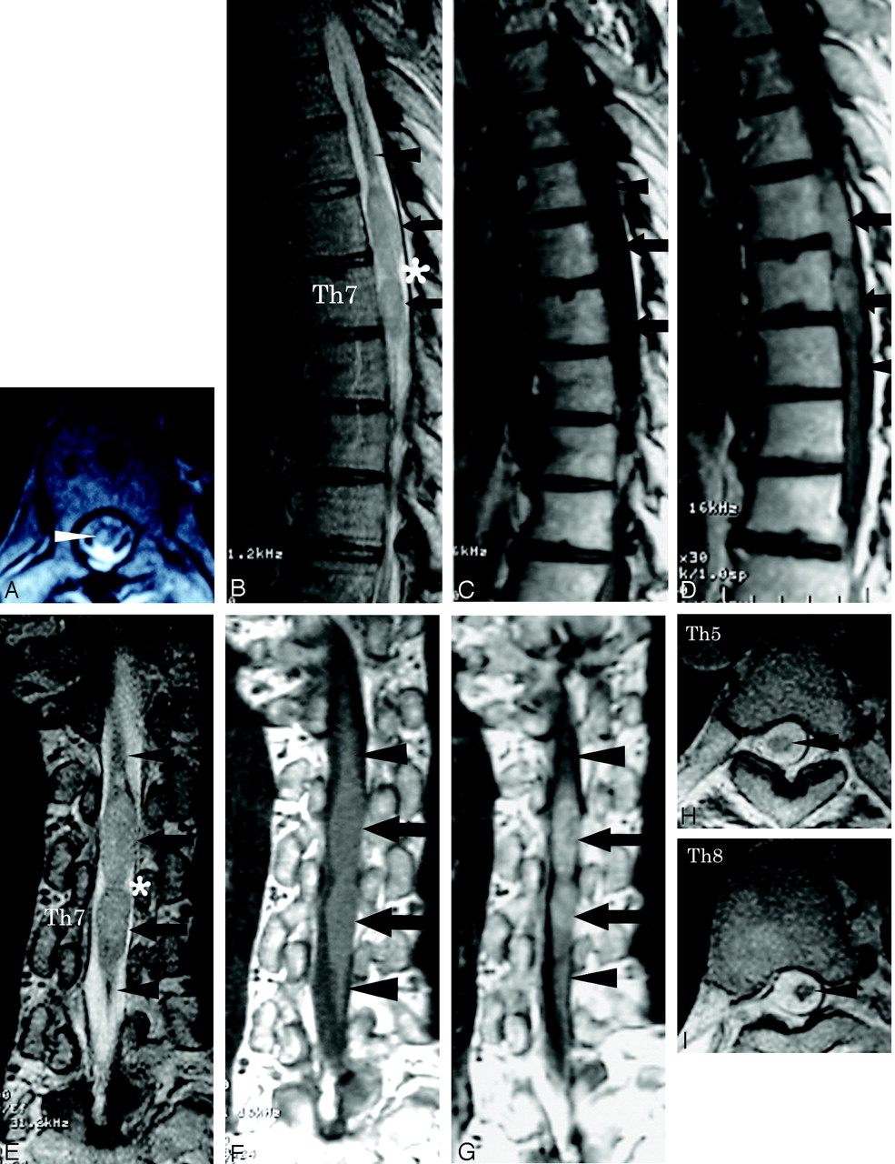
    A, T2-weighted axial image of the thoracic spine in 1999 revealed an area of increased signal intensity changes in the spinal cord at the level of the T7 vertebral body (arrow). Enlargement of the spinal cord was not observed. Good sagittal MR images were not obtained, because of scoliosis.

    B–G, Sagittal and coronal MR images of the thoracic spine in 2003 showed enlargement of the spinal cord between the level of T6 and T7 vertebral bodies, with hyperintensity on T2WI (B and E, arrows), isointensity on T1WI (C and F, arrows) and heterogeneous enhancement on T1WI obtained after an intravenous injection of contrast medium (D and G, arrows), which suggests an intramedullary tumor. Spinal cord atrophy was demonstrated at the upper and lower ends of the intramedullary tumor at the levels of T5 and T8 vertebral bodies on sagittal and coronal images (B–G, arrowheads). The tumor showed a “string of beads” appearance (B and E, asterisks).

    H and I, Spinal cord atrophy was also confirmed on T2-weighted axial images at the levels of T5 and T8 vertebral bodies (arrowheads).

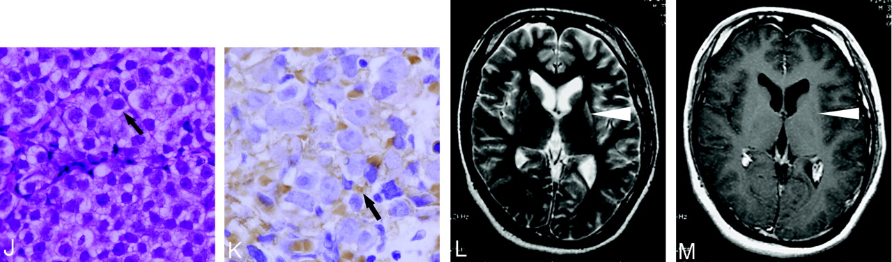
    J, Photomicrograph of a specimen of the intraspinal tumor showed large round cells with clear cytoplasm and central nuclei (arrow) along fibrovascular septa. Small lymphocytes were not apparent (hematoxylin-eosin stain; original magnification, ×66).

    K, Photomicrograph of a specimen of the intraspinal tumor showed that the cell membrane (arrow), which stains brown, was immunoreactive for PLAP. Cell membrane expression of PLAP was characteristic of germinoma (placental alkaline phosphatase stain; original magnification, ×132).

    L and M, The left putamen was atrophic and showed high signal intensity change on T2WI (L, arrowhead). No contrast enhancement was observed on T1WI obtained after an intravenous injection of contrast medium (M, arrowhead). The presence of germinoma was suggested in the left putamen.

  • Fig 2.
    • Download figure
    • Open in new tab
    • Download powerpoint
    Fig 2.

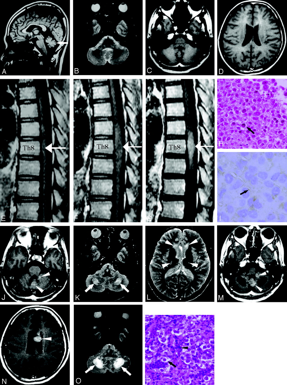
    Patient 2. MR images of the thoracic spine and brain. Because these images were obtained by using an old MR system with low magnetic field (0.5T), images were relatively poor in quality.

    A, T1-weighted sagittal image of the brain in 1989 showed cerebellar atrophy (arrow).

    B–D, No abnormal signal intensity changes suggesting the presence of tumors were detected on MR images, though contrast medium was not given.

    E, T1-weighted sagittal image of the thoracic spine in 1989 demonstrated a slight enlargement of the spinal cord at the T8 level (arrow).

    F and G, MR images of the thoracic spine in 1991 demonstrated further enlargement of the lesion in spinal cord between the level of T7 and T9 vertebral bodies, with hyperintensity on T2WI (not shown), isointensity on T1WI (F, arrow), and heterogeneous enhancement on T1WI obtained after an intravenous injection of contrast medium (G, arrow).

    H, Photomicrograph of a specimen of the intraspinal tumor showed large round cells with clear cytoplasm and central nuclei (arrow). Small lymphocytes were not apparent (hematoxylin-eosin stain; original magnification, ×66).

    I, Photomicrograph of a specimen of the intraspinal tumor showed that the cell membrane (arrow), which was stained brown slightly, was immunoreactive for PLAP. Cell membrane expression of PLAP was characteristic of germinoma (placental alkaline phosphatase stain; original magnification, ×132).

    J, MR images of the brain in 1992 revealed slight contrast enhancement in the cerebellum on T1WI obtained after an intravenous injection of the contrast medium (arrowheads).

    K and L, MR images of the brain in 1992 revealed lesions with increased signal intensity changes on T2WI in the corpus medullaris cerebelli (K, arrows), periventricular white matter around the anterior horn, and corticospinal tracts on both sides (L, arrowheads).

    M and N, MR images of the brain in 1994 demonstrated multiple tumors in the cerebellum and the left lateral ventricle on T1WI after an intravenous injection of contrast medium (M and N, arrowheads).

    O, MR images of the brain in 1994 demonstrated further enlargement of the lesions in the corpus medullaris cerebelli with increased signal intensity changes on T2WI (arrowheads). The lesions in the corpus medullaris cerebelli, periventricular white matter around anterior horn, and corticospinal tracts on both sides with increased signal intensity changes on T2WI remained stable.

    P, Photomicrograph of a specimen of the tumor in the vermis showed 2 distinct cell types: large round cells with clear cytoplasm and central nuclei (arrow) were admixed with small lymphocytes (arrowhead) along fibrovascular septa, indicating a germinoma (hematoxylin-eosin stain; original magnification, ×66).

Tables

  • Figures
    • View popup
    TABLE 1:

    Summary of previous cases of tumors in the central nervous system associated with Klinefelter syndrome

    Patient No./Age (y)/SexSeriesTumor LocationSurgeryHistologic Diagnosis
    1/16/MRubenstein, 19729Pineal regionNot describedGerminoma
    2/20/MAhagon et al, 198310Suprasellar regionBiopsyGerminoma
    3/12/MEllis et al, 198611Posterior hypothalamus, left frontal lobeRadical excisionGerminoma
    4/15/MArens et al, 198812Pineal regionBiopsyGerminoma
    5/40/MLiang et al, 199013Right frontoparietal regionPartial excisionNon-Hodgkin’s lymphoma
    6/19/MHashimoto, et al, 199214Medulla oblongataBiopsyGerminoma
    7/12/MPrall et al, 199515Pineal regionSubtotal resectionMalignant mixed germ cell tumor
    8/13/MWysocka et al, 199616CerebellumTotal resectionPilocytic astrocytoma
    9/19/MKaido et al, 20032Hypothalamus–temporal regionPartial removalGerminoma
    10/29/MGanslandt et al, 20006Spinal cord (T12–L4)Partial resectionGerminoma
    11/35/MOur patient 1Spinal cord (T6–T7), hypothalamic regionTotal resectionGerminoma
    12/27/MOur patient 2Spinal cord (T7–T9), cerebellum, intraventricular regionPartial resectionGerminoma
    • View popup
    TABLE 2:

    Summary of previous cases of primary intramedullary spinal cord germinoma

    Patient No./ Age (y)/SexSeriesTumor LocationSurgeryHistologic DiagnosisValues of hCG in Serum/CSF
    1/5/MHisa et al, 198519Th11–L3Biopsy, amputationGerminoma with syncitiotrophoblastic giant cellsHigh
    2/31/FMatsuoka et al, 199120Th12–L2BiopsyGerminomaNormal
    3/31/MNagasawa et al, 199117MidcervicalNot performedProbably germinomaNot described
    4/34/FHanafusa et al, 199321Th10–Th11Gross total resectionGerminomaNormal
    5/16/FSlagel et al, 199522Th11–L4Partial resectionGerminomaNot described
    6/34/FMatsuyama et al, 199523Th6–Th8Partial resectionGerminomaNormal
    7/24/MItoh et al, 19965Th11–Th12Gross total resectionGerminomaNegative
    8/24/MMiyauchi et al, 199624Th12–L3Partial resectionGerminomaNegative
    9/29/MGanslandt et al, 20006Th12–L4partial resectionGerminomaHigh
    10/33/MHata et al, 200218Th7–Th9Partial resectionGerminomaNormal
    11/7/MZhu et al, 200225Th12–L1Not describedGerminoma with syncitiotrophoblastic giant cellsHigh
    12/32/FSasaki et al, 200226Th3–Th4Not performeds/o germinoma with syncitiotrophoblastic giant cellsHigh
    13/18/MChute et al, 200327Th6–Th8BiopsyGerminomaHigh
    14/18/MHuang et al, 200428C3–C6Partial resectionGerminomaNot described
    15/33/FWatanabe et al, 200529Th1–Th3Partial resectionGerminomaNot described
    16/35/MOur patient 1Th6–Th7Total resectionGerminomaHigh
    17/27/MOur patient 2Th7–Th9Partial resectionGermiomaHigh
    • Note:—hCG indicates human chorionic gonadotropin; CSF, cerebrospinal fluid.

PreviousNext
Back to top

In this issue

American Journal of Neuroradiology: 27 (6)
American Journal of Neuroradiology
Vol. 27, Issue 6
June 2006
  • Table of Contents
  • Index by author
Advertisement
Print
Download PDF
Email Article

Thank you for your interest in spreading the word on American Journal of Neuroradiology.

NOTE: We only request your email address so that the person you are recommending the page to knows that you wanted them to see it, and that it is not junk mail. We do not capture any email address.

Enter multiple addresses on separate lines or separate them with commas.
Two Patients with Intraspinal Germinoma Associated with Klinefelter Syndrome: Case Report and Review of the Literature
(Your Name) has sent you a message from American Journal of Neuroradiology
(Your Name) thought you would like to see the American Journal of Neuroradiology web site.
CAPTCHA
This question is for testing whether or not you are a human visitor and to prevent automated spam submissions.
Cite this article
Y. Nakata, A. Yagishita, N. Arai
Two Patients with Intraspinal Germinoma Associated with Klinefelter Syndrome: Case Report and Review of the Literature
American Journal of Neuroradiology Jun 2006, 27 (6) 1204-1210;

Citation Manager Formats

  • BibTeX
  • Bookends
  • EasyBib
  • EndNote (tagged)
  • EndNote 8 (xml)
  • Medlars
  • Mendeley
  • Papers
  • RefWorks Tagged
  • Ref Manager
  • RIS
  • Zotero
0 Responses
Respond to this article
Share
Bookmark this article
Two Patients with Intraspinal Germinoma Associated with Klinefelter Syndrome: Case Report and Review of the Literature
Y. Nakata, A. Yagishita, N. Arai
American Journal of Neuroradiology Jun 2006, 27 (6) 1204-1210;
del.icio.us logo Twitter logo Facebook logo Mendeley logo
  • Tweet Widget
  • Facebook Like
  • Google Plus One

Jump to section

  • Article
    • Abstract
    • Case Presentation
    • Discussion
    • References
  • Figures & Data
  • Info & Metrics
  • Responses
  • References
  • PDF

Related Articles

  • No related articles found.
  • PubMed
  • Google Scholar

Cited By...

  • No citing articles found.
  • Crossref
  • Google Scholar

This article has not yet been cited by articles in journals that are participating in Crossref Cited-by Linking.

More in this TOC Section

  • Bern Score Validity for SIH
  • MP2RAGE 7T in MS Lesions of the Cervical Spine
  • Deep Learning for STIR Spine MRI Quality
Show more Spine

Similar Articles

Advertisement

Indexed Content

  • Current Issue
  • Accepted Manuscripts
  • Article Preview
  • Past Issues
  • Editorials
  • Editor's Choice
  • Fellows' Journal Club
  • Letters to the Editor
  • Video Articles

Cases

  • Case Collection
  • Archive - Case of the Week
  • Archive - Case of the Month
  • Archive - Classic Case

Special Collections

  • AJNR Awards
  • ASNR Foundation Special Collection
  • Most Impactful AJNR Articles
  • Photon-Counting CT
  • Spinal CSF Leak Articles (Jan 2020-June 2024)

More from AJNR

  • Trainee Corner
  • Imaging Protocols
  • MRI Safety Corner

Multimedia

  • AJNR Podcasts
  • AJNR Scantastics

Resources

  • Turnaround Time
  • Submit a Manuscript
  • Submit a Video Article
  • Submit an eLetter to the Editor/Response
  • Manuscript Submission Guidelines
  • Statistical Tips
  • Fast Publishing of Accepted Manuscripts
  • Graphical Abstract Preparation
  • Imaging Protocol Submission
  • Evidence-Based Medicine Level Guide
  • Publishing Checklists
  • Author Policies
  • Become a Reviewer/Academy of Reviewers
  • News and Updates

About Us

  • About AJNR
  • Editorial Board
  • Editorial Board Alumni
  • Alerts
  • Permissions
  • Not an AJNR Subscriber? Join Now
  • Advertise with Us
  • Librarian Resources
  • Feedback
  • Terms and Conditions
  • AJNR Editorial Board Alumni

American Society of Neuroradiology

  • Not an ASNR Member? Join Now

© 2025 by the American Society of Neuroradiology All rights, including for text and data mining, AI training, and similar technologies, are reserved.
Print ISSN: 0195-6108 Online ISSN: 1936-959X

Powered by HighWire