
Abstract
BACKGROUND AND PURPOSE: Reorganization of brain function may result in preservation of motor function in patients with brain tumors. The goal of the present study was to investigate whether function of the primary motor area (M1) was restored and whether motor function improved after brain tumor resection.
METHODS: Five patients with metastatic brain tumors located within or near M1 underwent awake surgery with intraoperative cortical mapping and continuous task monitoring. Preoperative and postoperative functional MR imaging (fMRI) was performed during hand clenching, and diffusion tensor imaging (DTI) was performed in 1 case to further characterize the area activated in fMRI.
RESULTS: Preoperative fMRI performed during hand clenching demonstrated reorganization of motor function. In patients with severe paresis (cases 3, 4, and 5), clenching of the affected hand induced a large blood oxygen level–dependent response in the right hemisphere, mainly in the anterior temporal lobe, despite the location site of the tumor. Postoperative fMRI during hand clenching demonstrated activation of the contralateral M1. Furthermore, in case 5, DTI detected tracts, possibly the inferior longitudinal fasciculus, arising from anterior temporal activated area as well as tracts connecting the premotor and M1 activated area. This patient demonstrated mirror movement of the hand during the course of motor function recovery.
CONCLUSIONS: Tumor resection resulted in restoration of M1 function and improved motor function in patients with preoperative reorganization of M1 function. Furthermore, the preoperative reorganization of motor function in cases with severe paresis may be related to changes in the right hemisphere, including the temporal lobe.
Functional MR imaging (fMRI) studies and intraoperative cortical mapping have demonstrated reorganization of the motor area in response to gradual compression of the primary motor cortex (M1) by a tumor.1–4 This reorganization allows for variable preservation of motor function and can occur in one of several patterns, including perilesional extension, shifts from primary to secondary motor areas, or shifts to homologous areas of the unaffected hemisphere.5–11 However, the status of this reorganization after tumor resection is not known. Thus, the goal of the present study was to investigate whether function of the M1 is restored and whether motor function improves after brain tumor resection.
Materials and Methods
Patient
The degree of paresis before and after surgery is indicated by using an objective scale as follows: 5 = normal, contraction against powerful resistance, 5− = between 5 and 4, 4 = good, contraction against gravity and some resistance, 3 = fair, contraction against gravity only, 2 = poor, movement only with gravity eliminated, 1 = trace, flicker of contraction, and 0 = complete paralysis.
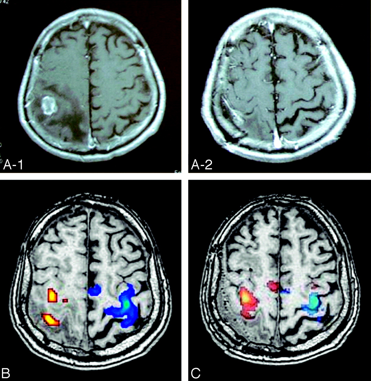
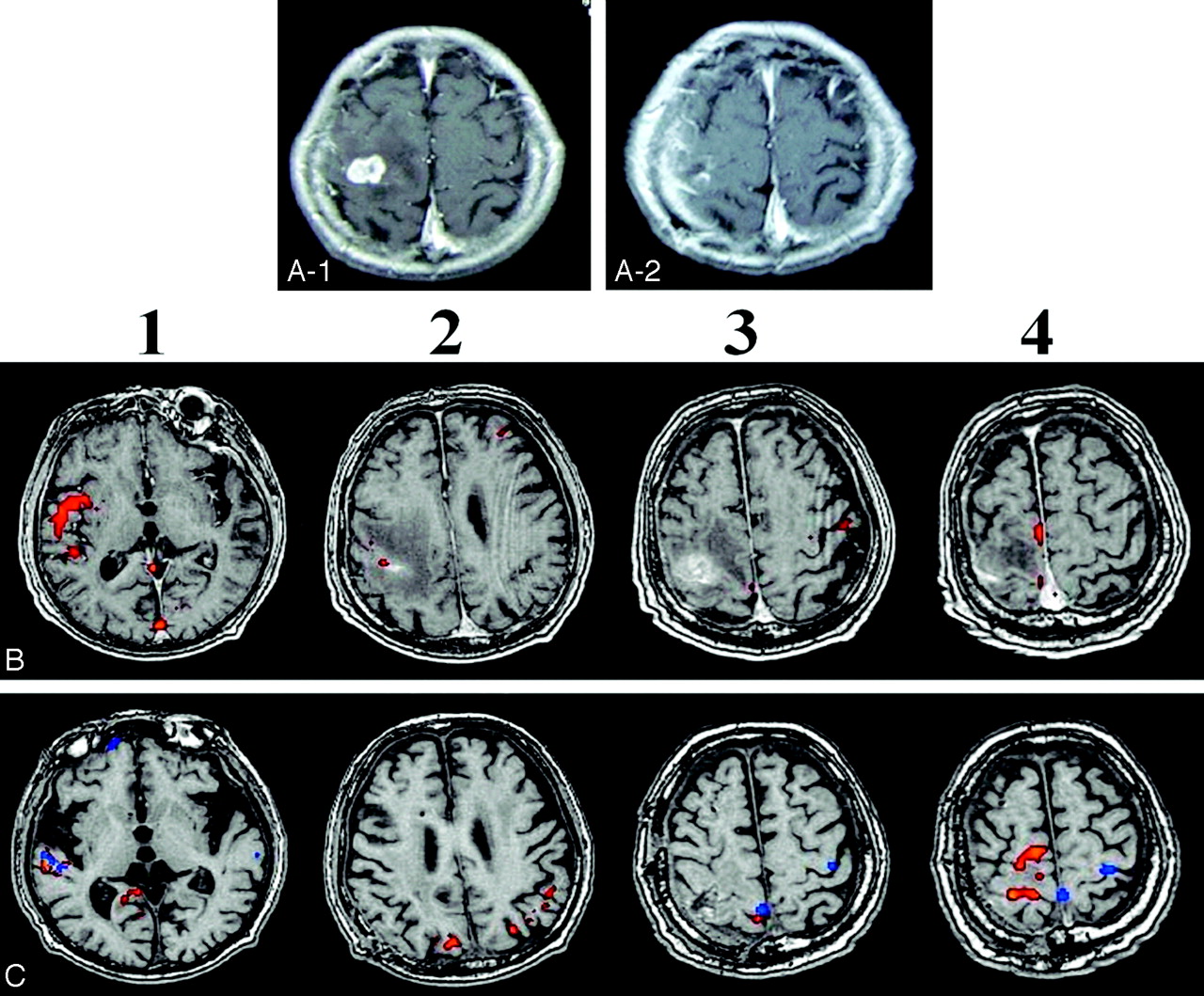
Case 1.
A 57-year-old woman with breast cancer experienced onset of right hand paresis (scale 5−) after developing epilepsy 1 month before surgery. MR imaging showed a heterogeneously enhancing tumor within a left frontal lesion (Fig 1A-1).

Case 1. A 57-year-old woman with metastatic brain tumor located inside the left-sided M1.
A-1, Preoperative MR imaging displays heterogeneously enhancing tumor in the M1.
A-2, Postoperative MR imaging shows ring enhancing lesion, but there was no residual tumor in the M1 according to the intraoperative observation for tumor cavity.
B, fMRI images activated by right hand clenching (B, orange) and right elbow flexion (B, blue) before surgery.
C, fMRI images activated by right hand clenching (orange) and right elbow flexion (blue) after surgery. A relatively small area of the contralateral M1 was activated by right hand clenching before surgery, whereas a large area of the contralateral M1 was activated by right hand clenching after surgery.
Case 2.
A 71-year-old man with lung cancer experienced onset of left-sided hemiparesis (scale 5−) 1 month before surgery. MR imaging demonstrated a heterogeneously enhancing tumor within a right parietal lesion (Fig 2A-1).
Case 2. A 71-year-old man with metastatic brain tumor located inside the right-sided S1.
A-1, Preoperative MR imaging displays heterogeneously enhancing tumor in the S1.
A-2, Postoperative MR imaging shows no residual tumor in the S1.
B, fMRI images activated by left hand clenching (B, orange) and right hand clenching (B, blue) before surgery.
C, fMRI images activated by left hand clenching (orange) and right hand clenching (blue) after surgery. Left hand clenching (orange area in B) activated the contralateral M1 and S1 areas separately before surgery and (orange area in C) activated the large area of the contralateral M1 and S1, and contralateral supplementary motor area after surgery.
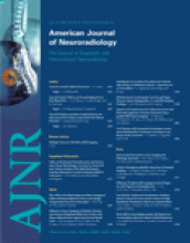
Case 3.
A 49-year-old man with colon cancer experienced onset of right-sided hemiparesis (upper extremity [scale 4] affected more than lower extremity [scale 5−]) and aphasia 1 month before surgery. MR imaging demonstrated a heterogeneously enhancing tumor within a left frontal lesion (Fig 3A-1).

Case 3. A 49-year-old man with metastatic brain tumor located inside the left-sided premotor area.
A-1, Preoperative MR imaging displays heterogeneously enhancing tumor in the premotor area.
A-2, Postoperative MR imaging shows no residual tumor in the premotor area.
B, fMRI images activated by right hand clenching (B-1, B-3; orange) and left hand clenching (B-2, B-4; blue) before surgery,
C, fMRI images activated by right hand clenching (orange) and left hand clenching (blue) after surgery. The ipsilateral and contralateral M1 and the anterior temporal lobe was activated by right hand clenching before surgery, whereas the contralateral M1 was activated by right hand clenching after surgery. The area in the anterior temporal lobe activated by right hand clenching before surgery (B-1) was not activated by right hand clenching after surgery (C-1).
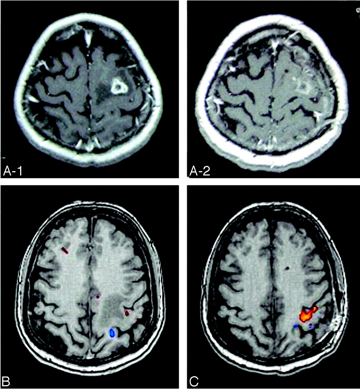
Case 4.
A 71-year-old woman with colon cancer experienced onset of left-sided hemiparesis (scale 4) 1 month before surgery. MR imaging demonstrated a heterogeneously enhancing tumor within a right frontal lesion (Fig 4A-1).

Case 4. A 71-year-old woman with metastatic brain tumor located inside the right-sided premotor area.
A-1, Preoperative MR imaging displays heterogeneously enhancing tumor in the premotor area.
A-2, Postoperative MR imaging shows no residual tumor in the premotor area.
B, fMRI images activated by right hand clenching (blue) and left hand clenching (red) before surgery,
C, fMRI images activated by right hand clenching (blue) and left hand clenching (orange) after surgery. Both left and right hand clenching activated the right temporal and frontal lobes before surgery. After surgery, left and right hand clenching activated the anterior temporal lobe (C-1,-2,-3), and left hand clenching activated a relatively small area of contralateral M1 (C-6). Because both preoperative and postoperative fMRI show similar atypical results such as no activation in the left hand M1 during right hand clenching, it seems that this result was not artifactual. It is not clear why there is no activation in the left M1 during right hand clenching, but the right hemisphere might be mainly activated by both right and left hand clenching in the case of severe paresis, possibly because of the preferential processing of spatial information in the right hemisphere.
Case 5.
A 76-year-old man with renal cancer experienced onset of left-sided hemiparesis 1 month before surgery (scale 4). Weakness of the left arm progressed after several episodes of partial seizures involving the left arm. MR imaging showed a heterogeneously enhancing mass within a right frontal lesion (Fig 5A-1).

Case 5. 76-year-old man with metastatic brain tumor located inside the right-sided M1.
A-1, Preoperative MR imaging displays heterogeneously enhancing tumor in the M1.
A-2, Postoperative MR imaging shows no residual tumor in the M1.
B, fMRI images activated by left hand clenching (orange) before surgery. fMRI by right hand clenching was not performed before surgery.
C, fMRI images activated by left hand clenching (orange) and right hand clenching (blue) after surgery. A relatively small area of the ipsilateral (B-3) and contralateral (B-2) M1 was activated by left hand clenching before surgery. A large portion of the right premotor and M1 were activated by left hand clenching after surgery (C-4, orange). The anterior temporal area was activated by left hand clenching before surgery (B-1) and by right or left hand clenching after surgery (C-1). The right medial M1 was activated by right or left hand clenching (C-3).
Preoperative paresis was relatively severe in cases 3, 4, and 5 compared with cases 1 and 2. Informed consent to perform fMRI, diffusion tensor imaging (DTI), and awake surgery was obtained from each patient.
fMRI and Image Analysis
The fMRI and image analysis was performed as described previously.12 In brief, the fMRI was performed with a 1.5T General Electric Signa Horizon LX imager (GE Yokokawa Medical System, Tokyo, Japan) with an acquisition sequence of: gradient-echo–echo-planar imaging (EPI) echo time [TE]/repetition time [TR] = 82.5/3000 ms, 128 × 128 matrix, with a 24 × 24-cm field of view. The reconstructed voxel size was 1.88 × 1.88 × 2.3 mm3. Five-millimeter thickness sections were obtained with 70 repetitions, and a block design of 30-second interval was used. fMRI was performed 1 week before surgery and 1 month after surgery, and the patient was asked to perform repetitive closing of the hand to a fist (hand clenching), because functional hand motor recovery is usually complete at this point in patients who had surgery for brain tumor. The fMRI acquisition time was approximately 3.5 minutes. Anatomic images were acquired with the use of a 3D fast- spoiled gradient echo sequence with TE of 2.4 ms, TR of 26.0 ms, flip angle of 30°, bandwidth of 31.25 kHz, image matrix of 256 × 256, and section thickness of 2.3 mm. After reconstruction, the EPI images were aligned to correct for head motion and were co-registered with anatomic images. EPI images were smoothed by using isotropic Gaussian kernels of 4 mm, and statistical analysis was performed by using the SPM program (London University, London, England).13 The significance of the activations (P value) was less than 0.05, depending on the degree of paresis of patients. When the degree of paresis was severe, we needed to increase P value to detect the activated area. The limitations of fMRI, especially when evaluating individual patients who might be uncomfortable to perform specific tasks because of their handicaps, should be noted.
DTI Image Analysis and Coregistration of fMRI and DTI Images
DTI and image analysis was performed as described previously.12 In brief, standard imaging gradients were used with a maximum strength of 22 mT/m and a slew rate of 77 mT/m/ms. All data were acquired by using a birdcage head coil. The DTI acquisition sequence was a single-shot, spin-echo EPI, with TE of 98.6 ms, 128 × 128 acquisition matrix, and a 24 × 24-cm field of view.14 Acquisitions of contiguous 5-mm sections were obtained, covering the whole brain, with a b value of 1000 mm2/s in 42 noncollinear directions. The reconstructed voxel size was 1.88 × 1.88 × 4.00 mm3. The DTI acquisition time for 61 images was approximately 10 minutes. The diffusion tensor eigenvalues (λ1, λ2, λ3) and eigenvectors (ε1, ε2, ε3) were calculated from DTI data, and fractional anisotropy (FA) maps were generated according to the Tensorlines algorithm,15,16 which was a combination of the tensor deflection algorithm at low FA and Streamlines tracking at high FA,17–19 with the use of DTI Analyzer (IDL v. 5.6; Research System, Boulder, Colo). Stopping criteria (threshold 0.1) were used for analysis.
Using the highest significantly activated fMRI voxel in each task, coregistration of fMRI and DTI images were used to select the seed points on the FA maps within the white matter adjacent to the activated cortex during fMRI. For the tractography of pathways, the following (regions of interest) ROIs were selected in case 5: the right premotor and M1, and the right anterior temporal lobe.
Mapping and Awake Tumor Resection
Mapping and awake tumor resection was performed as described previously.20 In brief, the patient was positioned in the supine position and given rigid head fixation (Sugita Headrest; Mizuho Medical Co, Tokyo, Japan) after administration of a local anesthetic agent at the pin sites and regional field block sites. Under general anesthesia with fentanyl and propofol by laryngeal mask airway, the skin was infiltrated with the same local anesthetic agent and incised, and a craniotomy and incision of the dura was performed.
The laryngeal airway was then removed and oxygen was administered via a nasal cannula. Cortical mapping was performed by stimulating the cortex with the modified Ojemann stimulator.21,22 To avoid an intraoperative seizure, a low-stimulus setting (3–5 mA, 60-Hz biphasic square wave pulse of 1 ms/phase for 4 seconds’ duration) was used. Electrocorticography was performed to monitor for after-discharges during stimulations. The patients were observed by the neurosurgeon, and movements of hand clenching were continuously monitored and reported to the operator.
Corticotomy was performed, avoiding the sites identified as eloquent cortex by cortical mapping, and the tumor was removed in the usual fashion. Continuous adequate task judging from the fMRI and DTI images was performed during the removal of tumor near the eloquent motor area responsible for the movement of hand. The patient was performing adequately tasks such as clenching the affected hand, bending the affected elbow, raising the affected arm, or bending the affected knee during the removal of the tumor that was located near the eloquent hand motor area as determined by fMRI and DTI. The fMRI and DTI data combined with a neuronavigation system was available to the neurosurgeon in the operating room, as described before.20 We have attempted to correlate preoperative fMRI and brain mapping data by using a neuronavigation system. To prevent deterioration of motor function, continuous task under awake anesthesia seems more useful in comparison with electrophysiologic monitoring of the motor strip during general anesthesia, because we could detect subtle deterioration of individual motor function by continuous task.20 After completion of the tumor resection, the patient was reintubated with a laryngeal airway, and general anesthesia with fentanyl and propofol was administered. After closure of dura, the bone flap was replaced, and the skin was closed in the usual manner.
Results
Cortical Mapping during Surgery and Postoperative Course
Case 1.
Cortical stimulation mapping detected the hand area of the M1 just beside the tumor. Corticotomy was performed in the caudal M1 area just beside the tumor. There was hemorrhage in the region of the tumor, and the right hand motor function worsened during tumor resection but improved by the end of awake surgery. It completely improved a week after surgery (scale 5). Postoperative MR imaging demonstrated complete removal of the tumor (Fig 1A-2).
Case 2.
Cortical stimulation mapping detected M1 rostral to the tumor. Corticotomy was performed in the caudal primary sensory area (S1) just beside the tumor. The patient did not develop new motor deficits at the end of awake surgery, and left-sided motor function improved by 1 week after surgery (scale 5). Postoperative MR imaging demonstrated complete removal of the tumor (Fig 2A-2).
Case 3.
Cortical stimulation mapping detected M1 caudal to the tumor. Corticotomy was performed in the rostral premotor area just beside the tumor. This patient did not develop new motor deficits at the end of awake surgery, and the right hemiparesis and aphasia that was present preoperatively improved within a week after surgery (scale 5). Postoperative MR imaging demonstrated complete removal of the tumor (Fig 3A-2).
Case 4.
Cortical stimulation mapping detected the M1 caudal to the tumor. Corticotomy was performed in the rostral premotor area just beside the tumor. The patient did not develop new motor deficits at the end of awake surgery. The left hemiparesis that was present preoperatively improved within a week after surgery (scale 5). Postoperative MR imaging demonstrated complete removal of the tumor (Fig 4A-2).
Case 5.
Cortical stimulation mapping identified the hand and arm area in the cortex just rostral to the M1, which was identified by fMRI and by intraoperative and postoperative neurologic observation, because the manipulation of the tumor located in M1 easily and frequently induced deterioration of paresis, which was observed by intraoperative and postoperative neurologic examination. Corticotomy was performed in the motor area facing the central sulcus just beside the tumor. The patient experienced frequent epileptiform phenomena during tumor removal and experienced agnosia of the right arm just after opening the dura. The patient demonstrated deficits in hand and arm muscle strength postoperatively, but these deficits resolved by postoperative day 14 (scale 5−). Postoperative MR imaging demonstrated complete removal of the tumor (Fig 5A-2). Postoperatively, the patient evinced mirror movement when trying to perform unilateral hand clenching on either side.
Preoperative and Postoperative fMRI
Case 1.
Left hand clenching (Fig 1B, orange area) activated a relatively small area of the contralateral M1 before surgery but activated a large area of the contralateral M1 after surgery (Fig 1C, orange area).
Case 2.
Right hand clenching (Fig 2B, orange area) activated the contralateral M1 and S1 areas separately before surgery but activated a large portion of the contralateral M1 and S1 and the contralateral supplementary motor area after surgery (Fig 2C, orange area).
Case 3.
Right hand clenching (Fig 3B-3, orange area) activated areas in both the ipsilateral and contralateral M1 and in the ipsilateral anterior temporal lobe (Fig 3B-1, orange area) before surgery. Further, left hand clenching (Fig 3B-4, orange area) only activated the contralateral M1 before surgery. After surgery, right hand clenching (Fig 3C-2, orange area) or left hand clenching (Fig 3C-2, blue area) produced activation only in the contralateral M1. The activated area in the anterior temporal lobe by right hand clenching before surgery was not activated by right hand clenching after surgery (Fig 3C-1).
Case 4.
Both left and right hand clenching (Fig 4B, red and blue areas, respectively) activated the right temporal and frontal lobes before surgery. After surgery, left and right hand clenching only activated the anterior temporal lobe (Fig 4C-1,-2,-3), and left hand clenching also activated a relatively small area of contralateral M1 (Fig 4C-6). There was an overlapping between the preoperative fMRI and the cortical mapping sites except case 4. In case 4, M1 site was determined by the postoperative fMRI and neurologic examination.
Case 5.
Left hand clenching activated relatively small areas in the ipsilateral and contralateral M1 (Fig 5B-2,-3, orange area) and relatively large areas in the ipsilateral anterior temporal lobe (Fig 5B-1, orange area) before surgery. After surgery, left hand clenching activated areas in the contralateral premotor and M1 (Fig 5C-4, orange area) and in the ipsilateral medial M1 close to the cortical rim (Fig 5C-3). Interestingly, left (Fig 5C-1, orange area) or right (Fig 5C-1, blue area) hand clenching activated the area in the right anterior temporal lobe.
In summary, the increase of the blood oxygen level–dependent (BOLD) response in the secondary and ipsilateral motor areas occurred in cases 3, 4, and 5, and it was completely reversible in case 3. Interestingly, paresis was relatively severe in cases 3, 4, and 5.
DTI of Activated Area in Case 5
To evaluate the connection between the activated area and surrounding cortex after surgery, tractography was performed with a focus on the highly activated area. In case 5, right premotor, M1, and anterior temporal areas were highly activated, and a tract connection was observed between premotor and M1 (Fig 6). The right anterior temporal areas were activated in response to right hand clenching but not left hand clenching before surgery and were activated by either right or left hand clenching after surgery (Fig 7). The tracts, possibly the inferior longitudinal fascicules (ILF), originated from the activated anterior temporal lobes (Fig 7).

Case 5. 3D reconstruction of fMRI and DTI images constructed during left hand (affected side) clenching after surgery. Fibers (blue) connecting the premotor area (yellow) and the M1 (white) were detected.

Case 5. 3D reconstruction of fMRI and DTI images constructed during left and right hand clenching after surgery. Fibers (blue), possibly the ILF, arise from the anterior temporal area activated by left or right hand clenching and connect between temporal and occipital lobes. At the current state of the art, the contribution of Figs. 6 and 7 is only demonstrative, not scientific.
Discussion
The present study demonstrated that tumor resection resulted in restoration of M1 function and improved motor function in patients with preoperative reorganization of M1 function. However, there are limitations to this study, such as small number of patients, lack of an objective correlation of fMRI with brain mapping, and intrinsic limitations to localize neuronal activity of the BOLD response. Previous studies using fMRI have demonstrated that the perilesional cortex, the ipsilateral primary motor area, and secondary motor areas (eg, supplementary motor area, premotor area, superior parietal cortex) that are not directly affected by tumor show activation in proportion to the size of lesion or degree of paresis.3,4,10,11,23 The signal intensity loss in lesions near the tumor may be related to tumor-induced hemodynamic change or to a loss of active neurons, resulting in a hemodynamic change by motor activation.4 These secondary motor areas represent the basic functional units of the motor system because of the attenuated connections between the different secondary motor areas and a convergence of connections on M1.24,25 Further, the shift of activation to secondary motor areas may mediate the compensatory responses of the brain and result in variable preservation of motor function.4 The reversible increase of the BOLD response in the secondary motor area including right temporal lobe or ipsilateral M1 can be explained by the compensatory responses of brain in the patients with relatively severe paresis of scale 4 (cases 3, 4, and 5; Table 1). On the other hand, the area activated by hand clenching increased after surgery in patients with relatively mild paresis of scale 5−, possibly because the degree of paresis was relatively mild and thus the compensatory mechanism did not work.
Tumor location, main activated area in pre- and postoperative fMRI, and brain mapping
In the present study, the ipsilateral and contralateral M1 were activated in cases 3 and 5. Ipsilateral pathways may act as a functional reserve after damage to contralateral routes.6,11 Interestingly, the amount of crossing corticospinal fibers varies considerably, ranging between total crossing and no crossing at all.26,27 Because the ipsilateral activated primary motor area in case 3 was much larger than that in case 5, the amount of uncrossed fibers may differ in a comparison of the 2 patients, though the amount of uncrossed fibers should be determined in future study.
Previous studies have demonstrated an increase in the activation of ipsilateral motor pathways, secondary motor areas, or cingulate cortex in patients recovering from stroke-induced hemiparesis.9,28,29 Short- and long-term reorganization of motor areas after surgery of brain tumor have also been reported by Duffau and colleagues,1,2 who used intraoperative brain mapping to demonstrated additional sites in M1 after removal of glioma.1 In patients with incomplete glioma resection who underwent a second operation, peritumorous functional reorganization was detected by cortical mapping.2 By contrast, the present study demonstrated that activation of M1 was restored after tumor resection in patients with preoperative reorganization of motor area in response to compression by the tumor. This observation underscores the reversible nature of motor function reorganization and suggests that these changes may occur secondary to changes in hemodynamics rather than loss or regeneration of neurons. In addition, both the M1 and premotor areas were activated after surgery in case 5, and fiber connections were demonstrated between both areas (Fig 6). This suggests that both the M1 and the premotor area were co-activated during hand clenching in the course of recovery as a result of the fiber connection of both areas, which is also demonstrated in healthy subjects. It is noteworthy that resection of tumor within or near primary motor area was highly challenging and difficult without inducing more severe paresis. However, this technical limitation seems to be overcome by using a combination of intraoperative cortical mapping, continuous motor task in awake surgery, and preoperative fMRI with tractography, which was co-registered with the patient head position and displayed in the operating room.20
In cases 3, 4, and 5, the paresis of affected hand was relatively severe, and the right hemisphere, especially the temporal lobe, was activated by the clenching of affected hand. Because processing of spatial information is preferentially handled by the right hemisphere,30–32 the right hemisphere, especially the temporal lobe, may play a role in the recovery of severe paresis by providing spatial information to the affected hand. Interestingly, mirror movements were observed after surgery in case 5. In acute stroke, mirror movement enhances ipsilateral cortical activity,33 and patients with persistent mirror movements show increased activity in the medial region of the ipsilateral M1 close to the cortical rim.34 Indeed, this patient also showed activation in the ipsilateral medial M1 close to the cortical rim (Fig 4B-3). The temporal lobe is associated with visual memory maintenance, and the ILF, which originated from this area, transfers signals from visual area to memory areas.35,36 Thus, mirror movement may be related to this temporal area, and patients may use the visual memory of hand movement for the recovery of paresis. Moreover, use of these visual images by looking at a mirror can result in improvement in motor function in patients recovering from paresis.37,38 Further investigation to elucidate the mechanism of mirror movement during recovery from paresis would be of benefit. In conclusion, tumor resection resulted in restoration of M1 function in patients and improved motor function in patients with preoperative reorganization of M1 function.
Acknowledgments
We thank H. Shinoura for assistance with manuscript preparation.
References
- Received July 18, 2005.
- Accepted after revision October 17, 2005.
- Copyright © American Society of Neuroradiology
In this issue


Jump to section
Related Articles
Cited By...
- No citing articles found.