Advertisement

Main menu

  • Home
  • Content
    • Current Issue
    • Accepted Manuscripts
    • Article Preview
    • Past Issue Archive
    • Video Articles
    • AJNR Case Collection
    • Case of the Week Archive
    • Case of the Month Archive
    • Classic Case Archive
  • Special Collections
    • AJNR Awards
    • ASNR Foundation Special Collection
    • Most Impactful AJNR Articles
    • Photon-Counting CT
    • Spinal CSF Leak Articles (Jan 2020-June 2024)
  • Multimedia
    • AJNR Podcasts
    • AJNR SCANtastic
    • Trainee Corner
    • MRI Safety Corner
    • Imaging Protocols
  • For Authors
    • Submit a Manuscript
    • Submit a Video Article
    • Submit an eLetter to the Editor/Response
    • Manuscript Submission Guidelines
    • Statistical Tips
    • Fast Publishing of Accepted Manuscripts
    • Graphical Abstract Preparation
    • Imaging Protocol Submission
    • Author Policies
  • About Us
    • About AJNR
    • Editorial Board
    • Editorial Board Alumni
  • More
    • Become a Reviewer/Academy of Reviewers
    • Subscribers
    • Permissions
    • Alerts
    • Feedback
    • Advertisers
    • ASNR Home

User menu

  • Alerts
  • Log in

Search

  • Advanced search
American Journal of Neuroradiology

American Journal of Neuroradiology

ASHNR American Society of Functional Neuroradiology ASHNR American Society of Pediatric Neuroradiology ASSR
  • Alerts
  • Log in

Advanced Search

  • Home
  • Content
    • Current Issue
    • Accepted Manuscripts
    • Article Preview
    • Past Issue Archive
    • Video Articles
    • AJNR Case Collection
    • Case of the Week Archive
    • Case of the Month Archive
    • Classic Case Archive
  • Special Collections
    • AJNR Awards
    • ASNR Foundation Special Collection
    • Most Impactful AJNR Articles
    • Photon-Counting CT
    • Spinal CSF Leak Articles (Jan 2020-June 2024)
  • Multimedia
    • AJNR Podcasts
    • AJNR SCANtastic
    • Trainee Corner
    • MRI Safety Corner
    • Imaging Protocols
  • For Authors
    • Submit a Manuscript
    • Submit a Video Article
    • Submit an eLetter to the Editor/Response
    • Manuscript Submission Guidelines
    • Statistical Tips
    • Fast Publishing of Accepted Manuscripts
    • Graphical Abstract Preparation
    • Imaging Protocol Submission
    • Author Policies
  • About Us
    • About AJNR
    • Editorial Board
    • Editorial Board Alumni
  • More
    • Become a Reviewer/Academy of Reviewers
    • Subscribers
    • Permissions
    • Alerts
    • Feedback
    • Advertisers
    • ASNR Home
  • Follow AJNR on Twitter
  • Visit AJNR on Facebook
  • Follow AJNR on Instagram
  • Join AJNR on LinkedIn
  • RSS Feeds

AJNR is seeking candidates for the AJNR Podcast Editor. Read the position description.

Research ArticleHEAD & NECK

Nasal Septal Abscess in Patients with Immunosuppression

J.M. Debnam, A.M. Gillenwater and L.E. Ginsberg
American Journal of Neuroradiology November 2007, 28 (10) 1878-1879; DOI: https://doi.org/10.3174/ajnr.A0708
J.M. Debnam
  • Find this author on Google Scholar
  • Find this author on PubMed
  • Search for this author on this site
A.M. Gillenwater
  • Find this author on Google Scholar
  • Find this author on PubMed
  • Search for this author on this site
L.E. Ginsberg
  • Find this author on Google Scholar
  • Find this author on PubMed
  • Search for this author on this site
  • Article
  • Figures & Data
  • Info & Metrics
  • Responses
  • References
  • PDF
Loading

Abstract

BACKGROUND AND PURPOSE: The purpose of this study was to review the imaging findings of nasal septal abscess in 2 patients with immunosuppression.

MATERIALS AND METHODS: Two patients with immunosuppression were identified as having a nasal septal abscess, and correlative CT imaging in both patients was evaluated.

RESULTS: The characteristic radiographic appearance of a nasal septal abscess included a fluid collection with thin rim enhancement, located within the cartilaginous nasal septum. After CT examination, incision and drainage was performed in both patients, and appropriate antibiotic coverage was initiated. Clinical and imaging follow-up demonstrated no signs of residual infection.

CONCLUSION: Nasal septal abscess has a characteristic appearance on CT examination. Prompt diagnosis and treatment, including incision and drainage and appropriate antibiotic coverage, are necessary to avoid serious complications.

Nasal septal abscess is defined as a collection of purulent material between the cartilaginous or bony septum and the mucoperichondrium or mucoperiosteum.1 Nasal septal abscesses occur more commonly after minor nasal trauma1 but may also result from iatrogenic causes2 and sinonasal3 or dental infection.4 After minor nasal trauma, a hematoma may form secondary to the rupture of small blood vessels in the nasal septum. The hematoma separates the mucoperichondrium from the septal cartilage and forms an ideal medium for the colonization and growth of bacteria, leading to the formation of an abscess.1

Serious complications may occur as a result of the nasal septal abscess, especially in patients with immunosuppression. The purpose of this study was to review the imaging findings of nasal septal abscess in 2 such patients to assist radiologists to identify this uncommon, but serious infection.

Materials and Methods

Two patients were identified with imaging consistent with a nasal septal abscess. Both patients had immunosuppression and were undergoing chemotherapy. The diagnosis of nasal septal abscess was suspected clinically by history and physical examination. The initial imaging, which was performed less than 1 day before the incision and drainage, included a CT examination in both patients. In patient 1, a CT examination of the head and neck was performed. In patient 2, a CT examination of the head and neck was requested, as per the physician's note; however, a CT of the brain was inadvertently performed. Follow-up imaging also included CT imaging in both patients.

Patient 1 was a 76-year-old man with acute myelogenous leukemia, which was diagnosed approximately 5 months before the development of the nasal septal abscess. He presented with a 3-week history of nasal stuffiness, difficulty with nasal breathing, headache, and facial pain. The patient's white blood cell count was 86.7 K/μL at presentation. Nasal soft tissue swelling was present on physical examination.

Anterior rhinoscopy was performed, which demonstrated fluctuant enlargement of the nasal septum bilaterally, suggesting a septal abscess. Aspiration of both the right and left nasal septa was performed with an 18-gauge needle; 8 mL of purulent discharge was obtained on both sides. CT scanning of the maxillofacial region was performed on a GE scanner (LightSpeed; GE Healthcare, Milwaukee, Wis), after intravenous administration of contrast material.

Patient 2 was a 17-year-old adolescent boy with a history of T-cell lymphoblastic lymphoma in complete remission. The patient was on chemotherapy consisting of modified hyper-CVAD (fractionated cyclophosphamide, vincristine, Adriamycin, and dexamethasone). The patient reported a recent history of minor nasal trauma and complained of frequent blowing of the nose secondary to allergic rhinitis, and nasal tenderness for the previous 2 to 3 days. On clinical examination, there was a mild amount of edema and erythema over the dorsum of the nose with mild tenderness and the presence of a saddle deformity. The white blood cell count was elevated to 13.1 K/UL. Fiberoptic examination of the nasal cavity was performed, which revealed no evidence of asymmetry, erythema, or nasal discharge. Septal abscess was suspected because of the saddle deformity and nasal tenderness. CT examination of the brain with contrast was performed on a GE scanner (GE Healthcare) after administration of intravenous contrast material the next day.

Results

In both patients, CT imaging revealed a fluid collection with thin rim enhancement, located within the cartilaginous nasal septum (Fig 1 and Fig 2A). The nasal septal abscess was not originally detected on the CT examination of the brain in patient 2; however, it was present in retrospect. There were no associated solid components with the abscesses. Patient 1 had swelling of the adjacent nasal soft tissues. There was no bony destruction or evidence of osteomyelitis involving the adjacent bony nasal septum in either patient.

Fig 1.
  • Download figure
  • Open in new tab
  • Download powerpoint
Fig 1.

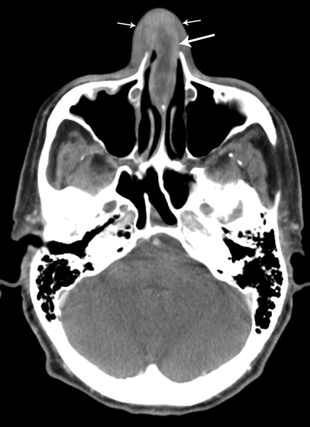
A 76-year-old man with acute myelogenous leukemia. CT imaging through the nasal cavity demonstrates a thin-walled, cystlike collection with peripheral enhancement involving the cartilaginous nasal septum (large arrow) consistent with a nasal abscess. There are no associated solid components. Note the swelling of the adjacent nasal soft tissues (small arrows).

Fig 2.
  • Download figure
  • Open in new tab
  • Download powerpoint
Fig 2.

A 17-year-old adolescent boy with T-cell lymphoblastic lymphoma. A, CT imaging of the brain demonstrates a subtle hypoattenuation of the cartilaginous nasal septum (arrows), found clinically to represent a nasal septal abscess. B, Resolution of nasal septal abscess after treatment.

After the CT examinations, an incision and drainage was performed on both patients. Collection of purulent material was sent to the laboratory for analysis and confirmed the diagnosis of an abscess. In patent 1, pathologic examination revealed evidence of acute inflammation and the presence of granulocytic sarcoma (chloroma). The patient was placed on Valacyclovir (Valtrex) and levofloxacin (Levaquin). Clinical and radiologic follow-up at approximately 2 months revealed no signs of residual infection. For patient 2, Gram stain and culture were performed, which identified the presence of Aspergillus flavus, which was treated with voriconazole. Clinical and radiologic follow-up at approximately 6 weeks revealed no signs of residual infection (Fig 2B).

Discussion

To our knowledge, no previous reports on the imaging appearance of nasal septal abscess in patients with immunosuppression have been published in the radiology literature. The imaging findings of nasal septal abscess, a thin-walled fluid collection with adjacent inflammatory change, are similar to those seen with abscesses located elsewhere within the body. Pang et al3 reported a case of nasal septal abscess as an unusual complication of acute spheno-ethmoiditis with similar imaging findings of a thin-walled, enhancing fluid collection involving the cartilaginous and bony septum.

Patients with a nasal septal abscess often present clinically with nasal obstruction. Less common symptoms include pain, rhinorrhea, fever, nasal fracture, bleeding, and cellulitis.5 Most patients have a history of at least minor trauma.2,5–7 Direct clinical inspection may reveal swelling and erythema of the dorsum of the nose and nasal septum as well as the presence of destroyed septal cartilage.2

Surgical drainage of the purulent contents of a nasal septal abscess is performed to relieve pressure and to provide a specimen for Gram stain, culture, and sensitivity. Nasal packing, the placement of a Penrose drain,3 reduction of nasal fractures, and delayed reconstruction of destroyed nasal cartilage2 may also be performed. The results of Gram stain and culture and sensitivity of the aspirate determine the appropriate systemic antibiotic coverage. The most common organism to be cultured is Streptococcus aureus.1,5 Other organisms include Streptococcus pneumoniae or Streptococcus viridans, Staphylococcus epidermidis, and Haemophilus influenzae.3,6

Early diagnosis and treatment are essential to prevent associated complications of the nasal septal abscess. These include sepsis, bacteremia, meningitis, brain abscess, cavernous sinus thrombosis, and maxillary hypoplasia.3,7,8 Destruction of cartilage may cause a depression of the nasal bridge from collapse of structural support leading to the formation of a saddle nose deformity.2,3,6 It should also be remembered that granulocytic sarcoma can both mimic and coexist with infection.9

Conclusion

A nasal septal abscess has a characteristic appearance on CT examination as a cystic collection of fluid with thin rim enhancement involving the nasal septum. It is important for the radiologist to be aware of this rare infectious complication of nasal trauma and sinus infection, especially in patients with immunosuppression, not only to provide the correct diagnosis, but also to ensure immediate treatment to avoid life-threatening complications.

References

  1. ↵
    Ambrus PS, Eavey RD, Baker AS, et al. Management of nasal septal abscess. Laryngoscope 1981;91:575–82
    CrossRefPubMed
  2. ↵
    Dispenza C, Saraniti C, Dispenza F, et al. Management of nasal septal abscess in childhood: our experience. Int J Pediatr Otorhinolaryngol 2004;68:1417–21
    CrossRefPubMed
  3. ↵
    Pang KP, Sethi DS. Nasal septal abscess: an unusual complication of acute spheno-ethmoiditis. J Laryngol Otol 2002;116:543–45
    CrossRefPubMed
  4. ↵
    da Silva M, Helman J, Eliachar I, et al. Nasal septal abscess of dental origin. Arch Otolaryngol 1982;108:380–81
    CrossRefPubMed
  5. ↵
    Canty PA Berkowitz RG. Hematoma and abscess of the nasal septum in children. Arch Otolaryngol Head Neck Surg 1996;122:1373–76
    CrossRefPubMed
  6. ↵
    Ehrlich A. Nasal septal abscess: an unusual complication of nasal trauma. Am J Emerg Med 1993;11:149–50
    CrossRefPubMed
  7. ↵
    Cuddihy PJ, Srinivasan V. An unusual presentation of nasal septal abscess. J Laryngol Otol 1998;112:775–76
    PubMed
  8. ↵
    Chukuezi AB. Nasal septal haematoma in Nigeria. J Laryngol Otol 1992;106:396–98
    PubMed
  9. ↵
    Singh SK, Ginsberg, LE. The spectrum of nonnodal acute lymphocytic leukemia in the head and neck. Annual Meeting of the American Society of Neuroradiology; April 23–27,2001 , Boston, Mass.
  • Received April 8, 2007.
  • Accepted after revision April 16, 2007.
  • Copyright © American Society of Neuroradiology
View Abstract
PreviousNext
Back to top

In this issue

American Journal of Neuroradiology: 28 (10)
American Journal of Neuroradiology
Vol. 28, Issue 10
November 2007
  • Table of Contents
  • Index by author
Advertisement
Print
Download PDF
Email Article

Thank you for your interest in spreading the word on American Journal of Neuroradiology.

NOTE: We only request your email address so that the person you are recommending the page to knows that you wanted them to see it, and that it is not junk mail. We do not capture any email address.

Enter multiple addresses on separate lines or separate them with commas.
Nasal Septal Abscess in Patients with Immunosuppression
(Your Name) has sent you a message from American Journal of Neuroradiology
(Your Name) thought you would like to see the American Journal of Neuroradiology web site.
CAPTCHA
This question is for testing whether or not you are a human visitor and to prevent automated spam submissions.
Cite this article
J.M. Debnam, A.M. Gillenwater, L.E. Ginsberg
Nasal Septal Abscess in Patients with Immunosuppression
American Journal of Neuroradiology Nov 2007, 28 (10) 1878-1879; DOI: 10.3174/ajnr.A0708

Citation Manager Formats

  • BibTeX
  • Bookends
  • EasyBib
  • EndNote (tagged)
  • EndNote 8 (xml)
  • Medlars
  • Mendeley
  • Papers
  • RefWorks Tagged
  • Ref Manager
  • RIS
  • Zotero
0 Responses
Respond to this article
Share
Bookmark this article
Nasal Septal Abscess in Patients with Immunosuppression
J.M. Debnam, A.M. Gillenwater, L.E. Ginsberg
American Journal of Neuroradiology Nov 2007, 28 (10) 1878-1879; DOI: 10.3174/ajnr.A0708
del.icio.us logo Twitter logo Facebook logo Mendeley logo
  • Tweet Widget
  • Facebook Like
  • Google Plus One
Purchase

Jump to section

  • Article
    • Abstract
    • Materials and Methods
    • Results
    • Discussion
    • Conclusion
    • References
  • Figures & Data
  • Info & Metrics
  • Responses
  • References
  • PDF

Related Articles

  • No related articles found.
  • PubMed
  • Google Scholar

Cited By...

  • Recurrent Nasal Septal Hematoma and Abscess: A Rare Manifestation of Leukemia
  • Crossref (26)
  • Google Scholar

This article has been cited by the following articles in journals that are participating in Crossref Cited-by Linking.

  • Nasal septal abscess in children: From diagnosis to management and prevention
    Nada Alshaikh, Stephen Lo
    International Journal of Pediatric Otorhinolaryngology 2011 75 6
  • Nasal tip abscess due to adverse skin reaction to Prolene: an unusual long term complication of rhinoplasty
    A Teymoortash, J A Fasunla, W Pfützner, S Steinbach-Hundt
    The Journal of Laryngology & Otology 2013 127 1
  • Orbital apex syndrome secondary to a fungal nasal septal abscess caused by Scedosporium apiospermum in a patient with uncontrolled diabetes: a case report
    Ippei Kishimoto, Shogo Shinohara, Tetsuhiro Ueda, Shoichi Tani, Hajime Yoshimura, Yukihiro Imai
    BMC Infectious Diseases 2017 17 1
  • An Unusual Case of a Pediatric Nasal Septal Abscess with Life-Threatening Complications in COVID-19 Pandemic
    Arvinder Singh Maan, Gurkiran Kaur, Rabia Arora, Jasmeet Kaur, Kamana Jyotsna Devi, Manjit Singh
    Indian Journal of Otolaryngology and Head & Neck Surgery 2022 74 S2
  • Spontaneous Nasal Septal Abscess Presenting as a Soft Tissue Mass in a Child
    Yi Cai, Anjali Saqi, Joseph Haddad
    The Journal of Emergency Medicine 2017 52 4
  • Isolated Nasal Septum Necrosis Caused by Aspergillus flavus in an Immunocompromised Child
    Fouzia Naeem, Jeffrey E. Rubnitz, Hana Hakim
    Pediatric Infectious Disease Journal 2011 30 7
  • Spontaneous Abscess of the Posterior Nasal Septum: An Unusual Cause of Nasal Obstruction in Children
    Marco Berlucchi, Michele Tomasoni, Roberta Bosio, Vittorio Rampinelli
    Annals of Otology, Rhinology & Laryngology 2021 130 8
  • Survival of an adult Quarter Horse gelding following bacterial meningitis caused by Escherichia coli
    K. L. Hepworth, D. M. Wong, B. A. Sponseller, C. J. Alcott, B. T. Sponseller, G. Ben‐Shlomo, R. D. Whitley
    Equine Veterinary Education 2014 26 10
  • A Case of Nasal Septal Abscess Caused by Combined Bacterial and Fungal Infection in the Immunocompromised Patient
    Il-Kwon Cho, Young-Jun Chung
    Korean Journal of Otorhinolaryngology-Head and Neck Surgery 2012 55 6
  • A Case of Nasal Septal Abscess Localized at Keystone Area, Suspected of Occurring Following Cauterization for Epistaxis
    Yong Seok Jo, Ick Soo Choi
    Journal of Clinical Otolaryngology Head and Neck Surgery 2020 31 1

More in this TOC Section

  • Chondrosarcoma vs Synovial Chondromatosis: Imaging
  • WHO Classification Update: Nasal&Skull Base Tumors
  • Peritumoral Signal in Vestibular Schwannomas
Show more HEAD & NECK

Similar Articles

Advertisement

Indexed Content

  • Current Issue
  • Accepted Manuscripts
  • Article Preview
  • Past Issues
  • Editorials
  • Editor's Choice
  • Fellows' Journal Club
  • Letters to the Editor
  • Video Articles

Cases

  • Case Collection
  • Archive - Case of the Week
  • Archive - Case of the Month
  • Archive - Classic Case

Special Collections

  • AJNR Awards
  • ASNR Foundation Special Collection
  • Most Impactful AJNR Articles
  • Photon-Counting CT
  • Spinal CSF Leak Articles (Jan 2020-June 2024)

More from AJNR

  • Trainee Corner
  • Imaging Protocols
  • MRI Safety Corner

Multimedia

  • AJNR Podcasts
  • AJNR Scantastics

Resources

  • Turnaround Time
  • Submit a Manuscript
  • Submit a Video Article
  • Submit an eLetter to the Editor/Response
  • Manuscript Submission Guidelines
  • Statistical Tips
  • Fast Publishing of Accepted Manuscripts
  • Graphical Abstract Preparation
  • Imaging Protocol Submission
  • Evidence-Based Medicine Level Guide
  • Publishing Checklists
  • Author Policies
  • Become a Reviewer/Academy of Reviewers
  • News and Updates

About Us

  • About AJNR
  • Editorial Board
  • Editorial Board Alumni
  • Alerts
  • Permissions
  • Not an AJNR Subscriber? Join Now
  • Advertise with Us
  • Librarian Resources
  • Feedback
  • Terms and Conditions
  • AJNR Editorial Board Alumni

American Society of Neuroradiology

  • Not an ASNR Member? Join Now

© 2025 by the American Society of Neuroradiology All rights, including for text and data mining, AI training, and similar technologies, are reserved.
Print ISSN: 0195-6108 Online ISSN: 1936-959X

Powered by HighWire