Advertisement

Main menu

  • Home
  • Content
    • Current Issue
    • Accepted Manuscripts
    • Article Preview
    • Past Issue Archive
    • Video Articles
    • AJNR Case Collection
    • Case of the Week Archive
    • Case of the Month Archive
    • Classic Case Archive
  • Special Collections
    • AJNR Awards
    • ASNR Foundation Special Collection
    • Most Impactful AJNR Articles
    • Photon-Counting CT
    • Spinal CSF Leak Articles (Jan 2020-June 2024)
  • Multimedia
    • AJNR Podcasts
    • AJNR SCANtastic
    • Trainee Corner
    • MRI Safety Corner
    • Imaging Protocols
  • For Authors
    • Submit a Manuscript
    • Submit a Video Article
    • Submit an eLetter to the Editor/Response
    • Manuscript Submission Guidelines
    • Statistical Tips
    • Fast Publishing of Accepted Manuscripts
    • Graphical Abstract Preparation
    • Imaging Protocol Submission
    • Author Policies
  • About Us
    • About AJNR
    • Editorial Board
    • Editorial Board Alumni
  • More
    • Become a Reviewer/Academy of Reviewers
    • Subscribers
    • Permissions
    • Alerts
    • Feedback
    • Advertisers
    • ASNR Home

User menu

  • Alerts
  • Log in

Search

  • Advanced search
American Journal of Neuroradiology

American Journal of Neuroradiology

ASHNR American Society of Functional Neuroradiology ASHNR American Society of Pediatric Neuroradiology ASSR
  • Alerts
  • Log in

Advanced Search

  • Home
  • Content
    • Current Issue
    • Accepted Manuscripts
    • Article Preview
    • Past Issue Archive
    • Video Articles
    • AJNR Case Collection
    • Case of the Week Archive
    • Case of the Month Archive
    • Classic Case Archive
  • Special Collections
    • AJNR Awards
    • ASNR Foundation Special Collection
    • Most Impactful AJNR Articles
    • Photon-Counting CT
    • Spinal CSF Leak Articles (Jan 2020-June 2024)
  • Multimedia
    • AJNR Podcasts
    • AJNR SCANtastic
    • Trainee Corner
    • MRI Safety Corner
    • Imaging Protocols
  • For Authors
    • Submit a Manuscript
    • Submit a Video Article
    • Submit an eLetter to the Editor/Response
    • Manuscript Submission Guidelines
    • Statistical Tips
    • Fast Publishing of Accepted Manuscripts
    • Graphical Abstract Preparation
    • Imaging Protocol Submission
    • Author Policies
  • About Us
    • About AJNR
    • Editorial Board
    • Editorial Board Alumni
  • More
    • Become a Reviewer/Academy of Reviewers
    • Subscribers
    • Permissions
    • Alerts
    • Feedback
    • Advertisers
    • ASNR Home
  • Follow AJNR on Twitter
  • Visit AJNR on Facebook
  • Follow AJNR on Instagram
  • Join AJNR on LinkedIn
  • RSS Feeds

AJNR is seeking candidates for the AJNR Podcast Editor. Read the position description.

Research ArticleBRAIN

Increased Signal in the Subarachnoid Space on Fluid-Attenuated Inversion Recovery Imaging Associated with the Clearance Dynamics of Gadolinium Chelate: A Potential Diagnostic Pitfall

J.M. Morris and G.M. Miller
American Journal of Neuroradiology November 2007, 28 (10) 1964-1967; DOI: https://doi.org/10.3174/ajnr.A0694
J.M. Morris
  • Find this author on Google Scholar
  • Find this author on PubMed
  • Search for this author on this site
G.M. Miller
  • Find this author on Google Scholar
  • Find this author on PubMed
  • Search for this author on this site
  • Article
  • Figures & Data
  • Info & Metrics
  • Responses
  • References
  • PDF
Loading

Abstract

BACKGROUND AND PURPOSE: Hyperintense CSF in the subarachnoid space (SAS) on fluid-attenuated inversion recovery (FLAIR) imaging has been reported in numerous pathologic conditions, including subarachnoid hemorrhage, meningitis, meningeal carcinomatosis, superior sagittal thrombosis, adjacent tumors, status epilepticus, and stroke. It has also been reported in otherwise healthy patients undergoing anesthesia with supplemental oxygen. We present a series of 11 patients with hyperintense CSF signal intensity in the SAS on FLAIR imaging after previous administration of gadolinium chelate.

MATERIALS AND METHODS: Head MR images of patients who had a prior gadolinium-enhanced body, spine, or brain MR imaging and who had increased signal intensity in the SAS on FLAIR images were prospectively and retrospectively reviewed. Correlation was made with the clinical and laboratory findings.

RESULTS: Eight of the 11 patients had negative findings on lumbar punctures. Seven patients had either chronic renal insufficiency or acute renal failure, but the remaining 4 had normal renal function. Nine patients had no other significant intracranial abnormalities, and 2 patients had acute infarcts remote from the CSF hyperintensity. One patient had follow-up studies at 24 and 48 hours, documenting resolution of the CSF hyperintensities.

CONCLUSION: Given the sharp rise in volume of contrast-enhanced MR imaging studies, it is inevitable that some patients will have undergone a contrast-enhanced MR imaging 24–48 hours before an MR imaging of the brain. The neuroradiologist should be aware that previous administration of gadolinium chelate can cause increased signal intensity in the SAS on FLAIR imaging in patients with or without a history of renal insufficiency and without abnormalities known to disrupt the blood-brain barrier.

Fluid-attenuated inversion recovery (FLAIR) is routinely used in brain imaging due to its high lesion-to-tissue contrast from T2 prolongation with nulling of normal CSF background. It is known to be particularly sensitive for pathologic entities that affect the CSF in the subarachnoid space (SAS). Increased signal intensity in the SAS on FLAIR imaging has been described in numerous conditions, including subarachnoid hemorrhage,1,2 meningitis,1,3–5 stroke,1,6–8 meningeal carcinomatosis,9 leptomeningeal metastasis,10 subdural hematoma,11 adjacent neoplasms,1,12 dural venous thrombosis,13 and status epilepticus.14 It has also been described as a diagnostic pitfall in patients undergoing an MR imaging with supplemental oxygen15 and in those patients who have previously received iodinated contrast material.16 It can be seen as a susceptibility artifact if there is adjacent metal. A few case reports have described increased signal intensity in the SAS on FLAIR imaging in patients with renal failure who had previously received gadolinium.17–19 These findings have also been described in vitro and in animal models.20 No reports to date have reported these findings in patients with normal renal function absent intracranial pathology.

Materials and Methods

We retrospectively and prospectively identified patients who underwent MR imaging of the brain and had a prior gadolinium-enhanced body, spine, or brain MR imaging. Patients with increased signal intensity in the SAS on FLAIR imaging were then reviewed. Patient characteristics, including age, renal function, milliliters per kilogram and rate of gadolinium administered, use of supplemental oxygen during the MR imaging, and any associated intracranial pathology, were then correlated with the MR images. Correlation was also made with the patients’ clinical and laboratory findings, including lumbar puncture.

Results

Eleven patients were identified with increased signal intensity in the SAS on FLAIR imaging who had been administered gadolinium between 1 and 6 days before their scanning (Figs 1–3). No patient received supplemental oxygen or was receiving steroids at the time of imaging. One patient had follow-up studies at 24 and 48 hours documenting resolution of the CSF hyperintensities (Fig 4). After the findings were reported, 8 of the 11 patients had lumbar punctures performed emergently and were negative for subarachnoid hemorrhage, infection, or malignant cells. Three patients had mildly elevated protein in their CSF (patients 3, 4, and 6; Table); however, this was not high enough to produce increased signal intensity in the SAS on FLAIR.21

Fig 1.
  • Download figure
  • Open in new tab
  • Download powerpoint
Fig 1.

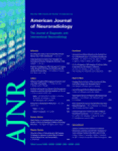
A 74-year-old man with normal renal function who underwent a gadolinium-enhanced MR imaging of the head and MR angiography (MRA) of the neck because of a transient ischemic attack and who had a follow-up head MR imaging the next day. A, Normal MRA findings of the neck. B, Precontrast axial FLAIR image shows no signal intensity in the SAS on the date of the gadolinium administration. C, Repeat noncontrast coronal FLAIR image 24 hours later shows increased signal intensity in the SAS. The patient had an emergent lumbar puncture, which was negative for subarachnoid hemorrhage, infection, or malignant cells.

Fig 2.
  • Download figure
  • Open in new tab
  • Download powerpoint
Fig 2.

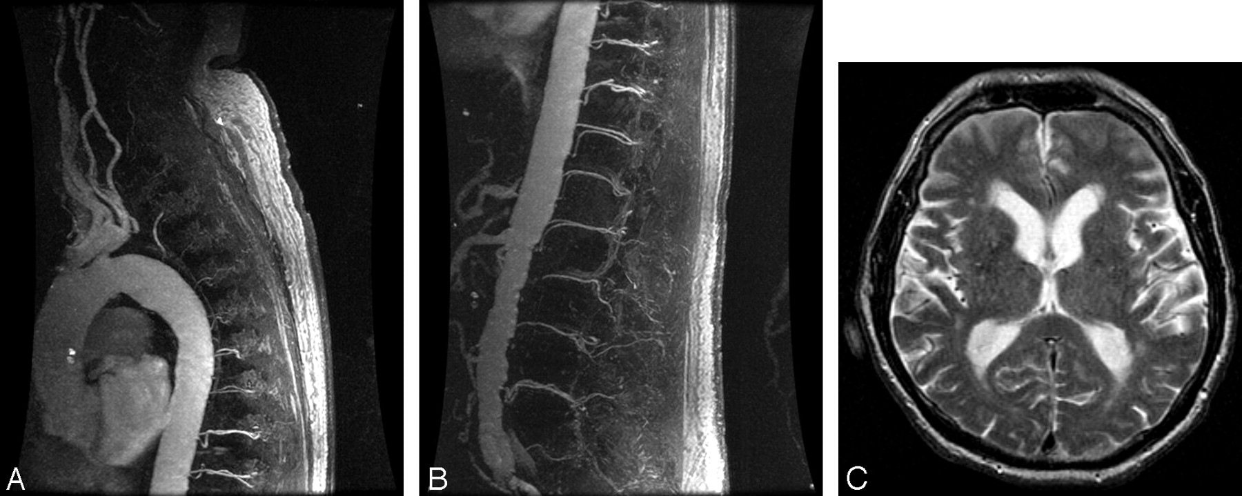
A 21-year-old man with chronic renal insufficiency who underwent gadolinium-enhanced MRA of the abdomen followed by MR imaging of the brain 6 days later to rule out causes of syncope. A, Normal findings on abdominal MRA. B, Precontrast FLAIR image shows diffuse increased signal intensity in the SAS 6 days after the gadolinium injection. The patient had an emergent lumbar puncture, which was as negative for subarachnoid hemorrhage, infection, or malignant cells.

Fig 3.
  • Download figure
  • Open in new tab
  • Download powerpoint
Fig 3.

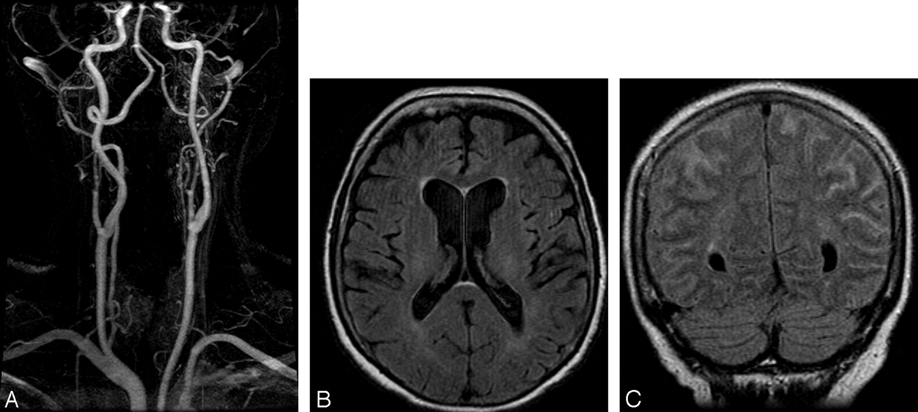
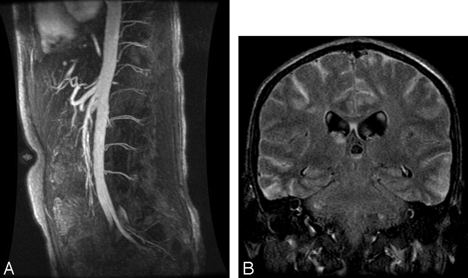
An 81-year-old man with normal renal function who underwent a triple-dose (60 mL) 3D MRA of the spine to exclude a dural arteriovenous fistula, followed by MR imaging of the brain 24 hours later for a syncopal episode. Upper thoracic (A) and thoracolumbar (B) spinal MRA images demonstrate no evidence of a dural arteriovenous fistula. C, Precontrast FLAIR image shows increased signal intensity in the SAS and in the lateral ventricles 24 hours after gadolinium injection. The patient returned to the department and was imaged a second time due to the T2 spin-echo appearance to ensure that there was not a technical error in the initial FLAIR sequence and was found to have similar results.

Fig 4.
  • Download figure
  • Open in new tab
  • Download powerpoint
Fig 4.

A 79-year-old man with normal renal function evaluated for a transient ischemic attack. A, Precontrast axial FLAIR image shows no abnormal signal intensity in the SAS. B, Forty-eight hours postgadolinium injection, repeat MR imaging shows diffuse increased signal intensity in the subarachnoid space (SAS) on coronal FLAIR images due to delayed clearance of the gadolinium chelate. C, Repeat MR imaging 72 hours postgadolinium injection shows resolution of the increased signal intensity in the SAS.

View this table:
  • View inline
  • View popup

Patient characteristics and time interval between gadolinium administration and increased signal in the subarachnoid space on FLAIR

Seven patients had either chronic renal insufficiency or acute renal failure, but the remaining 4 had normal renal function (Table). Nine patients had no other significant intracranial abnormalities seen on MR imaging. Postgadolinium T1-weighted imaging did not show corresponding enhancement in the SAS. Two patients had acute infarcts remote from the hyperintensity seen in the SAS on FLAIR imaging (Fig 3). The age range of the patients was 21–87 years. The rate of gadolinium administered varied between 1 and 2 mL/s, with total doses ranging from 20 to 60 mL/s. The average length of time between gadolinium administration and the increased signal intensity in the SAS seen on FLAIR was 2 days (range, 1–6 days), with the patients with normal renal function at the lower end of the spectrum. One patient had known prostate cancer. No other patient had a known malignancy, and none were found to have leptomeningeal metastasis or carcinomatosis.

Discussion

During the past decade, multiple investigators have shown FLAIR imaging to be sensitive but not specific in demonstrating varied pathologies involving the SAS, such as subarachnoid hemorrhage, meningitis, stroke, meningeal carcinomatosis, leptomeningeal metastasis, subdural hematoma, adjacent neoplasm, dural venous thrombosis, and status epilepticus.1–14 This relies on the fact that FLAIR imaging is designed to null the normal CSF signal intensity, and any alteration in the CSF results in an increased signal intensity in the CSF. Postgadolinium FLAIR imaging has also shown hyperintensity in the SAS in patients with pathologies that are not specific to the SAS but rather disrupt the blood-brain barrier.22,23 Alternatively, increased signal intensity in the SAS can be a diagnostic pitfall in patients receiving supplemental oxygen15 during MR imaging of the brain, in patients with renal insufficiency who have previously received gadolinium,17–19,23,24 and in patients immediately after carotid stent placement.25 Gadolinium has also been reported to diffuse across the choroid plexus and uveochoroid membrane in patients with impaired renal function, resulting in increased signal intensity in the ventricular system and in the vitreous and aqueous humor of the eye on FLAIR imaging.18

This CSF hyperintensity in the SAS and ventricles after prior administration of gadolinium has not been previously reported in patients with normal renal function without intracranial pathology that would otherwise cause a disruption in the blood-brain barrier. To our knowledge, this is the largest series to date of patients with increased signal intensity in the SAS after previous administration of gadolinium.

The direct mechanism by which CSF diffuses into the SAS in patients with or without renal insufficiency is not entirely known. It is postulated that in patients with renal insufficiency, the gadolinium may move across an osmotic gradient at the circumventricular organs in the setting of prolonged elevation of plasma concentrations,18 because it is primarily cleared through glomerular filtration.26

The normal half-life (t ½) of gadolinium chelates is 1.6 hours.26 Although patients with normal serum creatinine levels and renal function should not have prolonged clearance of gadolinium chelates, in those patients with renal insufficiency (glomerular filtration rate [GFR] <20 mL/min), the t ½ can increase up to 30 hours.27 Although the reported rate of gadolinium neurotoxicity in patients with renal insufficiency is <1%,28 this may, in fact, be higher than previously thought. It has been reported that the clinical correlation of increased signal intensity in the SAS on FLAIR imaging in patients with renal insufficiency also appears not to be associated with clinical neurotoxicity.11 This is likely not the case, given the reports of gadolinium-induced nephrogenic fibrosing dermopathy (NFD)29 and gadolinium-induced encephalopathy.19 For these very reasons, in our institution, we have recently instituted withholding gadolinium in patients with a GFR of less than 15 mL/min.

Conclusion

Given the sharp rise in volume of contrast-enhanced MR imaging studies, it is inevitable that some patients will have undergone contrast-enhanced MR imaging 24–48 hours before MR imaging of the brain. The neuroradiologist should be aware that delayed gadolinium chelate clearance can cause increased signal intensity in the SAS on FLAIR imaging in patients with or without a history of renal insufficiency and without abnormalities known to disrupt the blood-brain barrier. Although this represents a diagnostic pitfall, this finding could be clinically relevant in regards to encephalopathy and NFD.

Footnotes

  • Previously presented at: Annual Meeting of the American Society of Neuroradiology, April-May, 2005; Toronto, Ontario, Canada.

References

  1. ↵
    Adams JG, Melhem ER. Clinical usefulness of T2-weighted fluid-attenuated inversion recovery MR imaging of the CNS. AJR Am J Roentgenol 1999;172:529–36
    PubMed
  2. ↵
    Noguchi K, Oqawa T, Inugami A, et al. Acute subarachnoid hemorrhage: diagnosis with fluid-attenuated inversion-recovery MR imaging. Radiology 1995;196:773–77
    PubMed
  3. ↵
    Singer MB, Atlas SW, Drayer BP. Subarachnoid space disease: diagnosis with fluid–attenuated inversion-recovery MR imaging and comparisons with gadolinium-enhanced spin-echo MR imaging: blinded reader study. Radiology 1998;208:417–22
    PubMed
  4. Kamran S, Bener AB, Alper D, et al. Role of fluid–attenuated inversion-recovery in the diagnosis of meningitis: comparison with contrast-enhanced magnetic resonance imaging. J Comput Assist Tomogr 2004;28:68–72
    CrossRefPubMed
  5. ↵
    Mathews VP, Caldemeyer KS, Lowe MJ, et al. Brain: gadolinium-enhanced fast fluid-attenuated inversion-recovery MR imaging. Radiology 2000;215:922–24
    PubMed
  6. ↵
    Dechambre SD, Duprez T, Grandin CB, et al. High signal in cerebrospinal fluid mimicking subarachnoid haemorrhage on FLAIR following acute stroke and intravenous contrast medium. Neuroradiology 2000;42:608–11
    CrossRefPubMed
  7. Rumpel H, Chan LL. Serial FLAIR imaging after Gd-DTPA contrast: pitfalls in stroke trial magnetic resonance imaging. Stroke 2003;34:797–98
    Abstract/FREE Full Text
  8. ↵
    Warach S, Latour LL. Evidence of reperfusion injury, exacerbated by thrombolytic therapy, in human focal brain ischemia using a novel imaging marker of early blood-brain barrier disruption. Stroke 2004;35 (11 suppl 1):2659–61. Epub 2004 Oct 7
    Abstract/FREE Full Text
  9. ↵
    Tsuchiya K, Katase S, Yoshino A, et al. FLAIR MR imaging for diagnosing intracranial meningeal carcinomatosis. AJR Am J Roentgenol 2001;176:1585–98
    PubMed
  10. ↵
    Singh SK, Leeds NE, Ginsberg LE. MR imaging of leptomeningeal metastases: comparison of three sequences. AJNR Am J Neuroradiol 2002;23:817–21
    Abstract/FREE Full Text
  11. ↵
    Kanamalla US, Baker KB, Boyko OB. Gadolinium diffusion into subdural space: visualization with FLAIR MR imaging. AJR Am J Roentgenol 2001;176:1604–05
    PubMed
  12. ↵
    Kubota T, Yamada K, Kizu O, et al. Relationship between contrast enhancement on fluid-attenuated inversion recovery MR sequences and signal intensity on T2-weighted MR images: visual evaluation of brain tumors. J Magn Reson Imaging 2005;21:694–700
    CrossRefPubMed
  13. ↵
    Bozzao A, Bastianello S, Bozzao L. Superior sagittal sinus thrombosis with high-signal-intensity CSF mimicking subarachnoid hemorrhage on MR FLAIR images. AJR Am J Roentgenol 1997;169:1183–84
    PubMed
  14. ↵
    Villalobos-Chavez F, Rodriguez-Uranga JJ, Sanz-Fernandez G. Sequential changes in magnetic resonance in a limbic status epilepticus. Rev Neurol 2005;40:354–57
    PubMed
  15. ↵
    Frigon C, Jardine DS, Weinberger E, et al. Fraction of inspired oxygen in relation to cerebrospinal fluid hyperintensity on FLAIR MR imaging of the brain in children and young adults undergoing anesthesia. AJR Am J Roentgenol 2002;179:791–96
    PubMed
  16. ↵
    Kim EY, Kim SS, Na DG, et al. Sulcal hyperintensity on fluid-attenuated inversion recovery imaging in acute ischemic stroke patients treated with intra-arterial thrombolysis: iodinated contrast media as its possible cause and the association with hemorrhagic transformation. J Comput Assist Tomogr 2005;29:264–69
    CrossRefPubMed
  17. ↵
    Lev MH, Schaefer PW. Subarachnoid gadolinium enhancement mimicking subarachnoid hemorrhage on FLAIR MR images: fluid-attenuated inversion recovery. AJR Am J Roentgenol 1999;173:1414–15
    PubMed
  18. ↵
    Kanamalla US, Boyko OB. Gadolinium diffusion into orbital vitreous and aqueous humor, perivascular space, and ventricles in patients with chronic renal disease. AJR Am J Roentgenol 2002;179:1350–52
    PubMed
  19. ↵
    Maramattom BV, Manno EM, Wijdicks EF, et al. Gadolinium encephalopathy in a patient with renal failure. Neurology 2005;64:1276–78
    Abstract/FREE Full Text
  20. ↵
    Mamourian AC, Hoopes PJ, Lewis LD, et al. Visualization of intravenously administered contrast material in the CSF on fluid-attenuated inversion-recovery MR images: an in vitro and animal-model investigation. AJNR Am J Neuroradiol 2000;21:105–11
    Abstract/FREE Full Text
  21. ↵
    Melhem ER, Jara H, Eustace S. Fluid-attenuated inversion recovery MR imaging: identification of protein concentration thresholds for CSF hyperintensity. AJR Am J Roentgenol 1997;169:859–62
    CrossRefPubMed
  22. ↵
    Bozzao A, Floris R, Fasoli F, et al. Cerebral spinal fluid changes after intravenous injection of gadolinium chelate: assessment by FLAIR MR imaging. Eur Radiol 2003;13:592–97
    PubMed
  23. ↵
    Taoka T, Yuh WT, White M, et al. Sulcal hyperintensity on fluid-attenuated inversion recovery MR images in patients without apparent cerebrospinal fluid abnormality. AJR Am J Roentgenol 2001;176:519–24
    PubMed
  24. ↵
    Rai A, Hogg JP. Persistence of gadolinium in CSF: a diagnostic pitfall in patients with end-stage renal disease. AJNR Am J Neuroradiol 2001;22:1357–61
    Abstract/FREE Full Text
  25. ↵
    Wilkinson ID, Griffiths PD, Hoggard N, et al. Unilateral leptomeningeal enhancement after carotid stent detected by magnetic resonance imaging. Stroke 2000;31:848–51
    Abstract/FREE Full Text
  26. ↵
    Joffe P, Thomsen HS, Meusel M. Pharmacokinetics of gadodiamide injection in patients with severe renal insufficiency and patients undergoing hemodialysis or continuous ambulatory peritoneal dialysis. Acad Radiol 1998;5:491–502
    CrossRefPubMed
  27. ↵
    Shellock FG, Kanal E. Safety of magnetic resonance imaging contrast agents. J Magn Reson Imaging 1999;10:477–84
    CrossRefPubMed
  28. ↵
    Arsenault TM, King BF, Marsh JW, et al. Systemic gadolinium toxicity in patients with renal insufficiency and renal failure: retrospective analysis of an initial experience. Mayo Clin Proc 1996;71:1150–54
    CrossRefPubMed
  29. ↵
    Grobner, T. Gadolinium: a specific trigger for the development of nephrogenic fibrosing dermopathy and nephrogenic systemic fibrosis? Nephrol Dial Transplant 2006;21:1104–8
    FREE Full Text
  • Received January 19, 2007.
  • Accepted after revision April 4, 2007.
  • Copyright © American Society of Neuroradiology
View Abstract
PreviousNext
Back to top

In this issue

American Journal of Neuroradiology: 28 (10)
American Journal of Neuroradiology
Vol. 28, Issue 10
November 2007
  • Table of Contents
  • Index by author
Advertisement
Print
Download PDF
Email Article

Thank you for your interest in spreading the word on American Journal of Neuroradiology.

NOTE: We only request your email address so that the person you are recommending the page to knows that you wanted them to see it, and that it is not junk mail. We do not capture any email address.

Enter multiple addresses on separate lines or separate them with commas.
Increased Signal in the Subarachnoid Space on Fluid-Attenuated Inversion Recovery Imaging Associated with the Clearance Dynamics of Gadolinium Chelate: A Potential Diagnostic Pitfall
(Your Name) has sent you a message from American Journal of Neuroradiology
(Your Name) thought you would like to see the American Journal of Neuroradiology web site.
CAPTCHA
This question is for testing whether or not you are a human visitor and to prevent automated spam submissions.
Cite this article
J.M. Morris, G.M. Miller
Increased Signal in the Subarachnoid Space on Fluid-Attenuated Inversion Recovery Imaging Associated with the Clearance Dynamics of Gadolinium Chelate: A Potential Diagnostic Pitfall
American Journal of Neuroradiology Nov 2007, 28 (10) 1964-1967; DOI: 10.3174/ajnr.A0694

Citation Manager Formats

  • BibTeX
  • Bookends
  • EasyBib
  • EndNote (tagged)
  • EndNote 8 (xml)
  • Medlars
  • Mendeley
  • Papers
  • RefWorks Tagged
  • Ref Manager
  • RIS
  • Zotero
0 Responses
Respond to this article
Share
Bookmark this article
Increased Signal in the Subarachnoid Space on Fluid-Attenuated Inversion Recovery Imaging Associated with the Clearance Dynamics of Gadolinium Chelate: A Potential Diagnostic Pitfall
J.M. Morris, G.M. Miller
American Journal of Neuroradiology Nov 2007, 28 (10) 1964-1967; DOI: 10.3174/ajnr.A0694
del.icio.us logo Twitter logo Facebook logo Mendeley logo
  • Tweet Widget
  • Facebook Like
  • Google Plus One
Purchase

Jump to section

  • Article
    • Abstract
    • Materials and Methods
    • Results
    • Discussion
    • Conclusion
    • Footnotes
    • References
  • Figures & Data
  • Info & Metrics
  • Responses
  • References
  • PDF

Related Articles

  • No related articles found.
  • PubMed
  • Google Scholar

Cited By...

  • HARMless: Transient Cortical and Sulcal Hyperintensity on Gadolinium-Enhanced FLAIR after Elective Endovascular Coiling of Intracranial Aneurysms
  • Flat Detector Angio-CT following Intra-Arterial Therapy of Acute Ischemic Stroke: Identification of Hemorrhage and Distinction from Contrast Accumulation due to Blood-Brain Barrier Disruption
  • Elevated Cerebral Blood Volume Contributes to Increased FLAIR Signal in the Cerebral Sulci of Propofol-Sedated Children
  • Isolated Acute Nontraumatic Cortical Subarachnoid Hemorrhage
  • Crossref (36)
  • Google Scholar

This article has been cited by the following articles in journals that are participating in Crossref Cited-by Linking.

  • Biodistribution of gadolinium‐based contrast agents, including gadolinium deposition
    Silvio Aime, Peter Caravan
    Journal of Magnetic Resonance Imaging 2009 30 6
  • Isolated Acute Nontraumatic Cortical Subarachnoid Hemorrhage
    V. Cuvinciuc, A. Viguier, L. Calviere, N. Raposo, V. Larrue, C. Cognard, F. Bonneville
    American Journal of Neuroradiology 2010 31 8
  • Subarachnoid Hemorrhage: Beyond Aneurysms
    Carrie P. Marder, Vinod Narla, James R. Fink, Kathleen R. Tozer Fink
    American Journal of Roentgenology 2014 202 1
  • The Cerebrovascular-Chronic Kidney Disease Connection: Perspectives and Mechanisms
    Wei Ling Lau, Branko N. Huisa, Mark Fisher
    Translational Stroke Research 2017 8 1
  • Importance of Contrast-Enhanced Fluid-Attenuated Inversion Recovery Magnetic Resonance Imaging in Various Intracranial Pathologic Conditions
    Eun Kyoung Lee, Eun Ja Lee, Sungwon Kim, Yong Seok Lee
    Korean Journal of Radiology 2016 17 1
  • Colocalization of Gadolinium-Diethylene Triamine Pentaacetic Acid With High-Molecular-Weight Molecules After Intracerebral Convection-Enhanced Delivery in Humans
    John H Sampson, Martin Brady, Raghu Raghavan, Ankit I Mehta, Allan H Friedman, David A Reardon, Neil A Petry, Daniel P Barboriak, Terence Z Wong, Michael R Zalutsky, Denise Lally-Goss, Darell D Bigner
    Neurosurgery 2011 69 3
  • Retention of Gadolinium in Brain Parenchyma: Pathways for Speciation, Access, and Distribution. A Critical Review
    Marlène Rasschaert, Roy O. Weller, Josef A. Schroeder, Christoph Brochhausen, Jean‐Marc Idée
    Journal of Magnetic Resonance Imaging 2020 52 5
  • Methodological Aspects for Preclinical Evaluation of Gadolinium Presence in Brain Tissue
    Philippe Robert, Thomas Frenzel, Cécile Factor, Gregor Jost, Marlène Rasschaert, Gunnar Schuetz, Nathalie Fretellier, Janina Boyken, Jean-Marc Idée, Hubertus Pietsch
    Investigative Radiology 2018 53 9
  • Subarachnoid hemorrhage in ten questions
    M. Edjlali, C. Rodriguez-Régent, J. Hodel, R. Aboukais, D. Trystram, J.-P. Pruvo, J.-F. Meder, C. Oppenheim, J.-P. Lejeune, X. Leclerc, O. Naggara
    Diagnostic and Interventional Imaging 2015 96 7-8
  • Flat Detector Angio-CT following Intra-Arterial Therapy of Acute Ischemic Stroke: Identification of Hemorrhage and Distinction from Contrast Accumulation due to Blood-Brain Barrier Disruption
    T. Kau, M. Hauser, S. M. Obmann, M. Niedermayer, J. R. Weber, K. A. Hausegger
    American Journal of Neuroradiology 2014 35 9

More in this TOC Section

  • Evaluating the Effects of White Matter Multiple Sclerosis Lesions on the Volume Estimation of 6 Brain Tissue Segmentation Methods
  • Quiet PROPELLER MRI Techniques Match the Quality of Conventional PROPELLER Brain Imaging Techniques
  • Predictors of Reperfusion in Patients with Acute Ischemic Stroke
Show more BRAIN

Similar Articles

Advertisement

Indexed Content

  • Current Issue
  • Accepted Manuscripts
  • Article Preview
  • Past Issues
  • Editorials
  • Editor's Choice
  • Fellows' Journal Club
  • Letters to the Editor
  • Video Articles

Cases

  • Case Collection
  • Archive - Case of the Week
  • Archive - Case of the Month
  • Archive - Classic Case

Special Collections

  • AJNR Awards
  • ASNR Foundation Special Collection
  • Most Impactful AJNR Articles
  • Photon-Counting CT
  • Spinal CSF Leak Articles (Jan 2020-June 2024)

More from AJNR

  • Trainee Corner
  • Imaging Protocols
  • MRI Safety Corner

Multimedia

  • AJNR Podcasts
  • AJNR Scantastics

Resources

  • Turnaround Time
  • Submit a Manuscript
  • Submit a Video Article
  • Submit an eLetter to the Editor/Response
  • Manuscript Submission Guidelines
  • Statistical Tips
  • Fast Publishing of Accepted Manuscripts
  • Graphical Abstract Preparation
  • Imaging Protocol Submission
  • Evidence-Based Medicine Level Guide
  • Publishing Checklists
  • Author Policies
  • Become a Reviewer/Academy of Reviewers
  • News and Updates

About Us

  • About AJNR
  • Editorial Board
  • Editorial Board Alumni
  • Alerts
  • Permissions
  • Not an AJNR Subscriber? Join Now
  • Advertise with Us
  • Librarian Resources
  • Feedback
  • Terms and Conditions
  • AJNR Editorial Board Alumni

American Society of Neuroradiology

  • Not an ASNR Member? Join Now

© 2025 by the American Society of Neuroradiology All rights, including for text and data mining, AI training, and similar technologies, are reserved.
Print ISSN: 0195-6108 Online ISSN: 1936-959X

Powered by HighWire