Advertisement

Main menu

  • Home
  • Content
    • Current Issue
    • Accepted Manuscripts
    • Article Preview
    • Past Issue Archive
    • Video Articles
    • AJNR Case Collection
    • Case of the Week Archive
    • Case of the Month Archive
    • Classic Case Archive
  • Special Collections
    • AJNR Awards
    • ASNR Foundation Special Collection
    • Most Impactful AJNR Articles
    • Photon-Counting CT
    • Spinal CSF Leak Articles (Jan 2020-June 2024)
  • Multimedia
    • AJNR Podcasts
    • AJNR SCANtastic
    • Trainee Corner
    • MRI Safety Corner
    • Imaging Protocols
  • For Authors
    • Submit a Manuscript
    • Submit a Video Article
    • Submit an eLetter to the Editor/Response
    • Manuscript Submission Guidelines
    • Statistical Tips
    • Fast Publishing of Accepted Manuscripts
    • Graphical Abstract Preparation
    • Imaging Protocol Submission
    • Author Policies
  • About Us
    • About AJNR
    • Editorial Board
    • Editorial Board Alumni
  • More
    • Become a Reviewer/Academy of Reviewers
    • Subscribers
    • Permissions
    • Alerts
    • Feedback
    • Advertisers
    • ASNR Home

User menu

  • Alerts
  • Log in

Search

  • Advanced search
American Journal of Neuroradiology

American Journal of Neuroradiology

ASHNR American Society of Functional Neuroradiology ASHNR American Society of Pediatric Neuroradiology ASSR
  • Alerts
  • Log in

Advanced Search

  • Home
  • Content
    • Current Issue
    • Accepted Manuscripts
    • Article Preview
    • Past Issue Archive
    • Video Articles
    • AJNR Case Collection
    • Case of the Week Archive
    • Case of the Month Archive
    • Classic Case Archive
  • Special Collections
    • AJNR Awards
    • ASNR Foundation Special Collection
    • Most Impactful AJNR Articles
    • Photon-Counting CT
    • Spinal CSF Leak Articles (Jan 2020-June 2024)
  • Multimedia
    • AJNR Podcasts
    • AJNR SCANtastic
    • Trainee Corner
    • MRI Safety Corner
    • Imaging Protocols
  • For Authors
    • Submit a Manuscript
    • Submit a Video Article
    • Submit an eLetter to the Editor/Response
    • Manuscript Submission Guidelines
    • Statistical Tips
    • Fast Publishing of Accepted Manuscripts
    • Graphical Abstract Preparation
    • Imaging Protocol Submission
    • Author Policies
  • About Us
    • About AJNR
    • Editorial Board
    • Editorial Board Alumni
  • More
    • Become a Reviewer/Academy of Reviewers
    • Subscribers
    • Permissions
    • Alerts
    • Feedback
    • Advertisers
    • ASNR Home
  • Follow AJNR on Twitter
  • Visit AJNR on Facebook
  • Follow AJNR on Instagram
  • Join AJNR on LinkedIn
  • RSS Feeds

AJNR is seeking candidates for the AJNR Podcast Editor. Read the position description.

Research ArticleBRAIN

Bilateral Segmental Agenesis of the Vertebrobasilar Junction: Developmental and Angiographic Anatomy

I.M. Burger, F. Siclari, L. Gregg and P. Gailloud
American Journal of Neuroradiology November 2007, 28 (10) 2017-2022; DOI: https://doi.org/10.3174/ajnr.A0719
I.M. Burger
  • Find this author on Google Scholar
  • Find this author on PubMed
  • Search for this author on this site
F. Siclari
  • Find this author on Google Scholar
  • Find this author on PubMed
  • Search for this author on this site
L. Gregg
  • Find this author on Google Scholar
  • Find this author on PubMed
  • Search for this author on this site
P. Gailloud
  • Find this author on Google Scholar
  • Find this author on PubMed
  • Search for this author on this site
  • Article
  • Figures & Data
  • Info & Metrics
  • Responses
  • References
  • PDF
Loading

Article Figures & Data

Figures

  • Fig 1.
    • Download figure
    • Open in new tab
    • Download powerpoint
    Fig 1.

    A 69-year-old woman with pulsatile tinnitus. A, DSA, left common carotid injection, lateral view, showing a PTA opacifying the BA. Note the common origin of the left PCA and left AchoA from the distal ICA. B, DSA, left common carotid injection, right anterior oblique view, showing the basilar artery fed by the PTA (arrowhead) and the bilateral absence of connection between the basilar and posterior cerebral arteries. The BA ends into the superior cerebellar arteries distally and into prominent AICA proximally. The left AICA also supplies the left PICA territory. The right PICA territory is fed by the left VA (not shown). There is no visible connection between the VAs and the BA. C, DSA, right subclavian injection, showing opacification of the anterior spinal artery (arrowheads) via the right inferior thyroid artery.

  • Fig 2.
    • Download figure
    • Open in new tab
    • Download powerpoint
    Fig 2.

    A 65-year-old man with Wegener granulomatosis. A, DSA, right common carotid injection, lateral view, showing a prominent AchoA supplying the right PCA territory (arrowhead). Note a separate right PcomA feeding the BA (arrow). B, DSA, right common carotid injection, transfascial view, showing the BA fed by the right PcomA. The BA ends into the superior cerebellar arteries (SCAs) distally and into the AICA arteries proximally. Both VAs end as ipsilateral PICAs without connection to the BA (not shown).
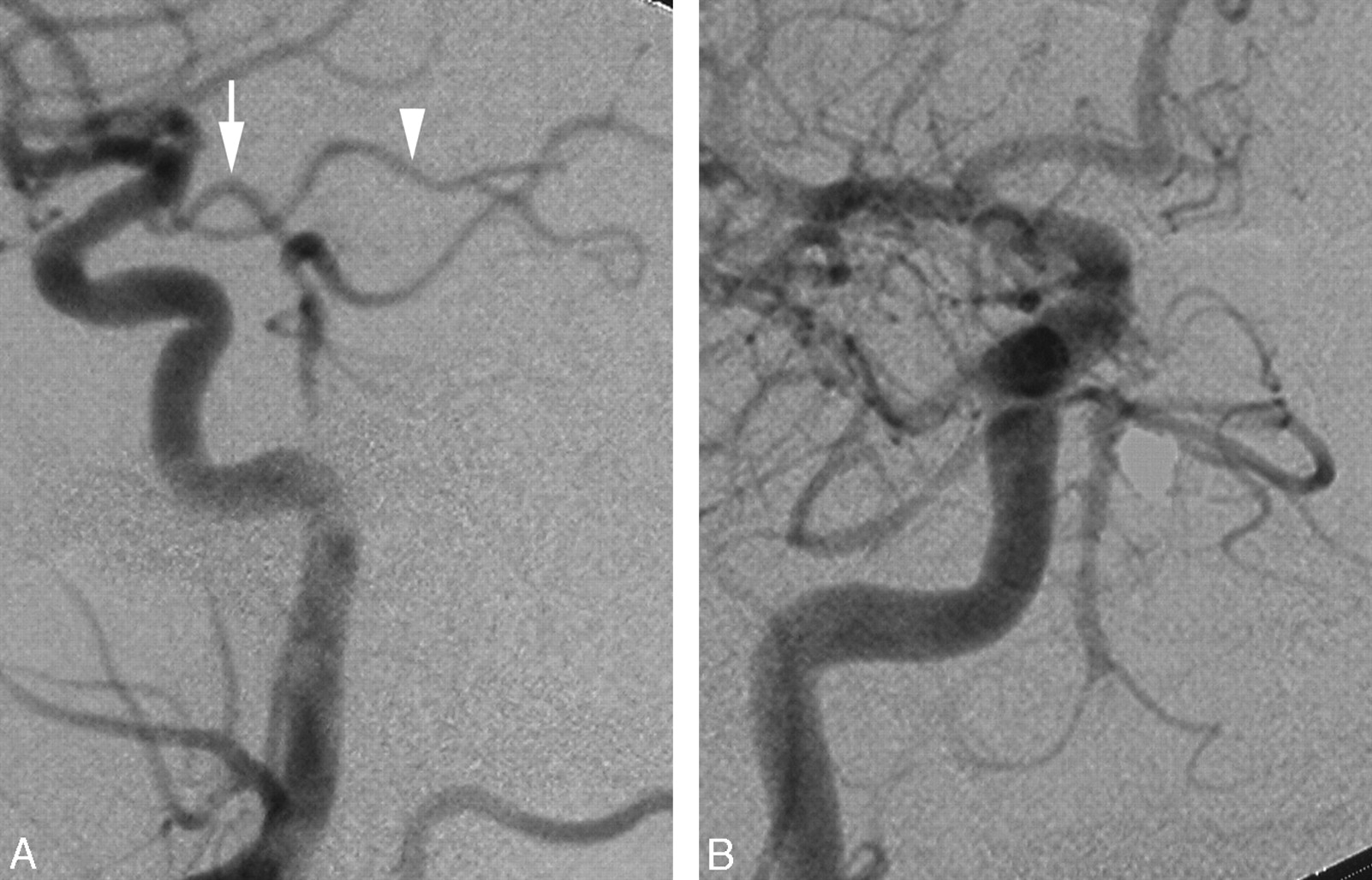
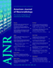
  • Fig 3.
    • Download figure
    • Open in new tab
    • Download powerpoint
    Fig 3.

    A 51-year-old man with intermittent dizziness. A, DSA, left VA injection, anteroposterior view, showing disease in the distal VA and proximal BA (arrowhead), including faint opacification of the right AICA (arrow). B, DSA, left common carotid artery injection, lateral view, showing a large PcomA supplying the BA. Note that the PcomA then continues as the left PCA (arrowhead). The hemispheric branch of the right PICA territory is fed through a connection with the BA. C, DSA, left common carotid injection, transfascial view, showing a large left PcomA supplying the BA and continuing its course as the left PCA. Note the opacification of the right AICA (arrow), which confirms the existence of a connection between the left VA and the distal BA.

  • Fig 4.
    • Download figure
    • Open in new tab
    • Download powerpoint
    Fig 4.

    Schematic representation of the vertebrobasilar circulation. Posterior view of the VAs and BA. On the left side, the segments derived from the radicular branch of the proatlantal artery are shown in gray. Ant. Spinal a. indicates anterior spinal artery; Post. Spinal a., posterior spinal artery.

  • Fig 5.
    • Download figure
    • Open in new tab
    • Download powerpoint
    Fig 5.

    Developmental anatomy of the distal BA. A, The anterior circulation supplies the developing posterior circulation (ie, the parallel longitudinal neural arteries [LNA]) via the trigeminal and the posterior communicating arteries (PcomAs). The terminal branches of the carotid axis are the AchoA, which provides the territory of the PcomAs and the anterior cerebral artery (ACA) and its developing branch, the middle cerebral artery (MCA). The VA is not yet connected to the LNA. The PcomA has 2 small branches, the PchoA and the diencephalic artery (Dienceph. A). A indicates artery. B, The VA connects to the proximal end of the LNA/BA. The trigeminal artery involutes, and the PcomA becomes the last of the carotid basilar anastomoses. C, A connection is formed between the AchoA/PCA and the PchoA. D, The connection between the PcomA and the PCA is now well established. The AchoA is not supplying the PCA territory anymore. The latter can be supplied equally by the posterior circulation via the distal segment of the PcomA or by the anterior circulation via the proximal segment of the PcomA. This configuration of equal contribution can persist at the adult stage. Other adult configurations depend on the relative regression of the PcomA, as described in E and F. Note that the diencephalic branch is now called the posterior medial choroidal artery (PMchoA), whereas the original PchoA is now called the posterior lateral choroidal artery (PLchoA).11 Although both arteries are originally derived from the PcomA, they are considered as branches of the PCA at the adult stage. The so-called “P1 segment of the PCA” is in fact the distal portion of the PcomA. E, In this second adult configuration, the distal segment of the PcomA has regressed and the anterior contribution to the PCA becomes dominant. This configuration is often mislabeled as a “fetal origin of the PCA.” The true “fetal origin of the PCA,” where the transfer of the PCA territory from the AchoA to the PcomA has not occurred, is not illustrated here but would be similar to B. It is illustrated in Fig 2A. F, In this third adult configuration, the proximal segment of the PcomA has regressed and the posterior contribution to the PCA becomes dominant.

PreviousNext
Back to top

In this issue

American Journal of Neuroradiology: 28 (10)
American Journal of Neuroradiology
Vol. 28, Issue 10
November 2007
  • Table of Contents
  • Index by author
Advertisement
Print
Download PDF
Email Article

Thank you for your interest in spreading the word on American Journal of Neuroradiology.

NOTE: We only request your email address so that the person you are recommending the page to knows that you wanted them to see it, and that it is not junk mail. We do not capture any email address.

Enter multiple addresses on separate lines or separate them with commas.
Bilateral Segmental Agenesis of the Vertebrobasilar Junction: Developmental and Angiographic Anatomy
(Your Name) has sent you a message from American Journal of Neuroradiology
(Your Name) thought you would like to see the American Journal of Neuroradiology web site.
CAPTCHA
This question is for testing whether or not you are a human visitor and to prevent automated spam submissions.
Cite this article
I.M. Burger, F. Siclari, L. Gregg, P. Gailloud
Bilateral Segmental Agenesis of the Vertebrobasilar Junction: Developmental and Angiographic Anatomy
American Journal of Neuroradiology Nov 2007, 28 (10) 2017-2022; DOI: 10.3174/ajnr.A0719

Citation Manager Formats

  • BibTeX
  • Bookends
  • EasyBib
  • EndNote (tagged)
  • EndNote 8 (xml)
  • Medlars
  • Mendeley
  • Papers
  • RefWorks Tagged
  • Ref Manager
  • RIS
  • Zotero
0 Responses
Respond to this article
Share
Bookmark this article
Bilateral Segmental Agenesis of the Vertebrobasilar Junction: Developmental and Angiographic Anatomy
I.M. Burger, F. Siclari, L. Gregg, P. Gailloud
American Journal of Neuroradiology Nov 2007, 28 (10) 2017-2022; DOI: 10.3174/ajnr.A0719
del.icio.us logo Twitter logo Facebook logo Mendeley logo
  • Tweet Widget
  • Facebook Like
  • Google Plus One
Purchase

Jump to section

  • Article
    • Abstract
    • Case Illustrations
    • Discussion
    • Conclusions
    • References
  • Figures & Data
  • Info & Metrics
  • Responses
  • References
  • PDF

Related Articles

  • No related articles found.
  • PubMed
  • Google Scholar

Cited By...

  • Ultrasound findings of bilateral hypoplasia of the vertebral arteries associated with a persistent carotid-hypoglossal artery
  • Bilateral vertebral artery occlusion with retrograde basilary flow in three cases of giant cell arteritis
  • Crossref (22)
  • Google Scholar

This article has been cited by the following articles in journals that are participating in Crossref Cited-by Linking.

  • A Functional Perspective on the Embryology and Anatomy of the Cerebral Blood Supply
    Khaled Menshawi, Jay P Mohr, Jose Gutierrez
    Journal of Stroke 2015 17 2
  • Normal and Abnormal Embryology and Development of the Intracranial Vascular System
    Charles Raybaud
    Neurosurgery Clinics of North America 2010 21 3
  • The persistent trigeminal artery: development, imaging anatomy, variants, and associated vascular pathologies
    Stephan Meckel, Bjoern Spittau, William McAuliffe
    Neuroradiology 2013 55 1
  • The Role of the Primitive Lateral Basilovertebral Anastomosis of Padget in Variations of the Vertebrobasilar Arterial System
    Lydia Gregg, Philippe Gailloud
    The Anatomical Record 2017 300 11
  • Basilar Artery Hypoplasia
    F. Caranci, M. Napoli, M. Cirillo, G. Briganti, L. Brunese, F. Briganti
    The Neuroradiology Journal 2012 25 6
  • Monolateral type I proatlantal artery with bilateral absence of vertebral arteries: description of a case and review of the literature
    M. Montechiari, A. Iadanza, A. Falini, L. S. Politi
    Surgical and Radiologic Anatomy 2013 35 9
  • Smaller Absolute Quantities but Greater Relative Densities of Microvessels Are Associated with Cerebellar Degeneration in Lurcher Mice
    Yaroslav Kolinko, Jan Cendelin, Milena Kralickova, Zbynek Tonar
    Frontiers in Neuroanatomy 2016 10
  • Bilateral Segmental Agenesis of Carotid and Vertebral Arteries with Rete Mirabile and the Prominent Anterior and Posterior Spinal Arteries as Compensations
    Jun Lu, Jiachun Liu, Lijun Wang, Peng Qi, Daming Wang
    Interventional Neuroradiology 2014 20 1
  • Bilateral Persistent Primitive Trigeminal Arteries Associated with Trigeminal Neuralgia
    B. Son, S. Yang, J. Sung, S. Lee
    Clinical Neuroradiology 2013 23 1
  • Bilateral vertebral artery occlusion with retrograde basilary flow in three cases of giant cell arteritis
    Markus Robert Boettinger, Schreglmann Robert Sebastian, Maria-Andreea Robert Gamulescu, Oliver Grauer, Markus Ritzka, Gerhard Robert Schuierer, Ulrich Robert Bogdahn, Andreas Steinbrecher, Felix Schlachetzki
    BMJ Case Reports 2009 2009

More in this TOC Section

  • Usefulness of Quantitative Susceptibility Mapping for the Diagnosis of Parkinson Disease
  • White Matter Alterations in the Brains of Patients with Active, Remitted, and Cured Cushing Syndrome: A DTI Study
  • Qualitative and Quantitative Analysis of MR Imaging Findings in Patients with Middle Cerebral Artery Stroke Implanted with Mesenchymal Stem Cells
Show more BRAIN

Similar Articles

Advertisement

Indexed Content

  • Current Issue
  • Accepted Manuscripts
  • Article Preview
  • Past Issues
  • Editorials
  • Editor's Choice
  • Fellows' Journal Club
  • Letters to the Editor
  • Video Articles

Cases

  • Case Collection
  • Archive - Case of the Week
  • Archive - Case of the Month
  • Archive - Classic Case

Special Collections

  • AJNR Awards
  • ASNR Foundation Special Collection
  • Most Impactful AJNR Articles
  • Photon-Counting CT
  • Spinal CSF Leak Articles (Jan 2020-June 2024)

More from AJNR

  • Trainee Corner
  • Imaging Protocols
  • MRI Safety Corner

Multimedia

  • AJNR Podcasts
  • AJNR Scantastics

Resources

  • Turnaround Time
  • Submit a Manuscript
  • Submit a Video Article
  • Submit an eLetter to the Editor/Response
  • Manuscript Submission Guidelines
  • Statistical Tips
  • Fast Publishing of Accepted Manuscripts
  • Graphical Abstract Preparation
  • Imaging Protocol Submission
  • Evidence-Based Medicine Level Guide
  • Publishing Checklists
  • Author Policies
  • Become a Reviewer/Academy of Reviewers
  • News and Updates

About Us

  • About AJNR
  • Editorial Board
  • Editorial Board Alumni
  • Alerts
  • Permissions
  • Not an AJNR Subscriber? Join Now
  • Advertise with Us
  • Librarian Resources
  • Feedback
  • Terms and Conditions
  • AJNR Editorial Board Alumni

American Society of Neuroradiology

  • Not an ASNR Member? Join Now

© 2025 by the American Society of Neuroradiology All rights, including for text and data mining, AI training, and similar technologies, are reserved.
Print ISSN: 0195-6108 Online ISSN: 1936-959X

Powered by HighWire