
Article Figures & Data
Figures
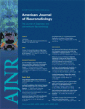
- Fig 1.
A 43-year-old man with an incidentally discovered tentorial dural fistula.
A, Lateral view of 3D right internal carotid artery angiogram demonstrates a tentorial dural fistula mainly supplied by the tentorial artery. Additional arterial supply was by lateral clival branches, the occipital artery, and the middle meningeal artery.
B, Angiogram via microcatheter in the tentorial artery during balloon occlusion of the internal carotid artery at the level of the tentorial artery. The balloon prevents reflux, stabilizes the microcatheter, and induces flow control. From this point, glue was injected with deep penetration into the veins.
C, Follow-up angiogram 6 weeks later demonstrates complete closure of the fistula.
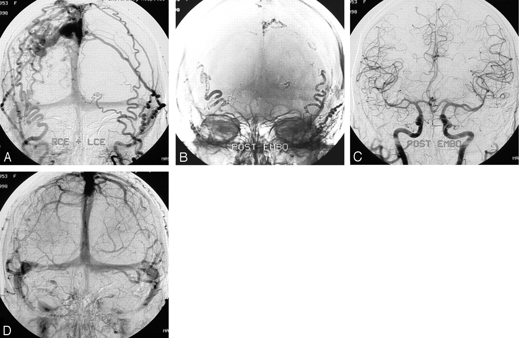
- Fig 2.
A 42-year-old woman with a right parietal hematoma. In this early case, distal access was not possible, and glue injection resulted in rather proximal occlusion of the feeders instead of occlusion of the draining vein. Although unusual, this resulted in complete cure.
A, Frontal view of bilateral external carotid angiogram shows a right parietal DAVF supplied by right and left occipital and middle meningeal arteries and draining via cortical veins to the superior sagittal sinus.
B, Glue cast after embolization shows that all 4 arterial feeders are occluded without occlusion of the draining veins.
C and D, Arterial (C) and venous (D) phases of bilateral common carotid artery angiograms after embolization show complete occlusion of the DAVF, confirmed at follow-up angiography 3 months later.
- Fig 3.
A 43-year-old woman presenting with a left parietal hematoma.
A and B, Frontal (A) and lateral (B) views of bilateral external carotid artery angiograms reveal a DAVF supplied by the right and left middle meningeal arteries with drainage on the cortical veins and subsequently into the superior sagittal sinus and deep venous system.
C, Glue cast after embolization shows occlusion of the draining veins.
D, Bilateral external carotid angiogram confirms complete obliteration of the DAVF.
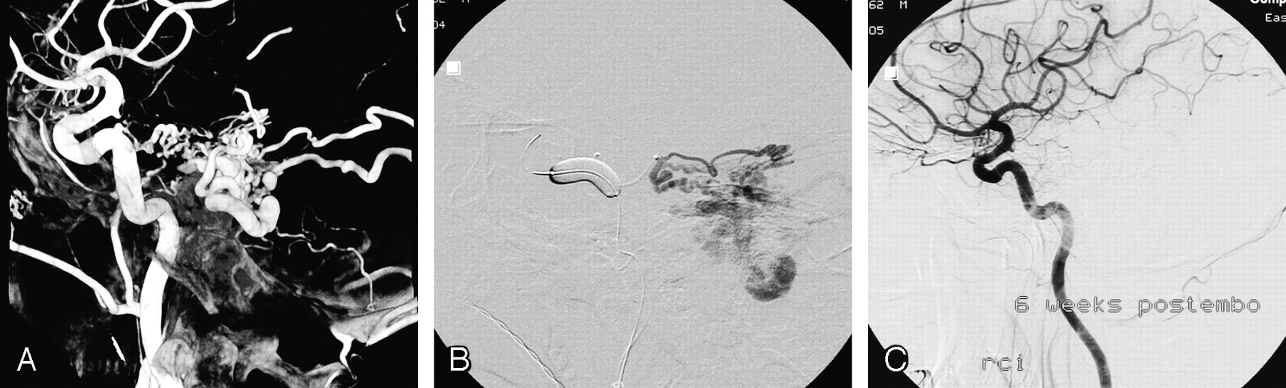
- Fig 4.
A 46-year-old man presenting with a seizure.
A, Lateral external carotid angiogram shows a DAVF on the left sigmoid sinus supplied by the middle meningeal and occipital arteries (single arrows), with drainage to the vein of Labee (double arrows).
B, Glue cast after embolization demonstrates occlusion of the draining vein of Labee (double arrows).


In this issue


Jump to section
Related Articles
Cited By...
- Engorged medullary vein on CT angiography in patients with dural arteriovenous fistula: prevalence, types, and comparison between regional and extensive types
- Resolution of bulbar and spinal symptoms postcranial dural arteriovenous fistula embolisation
- Dural arteriovenous fistula presenting with acute subdural haematoma showing impending cerebral herniation
- Quantifying the Cerebral Hemodynamics of Dural Arteriovenous Fistula in Transverse Sigmoid Sinus Complicated by Sinus Stenosis: A Retrospective Cohort Study
- Intracranial Dural Arteriovenous Fistulae: Clinical Presentation and Management Strategies
- Endovascular management of six simultaneous intracranial dural arteriovenous fistulas in a single patient
- Endovascular management of six simultaneous intracranial dural arteriovenous fistulas in a single patient
- Transarterial embolization with ONYX for treatment of intracranial non-cavernous dural arteriovenous fistula with or without cortical venous reflux
- Intraoperative Angiography for Cranial Dural Arteriovenous Fistula
- Curative Embolization with Onyx of Dural Arteriovenous Fistulas with Cortical Venous Drainage
- Cranial Dural Arteriovenous Fistula: Diagnosis and Classification with Time-Resolved MR Angiography at 3T
- Intracranial dural arterio-venous fistula
- Natural History of Dural Arteriovenous Shunts