
Article Figures & Data
Figures
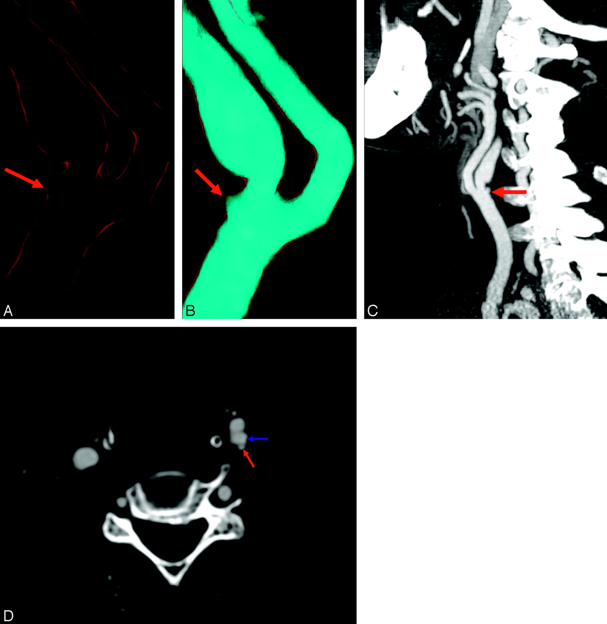
- Fig 1.
A 71-year-old man with persistent monocular visual loss. VR (A, B), MIP (C), and axial scans (D) show a severe and irregular ulcerated plaque of the internal carotid artery (ICA). In the left ICA, proximal to the point of maximum stenosis (60% NASCET), is clearly depicted an ulceration of 2 mm (red arrows). In the VR images, A is set without contrast media histogram and B is set with contrast media histogram. In C, there is visible hook morphology (ICA, blue arrow).
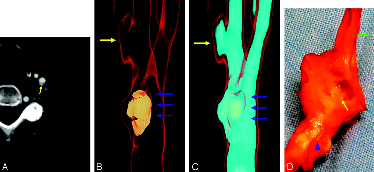
- Fig 2.
A 65-year-old woman with TIA. Ulcerated plaque of the left internal carotid artery (ICA); the typical button-of-shirt morphology is visible. Axial scan (A) illustrates ulcerations in a mixed plaque and displays a dissectionlike aspect. VR (B, C) clearly depicts the ulceration. Gross anatomic inspection confirms the presence of the ulcer (D). In this patient, the plaque is heavily calcified proximally and is not calcified at the location of the ulcer. This ulcer is distal to the point of maximum stenosis located in the bifurcation (80% NASCET), and could also have been a pseudoaneurysm from carotid dissection, but the surgical specimen confirmed the presence of ulceration. Note the big plaque calcification located in the bulbus/bifurcation (yellow arrows, ulceration; blue arrows, maximum stenosis point; green arrow, ICA; blue arrowhead, calcified plaque). The external carotid artery ostium is not visible because the cut plane passed through it.
- Fig 3.
A 78-year-old man with TIA. Ulcerated plaque of the left CCA. Axial scan (A) shows ulcerations in a calcified plaque; the atheromatous plaque involved the common carotid artery, the carotid bifurcation, and internal carotid artery origin. This ulcer is proximal to the point of maximum stenosis (90% NASCET). The ulceration was easily detected using VR technique with 1 histogram (B) and 2 histograms (C, D). Ulceration was less clearly visible using MIP and MPR reconstruction (E, F). Note: B is an internal visual, where the lumen is cut to clarify the ulcers. (red arrow in axial image and yellow arrows in VR, MIP, and MPR images, ulceration; green arrows, ICA; blue arrows, CCA)
- Fig 4.
Graph shows the sensitivity of SSD, MPR, MIP, VR, axial, and axial + VR. Mean values (▪) and 95% confidence intervals (error bars) are indicated
- Fig 5.
Graph shows the specificity of SSD, MPR, MIP, VR, axial, and axial + VR. Mean values (▪) and 95% confidence intervals (error bars) are indicated
- Fig 6.
Graph shows sensitivity, specificity, PPV, NPV of SSD, MPR, MIP, VR, axial, and axial + VR.


Tables
Method of Evaluation % Sensitivity (CI) % Specificity (CI) % PPV (CI) % NPV (CI) Axial scan 90.9 (81.1–100.7) 94.7 (89.7–99.8) 88.2 (77.4–99.1) 96.0 (91.6–100.4) MIP 78.8 (64.8–92.7) 92.1 (86.0–98.2) 81.3 (67.7–94.8) 90.9 (84.5–97.3) MPR 75.8 (61.1–90.4) 90.8 (84.3–97.3) 78.1 (63.8–92.4) 89.6 (82.8–96.4) SSD 39.4 (22.7–56.1) 88.2 (80.9–95.4) 59.1 (38.5–79.6) 77 (68.2–85.9) VR 87.9 (76.7–99) 97.4 (93.8–101) 93.5 (84.9–102.2) 94.9 (90–99.8) VR + axial scan 93.9 (85.8–102.1) 98.7 (96.1–101.2) 93.5 (84.9–102.2) 97.4 (93.8–101) Note:—CI indicates confidence interval; PPV, positive predictive value; NPV, negative predictive value; MIP, maximum intensity projection; MPR, multiplanar reconstruction; SSD, shaded surface display; VR, volume rendering.
Stenosis Degree (NASCET) Patients with Plaque Ulceration Patients without Ulceration % with Plaque Ulceration 50–69 % 1 6 14.29 70–84 % 8 26 23.54 85–99 % 22 46 32.35 Note:—NASCET indicates North American Symptomatic Carotid Endarterectomy Trial.
Plaque Type Patients with Plaque Ulceration Patients without Ulceration % with Plaque Ulceration Fatty 19 16 54.3 Mixed 8 34 19.1 Calcified 4 28 12.5
In this issue


Jump to section
Related Articles
Cited By...
- Carotid Vessel Wall Imaging on CTA
- Carotid Artery Wall Imaging: Perspective and Guidelines from the ASNR Vessel Wall Imaging Study Group and Expert Consensus Recommendations of the American Society of Neuroradiology
- Imaging Carotid Atherosclerosis Plaque Ulceration: Comparison of Advanced Imaging Modalities and Recent Developments
- Intraplaque Hemorrhage and the Plaque Surface in Carotid Atherosclerosis: The Plaque At RISK Study (PARISK)
- CTA for Screening of Complicated Atherosclerotic Carotid Plaque--American Heart Association Type VI Lesions as Defined by MRI
- Carotid Artery Wall Thickness Measured Using CT: Inter- and Intraobserver Agreement Analysis
- Vascular Wall Imaging of Vulnerable Atherosclerotic Carotid Plaques: Current State of the Art and Potential Future of Endovascular Optical Coherence Tomography
- Association Between Carotid Artery Plaque Ulceration and Plaque Composition Evaluated With Multidetector CT Angiography
- Characterization of Carotid Plaque Hemorrhage: A CT Angiography and MR Intraplaque Hemorrhage Study
- Atherosclerotic Plaque Ulceration in the Symptomatic Internal Carotid Artery Is Associated With Nonlacunar Ischemic Stroke
- Assessment of Intracranial Arterial Stenosis with Multidetector Row CT Angiography: A Postprocessing Techniques Comparison
- Window Settings for the Study of Calcified Carotid Plaques with Multidetector CT Angiography
- Contrast-Enhanced MR Angiography Is Not More Accurate Than Unenhanced 2D Time-of-Flight MR Angiography for Determining >=70% Internal Carotid Artery Stenosis
- Atherosclerotic Plaque Surface Morphology in the Carotid Bifurcation Assessed With Multidetector Computed Tomography Angiography