Advertisement

Main menu

  • Home
  • Content
    • Current Issue
    • Accepted Manuscripts
    • Article Preview
    • Past Issue Archive
    • Video Articles
    • AJNR Case Collection
    • Case of the Week Archive
    • Case of the Month Archive
    • Classic Case Archive
  • Special Collections
    • AJNR Awards
    • ASNR Foundation Special Collection
    • Most Impactful AJNR Articles
    • Photon-Counting CT
    • Spinal CSF Leak Articles (Jan 2020-June 2024)
  • Multimedia
    • AJNR Podcasts
    • AJNR SCANtastic
    • Trainee Corner
    • MRI Safety Corner
    • Imaging Protocols
  • For Authors
    • Submit a Manuscript
    • Submit a Video Article
    • Submit an eLetter to the Editor/Response
    • Manuscript Submission Guidelines
    • Statistical Tips
    • Fast Publishing of Accepted Manuscripts
    • Graphical Abstract Preparation
    • Imaging Protocol Submission
    • Author Policies
  • About Us
    • About AJNR
    • Editorial Board
    • Editorial Board Alumni
  • More
    • Become a Reviewer/Academy of Reviewers
    • Subscribers
    • Permissions
    • Alerts
    • Feedback
    • Advertisers
    • ASNR Home

User menu

  • Alerts
  • Log in

Search

  • Advanced search
American Journal of Neuroradiology

American Journal of Neuroradiology

ASHNR American Society of Functional Neuroradiology ASHNR American Society of Pediatric Neuroradiology ASSR
  • Alerts
  • Log in

Advanced Search

  • Home
  • Content
    • Current Issue
    • Accepted Manuscripts
    • Article Preview
    • Past Issue Archive
    • Video Articles
    • AJNR Case Collection
    • Case of the Week Archive
    • Case of the Month Archive
    • Classic Case Archive
  • Special Collections
    • AJNR Awards
    • ASNR Foundation Special Collection
    • Most Impactful AJNR Articles
    • Photon-Counting CT
    • Spinal CSF Leak Articles (Jan 2020-June 2024)
  • Multimedia
    • AJNR Podcasts
    • AJNR SCANtastic
    • Trainee Corner
    • MRI Safety Corner
    • Imaging Protocols
  • For Authors
    • Submit a Manuscript
    • Submit a Video Article
    • Submit an eLetter to the Editor/Response
    • Manuscript Submission Guidelines
    • Statistical Tips
    • Fast Publishing of Accepted Manuscripts
    • Graphical Abstract Preparation
    • Imaging Protocol Submission
    • Author Policies
  • About Us
    • About AJNR
    • Editorial Board
    • Editorial Board Alumni
  • More
    • Become a Reviewer/Academy of Reviewers
    • Subscribers
    • Permissions
    • Alerts
    • Feedback
    • Advertisers
    • ASNR Home
  • Follow AJNR on Twitter
  • Visit AJNR on Facebook
  • Follow AJNR on Instagram
  • Join AJNR on LinkedIn
  • RSS Feeds

AJNR is seeking candidates for the AJNR Podcast Editor. Read the position description.

LetterLetter

Congenital Glioependymal Cyst Presenting with Severe Proptosis

Ruby E. Obaldo, Lei Shao and Lisa H. Lowe
American Journal of Neuroradiology June 2007, 28 (6) 999-1000; DOI: https://doi.org/10.3174/ajnr.A0533
Ruby E. Obaldo
  • Find this author on Google Scholar
  • Find this author on PubMed
  • Search for this author on this site
Lei Shao
  • Find this author on Google Scholar
  • Find this author on PubMed
  • Search for this author on this site
Lisa H. Lowe
  • Find this author on Google Scholar
  • Find this author on PubMed
  • Search for this author on this site
  • Article
  • Figures & Data
  • Info & Metrics
  • Responses
  • References
  • PDF
Loading

We discuss a unique case of a congenital glioependymal cyst arising from the left middle cranial fossa with extension into the left orbit causing severe proptosis, a presentation that has not been reported previously. A female neonate at 38 weeks’ gestational age was diagnosed with severe left proptosis on a prenatal sonography at 35 weeks’ gestation. The sonogram was performed to determine the size of the infant due to a maternal history of macrosomia in a previous pregnancy. The infant's prenatal history was otherwise unremarkable. The patient was delivered by an uncomplicated cesarean birth.

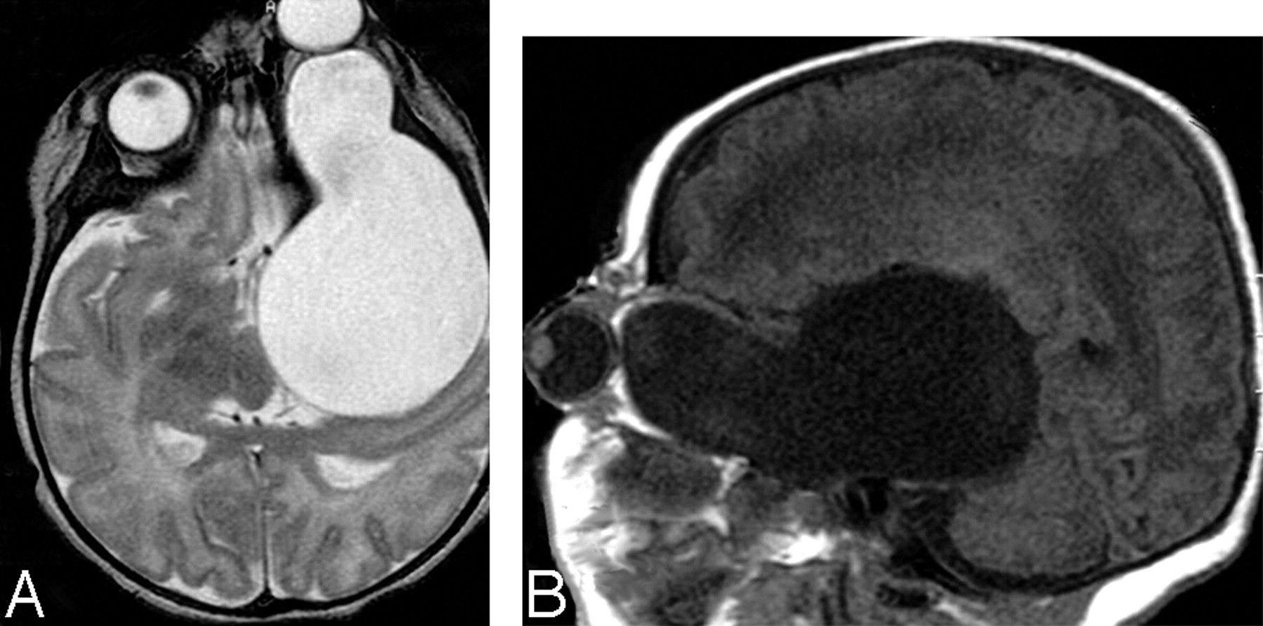
Physical examination showed severe left proptosis with intact extraocular movements. The right eye and findings from the remaining physical examination were unremarkable. The results of chromosome studies were normal. Noncontrast enhanced CT demonstrated a cystic structure occupying the left middle cranial fossa and extending into the left orbit. MR imaging verified the CT findings and showed no associated brain abnormalities (Fig 1).

On the patient's second day of life, a cyst-to-peritoneum shunt was placed without complications. The left proptosis rapidly improved, and a sonogram showed only a small cyst on the fifth day of life. The patient was discharged but was returned on the 15th day of life because of recurrent left proptosis. After a failed shunt revision, a left temporal craniotomy was performed with removal of the lateral wall of the cyst and fenestration of the cyst to the subarachnoid space. No brain parenchymal abnormalities were identified. Since that time, the child has had an uneventful clinical course, and a noncontrast CT 1 year later showed that only a small cyst remained.

Upon gross examination, the pathologic specimen contained multiple fragments of fibrous tissue in loose aggregates (not photographed). Microscopic examination revealed that the specimen consisted of a thick layer of glial/connective tissue lined by a single layer of cuboidal or columnar epithelium with cilia. In certain areas, papillary structures resembling the choroid plexus were identified. No goblet cells were seen in the specimen. The epithelium was diffusely positive for S-100 protein and glial fibrillary acidic protein, and focally positive for cytokeratin in the areas resembling the choroid plexus. The epithelial cells were negative for epithelial membrane antigen and carcinoembryonic antigen. Focal acute inflammation was noted, consistent with previous surgical intervention. The histologic and immunophenotypic findings were most consistent with a glioependymal cyst.

Based on the imaging features, the differential diagnoses included a glioependymal cyst, ependymal cyst, arachnoid cyst, enterogenous cyst, dermoid cyst, encephalocele, and congenital dysplasia of the sphenoid wing in neurofibromatosis type I. The diagnosis of a glioependymal cyst requires correlation of clinical, radiologic, and histologic findings. Glioependymal cysts, also known as neuroepithelial cysts, are thought to arise from ectopic rests of primitive neuroglial tissue and therefore can arise anywhere in the neuraxis.1 On imaging, they appear as nonenhancing CSF containing thin-walled cysts.2,3 Our case is unique because of the unusual clinical presentation of severe proptosis. To our knowledge, neither this presentation nor the imaging appearance has been reported previously in a neonate with a glioependymal cyst.

  Fig 1.
  • Download figure
  • Open in new tab
  • Download powerpoint
Fig 1.

Noncontrast MR imaging of axial T2 (A) and sagittal T1 (B) images show the cystic structure after CSF signal intensity on all sequences. Note proptotic left globe.

References

  1. ↵
    Osborn AG, Preece MT. Intracranial cysts: radiologic-pathologic correlation and imaging approach. Radiology 2006;239:650–64
    CrossRefPubMed
  2. ↵
    Shields JA, Shields CL. Orbital cysts of childhood–classification, clinical features, and management. Surv Ophthalmol 2004;49:281–99
    CrossRefPubMed
  3. ↵
    Guermazi A, Miaux Y, Majoulet JF, et al. Imaging findings of central nervous system neuroepithelial cysts. Eur Radiol 1998;8:618–23
    CrossRefPubMed
  • Copyright © American Society of Neuroradiology
PreviousNext
Back to top

In this issue

American Journal of Neuroradiology: 28 (6)
American Journal of Neuroradiology
Vol. 28, Issue 6
June 2007
  • Table of Contents
  • Index by author
Advertisement
Print
Download PDF
Email Article

Thank you for your interest in spreading the word on American Journal of Neuroradiology.

NOTE: We only request your email address so that the person you are recommending the page to knows that you wanted them to see it, and that it is not junk mail. We do not capture any email address.

Enter multiple addresses on separate lines or separate them with commas.
Congenital Glioependymal Cyst Presenting with Severe Proptosis
(Your Name) has sent you a message from American Journal of Neuroradiology
(Your Name) thought you would like to see the American Journal of Neuroradiology web site.
CAPTCHA
This question is for testing whether or not you are a human visitor and to prevent automated spam submissions.
Cite this article
Ruby E. Obaldo, Lei Shao, Lisa H. Lowe
Congenital Glioependymal Cyst Presenting with Severe Proptosis
American Journal of Neuroradiology Jun 2007, 28 (6) 999-1000; DOI: 10.3174/ajnr.A0533

Citation Manager Formats

  • BibTeX
  • Bookends
  • EasyBib
  • EndNote (tagged)
  • EndNote 8 (xml)
  • Medlars
  • Mendeley
  • Papers
  • RefWorks Tagged
  • Ref Manager
  • RIS
  • Zotero
0 Responses
Respond to this article
Share
Bookmark this article
Congenital Glioependymal Cyst Presenting with Severe Proptosis
Ruby E. Obaldo, Lei Shao, Lisa H. Lowe
American Journal of Neuroradiology Jun 2007, 28 (6) 999-1000; DOI: 10.3174/ajnr.A0533
del.icio.us logo Twitter logo Facebook logo Mendeley logo
  • Tweet Widget
  • Facebook Like
  • Google Plus One
Purchase

Jump to section

  • Article
    • References
  • Figures & Data
  • Info & Metrics
  • Responses
  • References
  • PDF

Related Articles

  • No related articles found.
  • PubMed
  • Google Scholar

Cited By...

  • No citing articles found.
  • Crossref (13)
  • Google Scholar

This article has been cited by the following articles in journals that are participating in Crossref Cited-by Linking.

  • Intracranial glioependymal (neuroglial) cysts: a systematic review
    Luis A. Robles, Jesus M. Paez, Dafne Ayala, Bernardo Boleaga-Duran
    Acta Neurochirurgica 2018 160 7
  • Glioependymal cyst in children: A case report
    Song-Ping Zheng, Yan Ju, Chao You
    Clinical Neurology and Neurosurgery 2013 115 10
  • Management of neonatal proptosis: A systematic review
    Benjamin P. Erickson, David T. Tse
    Survey of Ophthalmology 2014 59 4
  • Intracranial ependymal cyst with unusual presentation: Case report and review of literature
    Gbètoho Fortuné Gankpé, Geoffrey Jean Ndekha, Naama Okacha, Khalid Chakour, Mohammed El Faiz Chaoui, Mohammed Benzagmout
    Interdisciplinary Neurosurgery 2019 17
  • A large interhemispheric glioependymal cyst associated with partial defect of the corpus callosum in an elderly man
    Mio Takayama, Masayuki Kashiwagi, Kenji Hara, Aya Matsusue, Brian Waters, Shin‐ichi Kubo
    Neuropathology 2020 40 5
  • Combined neuroendoscopic cyst wall fenestration and cyst-peritoneal shunt in an infant with glioependymal cyst
    Kohei Irie, Takafumi Shimogawa, Nobutaka Mukae, Daisuke Kuga, Toru Iwaki, Masahiro Mizoguchi, Koji Yoshimoto
    Surgical Neurology International 2022 13
  • Giant supra and retrosellar glioependymal cyst presenting with only precocious puberty. Clinical study and review of the literature
    Hassan Kadri, Mazen Dughly, Mohamad Shehadeh Agha, Ghiath Hamed, Raed Abouharb, Rostom Mackieh
    International Journal of Surgery Case Reports 2024 116
  • Occipital lobe ependymal cyst with unusual presentation
    O Naama, A Idir, O Boulahroud
    Journal of Clinical Medical and Experimental Images 2019 3 1
  • Contralateral Endoscopic Approach for a Rare Case of Neuroglial Cyst
    Deepak Kumar Singh, Kshitij Sinha, Rakesh Kumar Singh, Vipin Kumar Chand, Neha Singh, Arun Kumar Singh, Krishan Kumar
    Indian Journal of Neurosurgery 2025 14 01
  • Diagnostic Imaging: Obstetrics
    2016

More in this TOC Section

  • Letter to the Editor regarding “Automated Volumetric Software in Dementia: Help or Hindrance to the Neuroradiologist?”
  • Reply:
  • Brain AVM’s Nidus: What if We Hadn’t Understood Anything?
Show more Letters

Similar Articles

Advertisement

Indexed Content

  • Current Issue
  • Accepted Manuscripts
  • Article Preview
  • Past Issues
  • Editorials
  • Editor's Choice
  • Fellows' Journal Club
  • Letters to the Editor
  • Video Articles

Cases

  • Case Collection
  • Archive - Case of the Week
  • Archive - Case of the Month
  • Archive - Classic Case

Special Collections

  • AJNR Awards
  • ASNR Foundation Special Collection
  • Most Impactful AJNR Articles
  • Photon-Counting CT
  • Spinal CSF Leak Articles (Jan 2020-June 2024)

More from AJNR

  • Trainee Corner
  • Imaging Protocols
  • MRI Safety Corner

Multimedia

  • AJNR Podcasts
  • AJNR Scantastics

Resources

  • Turnaround Time
  • Submit a Manuscript
  • Submit a Video Article
  • Submit an eLetter to the Editor/Response
  • Manuscript Submission Guidelines
  • Statistical Tips
  • Fast Publishing of Accepted Manuscripts
  • Graphical Abstract Preparation
  • Imaging Protocol Submission
  • Evidence-Based Medicine Level Guide
  • Publishing Checklists
  • Author Policies
  • Become a Reviewer/Academy of Reviewers
  • News and Updates

About Us

  • About AJNR
  • Editorial Board
  • Editorial Board Alumni
  • Alerts
  • Permissions
  • Not an AJNR Subscriber? Join Now
  • Advertise with Us
  • Librarian Resources
  • Feedback
  • Terms and Conditions
  • AJNR Editorial Board Alumni

American Society of Neuroradiology

  • Not an ASNR Member? Join Now

© 2025 by the American Society of Neuroradiology All rights, including for text and data mining, AI training, and similar technologies, are reserved.
Print ISSN: 0195-6108 Online ISSN: 1936-959X

Powered by HighWire