
Article Figures & Data
Figures
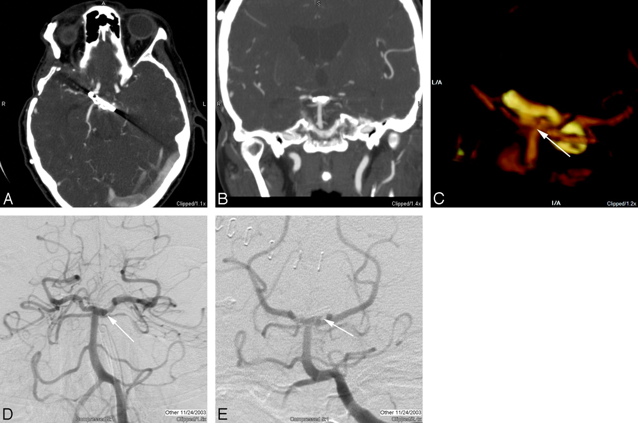
- Fig 1.
A, 3D volume rendering of CTA data viewed from the anterior and to patient's left side demonstrates a straight and right-angle clip at the anterior communicating artery. B, The combination of this view and a projection viewed from the posterior and slightly to the patient's right side demonstrates patency of the anterior communicating artery and adjacent vessels, with no evidence of residual aneurysm. A view from above (C) and from the patient's right side (D) demonstrates an additional aneurysm (arrowhead) arising in the medial clinoid segment, also referred to as the “carotid cave” with growth of the dome directed posteriorly and inferiorly toward the sella. A frontal projection angiogram (E) shows the region of the clips (arrow), which are subtracted out on subtracted conventional angiography and also demonstrates the medially directed “cave” aneurysm (arrowhead).
- Fig 2.
A, Axial source image through a surgical clip at the basilar tip demonstrates artifacts directed along the long axis of the clip as the result of the increased attenuation through that axis. B, The coronal multiplanar reconstruction image does not demonstrate many artifacts, despite the degradation of the axial source image, but the area immediately adjacent to the clip is not optimally imaged. C, With optimal threshold settings, in a 3D volume-rendered image viewed from the posterior and inferior aspect clips at the basilar tip, a small residual is evident (arrow), projecting off the left and posterior aspect of the aneurysm neck. Note that the orientation of this image is close to the projection for a Towne view routinely used for diagnostic evaluation of the posterior circulation, except that the CTA is being viewed from behind. D, Frontal “transfascial” view from catheter-based DSA demonstrates a double attenuation (arrow) that is suggestive of a residual aneurysm remnant. E, Towne view similar to the projection from the volume-rendered CTA, but now viewed from the front, demonstrates the small “dog ear” residual (arrow) on the left, measuring approximately 2 mm.
- Fig 3.
A, Axial source image at the level of 2 cobalt clips demonstrates significant artifacts that obscure the surrounding brain and vessels. Wider windows were used to minimize artifacts but resulted in poor vessel contrast differentiation. B, Axial source image through a series of 3 titanium aneurysm clips, with a fourth clip partially imaged, demonstrates minimal artifacts related to the titanium, and the parent vessel can easily be seen (arrow) with no evidence of residual aneurysm. C, 3D volume rendering demonstrates the 4 titanium clips and adjacent vessels without significant degradation by artifacts. D, Catheter angiogram demonstrates the original aneurysm before clip reconstruction.
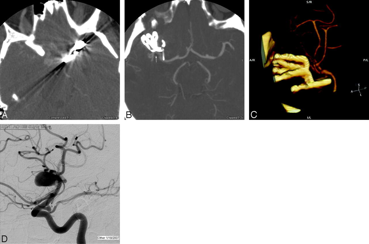
- Fig 4.
A, 3D volume reformat of CTA demonstrates a bilobed anterior communicating aneurysm. B, Axial MPR image obtained in the perioperative period demonstrates a round contrast collection consistent with a residual aneurysm remnant (arrow) of the posterior lobe of the bilobed aneurysm. C, Sagittal reformat also demonstrates a similar finding (arrow) and better shows the relation to the aneurysm clips. D, 3D volume rendering of the same CTA data, viewed from the anterior and below, gives an easily identifiable angiographic appearance that shows the residual aneurysm remnant (arrow) and the relation to the parent vessel and clips. E, Follow-up angiogram of the right internal carotid artery obtained 1 week post-subarachnoid hemorrhage demonstrates the residual aneurysm lumen (arrow) as well as a vasospasm involving the supraclinoid internal carotid artery and A1 segment. F, After coil embolization of the aneurysm remnant (arrow), the angiogram shows no residual aneurysm.
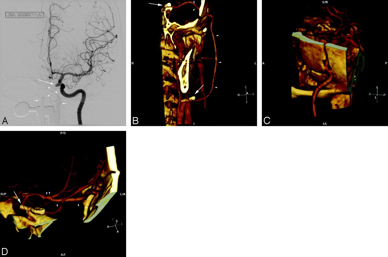
- Fig 5.
A, Frontal projection radiograph of a patient sent to our institution for treatment of a pseudoaneurysm (arrow) in the cavernous segment of the left internal carotid artery resulting from transsphenoidal surgery. Packing material (arrowheads) and a speculum (small arrows) were left in place from surgery. B, Postoperative CTA with 3D volume rendering demonstrates a clip occluding the proximal left internal carotid artery (arrow). A patent radial artery bypass (arrowheads) has a proximal anastomosis site to the left internal carotid artery just proximal to the clip. C, A second view demonstrates that the bypass is patent through the craniotomy site. D, Intracranial view shows the distal clip (arrow) effectively trapping the cavernous carotid pseudoaneurysm. The bypass (single arrowheads) is intact through the intracranial course, and the distal anastomosis (double arrowheads) to the middle cerebral artery is intact.


Tables
CTA protocol
CTA Routine CTA Postclipping Pre-CTA Routine head precontrast Routine head precontrast Coverage C1–2 to 2 cm below the inner table of the vertex C1–2 to 2 cm below the inner table of the vertex Acquisition Helical Helical Detectors 16i (64i) 16i (64i) Rotation time 0.6 seconds 0.6 seconds Section thickness 0.625 mm 0.625 mm Pitch 0.938:1 (0.969:1) 0.562:1 (0.531:1) Table speed 9.38 cm/s (19.37 cm/s) 5.62 cm/s (10.62 cm/s) Scanning FOV 25 cm (32 cm) 25 cm (32 cm) kV(p) 120 120 mA 750 750 Display FOV 15 cm 15 cm Algorithm Standard Standard Contrast injection Dual injector: 50-mL Omnipaque 300,* 4 mL/s; 40 mL saline, 2.5 mL/s Dual injector: 50 mL Omnipaque 300,* 4 mL/s; 40 mL saline, 2.5 mL/s Reconstruction Sagittal and coronal MPRs, 2.5 mm thick × 2.0-mm spacing Sagittal and coronal MPRs, 2.5 mm thick × 2.0-mm spacing Note:—MPR indicates multiplanar reconstruction image; FOV, field of view.
* Omnipaque 300 (iohexol), GE Health, Piscataway, NJ.
In this issue


Jump to section
- Article
- Abstract
- Available Noninvasive Imaging Techniques and Their Utility in the Assessment of Clipped Aneurysms
- CTA Techniques for the Evaluation of Clipped Aneurysms: Efficacy
- Utility of CTA for the Evaluation of Clipped Aneurysms
- CTA Techniques for the Evaluation of Aneurysms after Surgical Clipping: Limitations and Technical Points
- Image Interpretation
- Conclusions
- References
- Figures & Data
- Info & Metrics
- Responses
- References
Related Articles
Cited By...
- Noninvasive Angiographic Results of Clipped or Coiled Intracranial Aneurysms: An Inter- and Intraobserver Reliability Study
- Anatomical results, rebleeding and factors that affect the degree of occlusion in ruptured cerebral aneurysms after endovascular therapy
- Guidelines for the Management of Patients With Unruptured Intracranial Aneurysms: A Guideline for Healthcare Professionals From the American Heart Association/American Stroke Association
- Use of CT Angiography in Comparison with Other Imaging Techniques for the Determination of Embolus and Remnant Size in Experimental Aneurysms Embolized with Hydrogel Filaments