Advertisement

Main menu

  • Home
  • Content
    • Current Issue
    • Publication Preview--Ahead of Print
    • Past Issue Archive
    • Case of the Week Archive
    • Classic Case Archive
    • Case of the Month Archive
    • COVID-19 Content and Resources
  • For Authors
  • About Us
    • About AJNR
    • Editors
    • American Society of Neuroradiology
  • Submit a Manuscript
  • Podcasts
    • Subscribe on iTunes
    • Subscribe on Stitcher
  • More
    • Subscribers
    • Permissions
    • Advertisers
    • Alerts
    • Feedback
  • Other Publications
    • ajnr

User menu

  • Subscribe
  • Alerts
  • Log in

Search

  • Advanced search
American Journal of Neuroradiology

American Journal of Neuroradiology

  • Subscribe
  • Alerts
  • Log in

Advanced Search

  • Home
  • Content
    • Current Issue
    • Publication Preview--Ahead of Print
    • Past Issue Archive
    • Case of the Week Archive
    • Classic Case Archive
    • Case of the Month Archive
    • COVID-19 Content and Resources
  • For Authors
  • About Us
    • About AJNR
    • Editors
    • American Society of Neuroradiology
  • Submit a Manuscript
  • Podcasts
    • Subscribe on iTunes
    • Subscribe on Stitcher
  • More
    • Subscribers
    • Permissions
    • Advertisers
    • Alerts
    • Feedback
  • Follow AJNR on Twitter
  • Visit AJNR on Facebook
  • Follow AJNR on Instagram
  • Join AJNR on LinkedIn
  • RSS Feeds
OtherBRAIN

CT and MR Imaging of Progressive Dural Involvement by Nephrogenic Systemic Fibrosis

S. Zelasko, M. Hollingshead, M. Castillo and T.W. Bouldin
American Journal of Neuroradiology November 2008, 29 (10) 1880-1882; DOI: https://doi.org/10.3174/ajnr.A1225
S. Zelasko
  • Find this author on Google Scholar
  • Find this author on PubMed
  • Search for this author on this site
M. Hollingshead
  • Find this author on Google Scholar
  • Find this author on PubMed
  • Search for this author on this site
M. Castillo
  • Find this author on Google Scholar
  • Find this author on PubMed
  • Search for this author on this site
T.W. Bouldin
  • Find this author on Google Scholar
  • Find this author on PubMed
  • Search for this author on this site
  • Article
  • Figures & Data
  • Info & Metrics
  • References
  • PDF
Loading

Abstract

SUMMARY: We present a patient with progressive dural calcifications, thickening, and enhancement presumably related to the development of nephrogenic systemic fibrosis (NSF). Head CT demonstrated progressive dural calcifications, whereas MR imaging demonstrated progressive dural thickening and enhancement during a 3-year period in which the patient received several gadolinium-enhanced MR imaging studies. To the best of our knowledge, dural calcifications are the only described intracranial finding of NSF.

Nephrogenic systemic fibrosis (NSF), previously referred to as nephrogenic fibrosing dermopathy, is a relatively new, rare systemic condition first described in the literature in 2000.1 NSF occurs only in people with renal disease, with more than 215 documented cases in the NSF Registry at Yale University.2 All but a few of the known cases have been associated with intravenous administration of gadolinium-based contrast material.3 Manifestations of the disease are primarily cutaneous, but multiorgan system involvement has been described.4–7 Dural involvement has been described at autopsy; however, the depiction of the progressive imaging changes has not been documented.4,6,8 We report a patient with progressive dural calcification, thickening, and enhancement as a manifestation of NSF.

Case Report

A 52-year-old woman with a medical history of end stage renal disease secondary to adult polycystic kidney disease required hemodialysis starting in 2004. She had 8 gadolinium-enhanced MR imaging studies of the brain and abdomen between September 2004 and November 2006, all performed with gadodiamide.

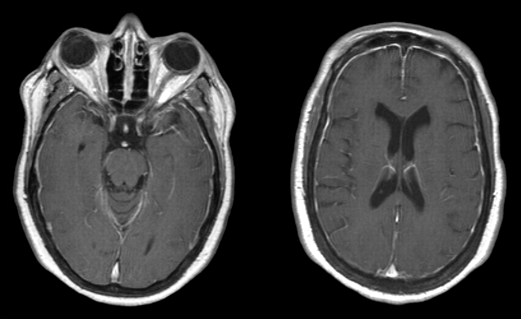
The initial brain MR imaging examination in September 2004 was performed for elevated serum prolactin levels. A pituitary microadenoma was seen, but results of the study were otherwise normal. A follow-up study in October 2005 demonstrated an interval decrease in adenoma size as well as new dural thickening and enhancement (Fig 1). The dural thickening and enhancement progressed on the gadolinium-enhanced MR imaging study obtained in March 2006.

Fig 1.
  • Download figure
  • Open in new tab
  • Download powerpoint
Fig 1.

October 2005. Axial T1-weighted postgadolinium MR imaging examination demonstrates diffuse dural enhancement. Changes in bone marrow are likely secondary to anemia.

Because of the uncommon nature of the meningeal abnormalities, the patient had a right frontal craniotomy and dural biopsy in May 2006. The dura mater contained multiple areas of fibrosis and calcification with rare, multinucleated giant cells adjacent to the foci of calcification (Fig 2).

Fig 2.
  • Download figure
  • Open in new tab
  • Download powerpoint
Fig 2.

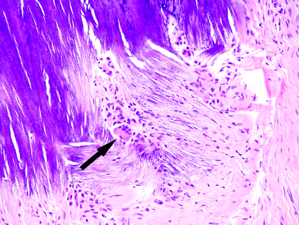
May 2006. Section of dura mater from the right frontal lobe shows fibrosis and blue-staining area of calcification. A multinucleated giant cell (arrow) is in the area of fibrosis next to the focus of calcification (hematoxylin-eosin stain).

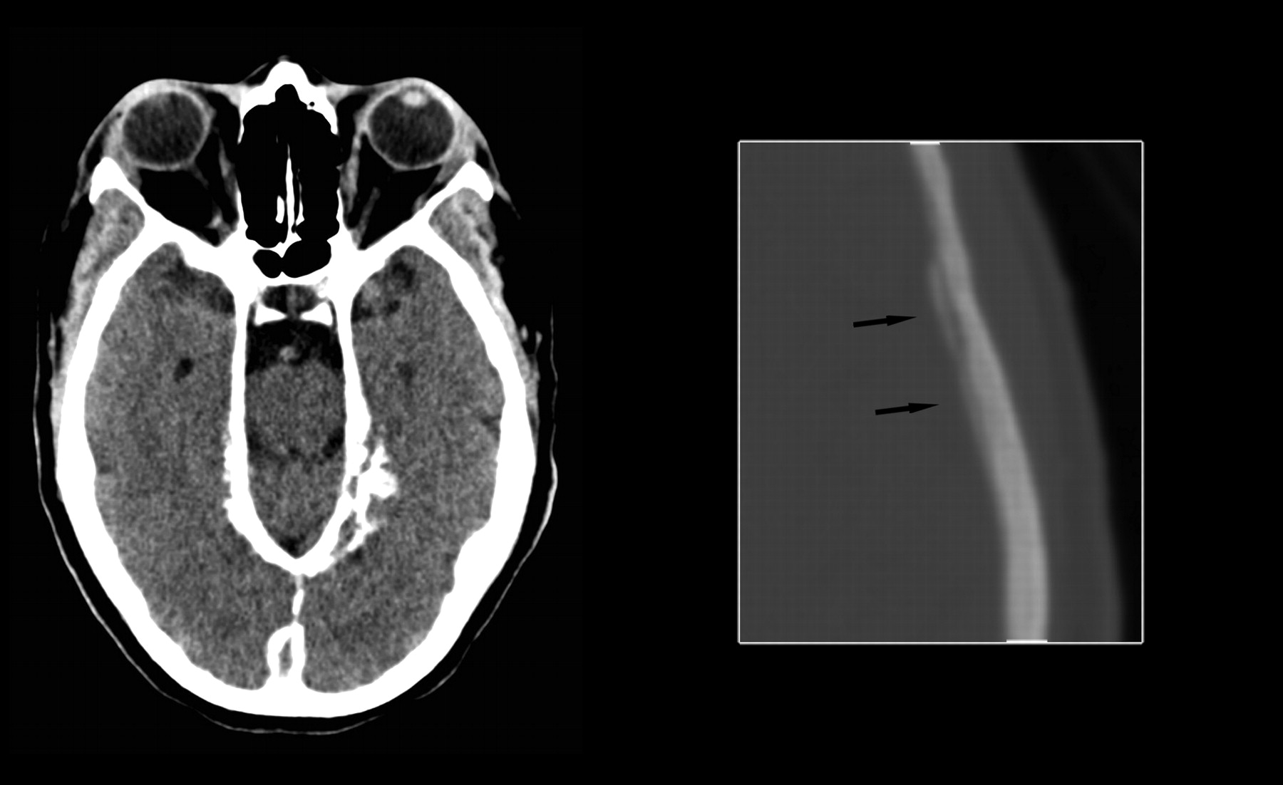
Head CT scan obtained in June 2006 demonstrated diffuse dural calcification (Fig 3). Follow-up gadolinium-enhanced MR imaging examination in November 2006 showed further progression of dural thickening and enhancement (Fig 4). Head CT scan in March 2008 demonstrated progressive calcification (Fig 5).

Fig 3.
  • Download figure
  • Open in new tab
  • Download powerpoint
Fig 3.

June 2006. Axial noncontrast CT image at the level of the tentorium in brain windows and magnified image at the level of the lateral ventricles in bone windows demonstrate diffuse dural calcification (arrows on magnified image).

Fig 4.
  • Download figure
  • Open in new tab
  • Download powerpoint
Fig 4.

November 2006. Axial T1-weighted postgadolinium MR imaging examination demonstrates progressive thickening and enhancement of the dura.

Fig 5.
  • Download figure
  • Open in new tab
  • Download powerpoint
Fig 5.

March 2008. Axial noncontrast CT image at the level of the tentorium in brain windows and magnified image at the level of the lateral ventricles in bone windows demonstrate progressive dural calcification (arrows on magnified image).

In April 2007, the patient presented with progressive skin thickening and decreased range of motion of her hands during a 4-month period. Results of skin biopsy from the right palm revealed fibrocellular thickening of dermal collagen, patchy calcification of blood vessel walls and dermal collagen, and increased Alcian-blue–positive mucin staining consistent with NSF.

Discussion

NSF is a rare, systemic disease linked to patients with renal failure who are exposed to intravenous gadolinium contrast. In 2006, Grobner9 suggested gadolinium as the cause of NSF. The disease was first described in the literature as skin thickening with “brawny hyperpigmentation” in 15 patients.1 Typical presenting symptoms are skin and subcutaneous tissue fibrosis in the extremities, then the trunk.7 Symptoms mimic scleroderma and other systemic fibrosing disorders.7 Serologic markers of scleroderma including antinuclear antibodies, anti-Scl 70 antibodies, and anticentromere antibodies are absent in NSF.10 Initially, NSF was believed to involve the skin and subcutaneous tissues, but subsequent reports documented involvement of the myocardium, muscle, lung, pericardium, pleura, kidney, bone, testis, and dura.4–8

The diagnosis of NSF is made by a combination of clinical, laboratory, and histopathologic findings.2,7 Prognosis is variable, but NSF may be fatal.7,11 Treatment leading to improved renal function may lead to relief of symptoms and may halt progression.7,11,12

Dural fibrosis from NSF was first reported in 2006.4,6 Both spinal and intracranial dural involvement have been described.4,6,8 Microscopic examination demonstrates fibrosis with a spindle-cell proliferation, areas of calcification, collections of CD68-positive mononuclear cells, and occasional multinucleated giant cells.4 The histologic changes in the dura mater of our patient were similar to those previously reported.4,6,8

The differential diagnosis for dural thickening and enhancement is extensive and includes sarcoidosis, tuberculosis, Wegener granulomatosis, intracranial hypotension, lymphoma, and metastatic disease. The differential diagnosis for dural calcifications includes physiologic calcifications, previous hemorrhage, previous infection, pseudoxanthoma elasticum, hyperparathyroidism, basal cell nevus syndrome, and idiopathic calcification.13

Virtually all patients with endstage renal disease have resultant hyperparathyroidism, and “metastatic calcification” may develop, with calcium deposition in the soft tissues and dura mater. However, dural thickening and enhancement are not typically seen in these patients. The patient in this case report had serum calcium values within normal limits and did not have other foci of soft tissue calcification. Her serum phosphorus levels ranged from low to elevated but were predominantly within normal limits. Although hyperparathyroidism cannot be excluded as the cause of the dural calcification because of the dural thickening and enhancement, the lack of other evidence of metastatic calcification, and her normal serum calcium levels, it is believed that the dural disease most likely represents a manifestation of NSF.

We suggest that dural calcification, thickening, and enhancement in patients with renal disease and a history of exposure to gadolinium may represent an early manifestation of NSF. Neuroradiologists must recognize these findings as a part of the spectrum of disease in NSF and may be the first to suggest the diagnosis if these radiologic findings precede cutaneous manifestations.

References

  1. ↵
    Cowper SE, Robin HS, Steinberg HM, et al. Scleromyxedema-like cutaneous disease in renal-dialysis patients. Lancet 2000;356:1000–01
    CrossRefPubMed
  2. ↵
    Cowper SE. The International Center for Nephrogenic Fibrosing Dermopathy Research (ICNFDR). Available at http://www.icnfdr.org. Accessed March 13,2008
  3. ↵
    Broome DR. Nephrogenic systemic fibrosis associated with gadolinium based contrast agents: A summary of the medical literature reporting. Eur J Radiol 2008;66:230–34
    CrossRefPubMed
  4. ↵
    Gibson SE, Farver CF, Prayson RA. Multiorgan involvement in nephrogenic fibrosing dermopathy: an autopsy case and review of the literature. Arch Pathol Lab Med 2006;130:209–12
    PubMed
  5. Kucher C, Steere J, Elenitsas R, et al. Nephrogenic fibrosing dermopathy/nephrogenic systemic fibrosis with diaphragmatic involvement in a patient with respiratory distress. J Am Acad Dermatol 2006;54:S31–34
    CrossRefPubMed
  6. ↵
    Saenz A, Mandal R, Kradin R, et al. Nephrogenic fibrosing dermopathy with involvement of the dura mater. Virchows Arch 2006;449:389–91
    CrossRefPubMed
  7. ↵
    Cowper SE, Rabach M, Girardi M. Clinical and histological findings in nephrogenic systemic fibrosis. Eur J Radiol 2008;66:191–99
    CrossRefPubMed
  8. ↵
    Krous HF, Breisch E, Chadwick AE, et al. Nephrogenic systemic fibrosis with multiorgan involvement in a teenage male after lymphoma, Ewing's sarcoma, end-stage renal disease, and hemodialysis. Pediatr Dev Pathol 2007;10:395–402
    CrossRefPubMed
  9. ↵
    Grobner T. Gadolinium–a specific trigger for the development of nephrogenic fibrosing dermopathy and nephrogenic systemic fibrosis [published erratum appears in Nephrol Dial Transplant 2006; 21:1745]. Nephrol Dial Transplant 2006;21:1104–08
    FREE Full Text
  10. ↵
    Broome DR, Girgus MS, Baron PW, et al. Gadodiamide-associated nephrogenic systemic fibrosis: why radiologists should be concerned. AJR Am J Roentgenol 2007;188:586–92
    CrossRefPubMed
  11. ↵
    Wiginton CD, Kelly B, Oto A, et al. Gadolinium-based contrast exposure, nephrogenic systemic fibrosis, and gadolinium detection in tissue. AJR Am J Roentgenol 2008;190:1060–68
    CrossRefPubMed
  12. ↵
    Knopp EA, Cowper SE. Nephrogenic systemic fibrosis: early recognition and treatment. Semin Dial 2008;21:123–28
    CrossRefPubMed
  13. ↵
    Dorenbeck U, Leingärtner T, Bretschneider T, et al. Tentorial and dural calcification with tertiary hyperparathyroidism: a rare entity in chronic renal failure. Eur Radiol 2002;12:S11–13
    CrossRef
  • Received April 28, 2008.
  • Accepted after revision May 28, 2008.
  • Copyright © American Society of Neuroradiology
View Abstract
PreviousNext
Back to top

In this issue

American Journal of Neuroradiology: 29 (10)
American Journal of Neuroradiology
Vol. 29, Issue 10
November 2008
  • Table of Contents
  • Index by author
Advertisement
Print
Download PDF
Email Article

Thank you for your interest in spreading the word on American Journal of Neuroradiology.

NOTE: We only request your email address so that the person you are recommending the page to knows that you wanted them to see it, and that it is not junk mail. We do not capture any email address.

Enter multiple addresses on separate lines or separate them with commas.
CT and MR Imaging of Progressive Dural Involvement by Nephrogenic Systemic Fibrosis
(Your Name) has sent you a message from American Journal of Neuroradiology
(Your Name) thought you would like to see the American Journal of Neuroradiology web site.
CAPTCHA
This question is for testing whether or not you are a human visitor and to prevent automated spam submissions.
Citation Tools
CT and MR Imaging of Progressive Dural Involvement by Nephrogenic Systemic Fibrosis
S. Zelasko, M. Hollingshead, M. Castillo, T.W. Bouldin
American Journal of Neuroradiology Nov 2008, 29 (10) 1880-1882; DOI: 10.3174/ajnr.A1225

Citation Manager Formats

  • BibTeX
  • Bookends
  • EasyBib
  • EndNote (tagged)
  • EndNote 8 (xml)
  • Medlars
  • Mendeley
  • Papers
  • RefWorks Tagged
  • Ref Manager
  • RIS
  • Zotero
Share
CT and MR Imaging of Progressive Dural Involvement by Nephrogenic Systemic Fibrosis
S. Zelasko, M. Hollingshead, M. Castillo, T.W. Bouldin
American Journal of Neuroradiology Nov 2008, 29 (10) 1880-1882; DOI: 10.3174/ajnr.A1225
Reddit logo Twitter logo Facebook logo Mendeley logo
  • Tweet Widget
  • Facebook Like
  • Google Plus One
Purchase

Jump to section

  • Article
    • Abstract
    • Case Report
    • Discussion
    • References
  • Figures & Data
  • Info & Metrics
  • References
  • PDF

Related Articles

  • No related articles found.
  • PubMed
  • Google Scholar

Cited By...

  • Nephrogenic Systemic Fibrosis: Review of 370 Biopsy-Confirmed Cases
  • Crossref
  • Google Scholar

This article has not yet been cited by articles in journals that are participating in Crossref Cited-by Linking.

More in this TOC Section

  • Predictors of Reperfusion in Patients with Acute Ischemic Stroke
  • Enhanced Axonal Metabolism during Early Natalizumab Treatment in Relapsing-Remitting Multiple Sclerosis
  • Progression of Microstructural Damage in Spinocerebellar Ataxia Type 2: A Longitudinal DTI Study
Show more BRAIN

Similar Articles

Advertisement

News and Updates

  • Lucien Levy Best Research Article Award
  • Thanks to our 2022 Distinguished Reviewers
  • Press Releases

Resources

  • Evidence-Based Medicine Level Guide
  • How to Participate in a Tweet Chat
  • AJNR Podcast Archive
  • Ideas for Publicizing Your Research
  • Librarian Resources
  • Terms and Conditions

Opportunities

  • Share Your Art in Perspectives
  • Get Peer Review Credit from Publons
  • Moderate a Tweet Chat

American Society of Neuroradiology

  • Neurographics
  • ASNR Annual Meeting
  • Fellowship Portal
  • Position Statements

© 2023 by the American Society of Neuroradiology | Print ISSN: 0195-6108 Online ISSN: 1936-959X

Powered by HighWire