Advertisement

Main menu

  • Home
  • Content
    • Current Issue
    • Publication Preview--Ahead of Print
    • Past Issue Archive
    • Case of the Week Archive
    • Classic Case Archive
    • Case of the Month Archive
    • COVID-19 Content and Resources
  • For Authors
  • About Us
    • About AJNR
    • Editors
    • American Society of Neuroradiology
  • Submit a Manuscript
  • Podcasts
    • Subscribe on iTunes
    • Subscribe on Stitcher
  • More
    • Subscribers
    • Permissions
    • Advertisers
    • Alerts
    • Feedback
  • Other Publications
    • ajnr

User menu

  • Subscribe
  • Alerts
  • Log in

Search

  • Advanced search
American Journal of Neuroradiology

American Journal of Neuroradiology

  • Subscribe
  • Alerts
  • Log in

Advanced Search

  • Home
  • Content
    • Current Issue
    • Publication Preview--Ahead of Print
    • Past Issue Archive
    • Case of the Week Archive
    • Classic Case Archive
    • Case of the Month Archive
    • COVID-19 Content and Resources
  • For Authors
  • About Us
    • About AJNR
    • Editors
    • American Society of Neuroradiology
  • Submit a Manuscript
  • Podcasts
    • Subscribe on iTunes
    • Subscribe on Stitcher
  • More
    • Subscribers
    • Permissions
    • Advertisers
    • Alerts
    • Feedback
  • Follow AJNR on Twitter
  • Visit AJNR on Facebook
  • Follow AJNR on Instagram
  • Join AJNR on LinkedIn
  • RSS Feeds
OtherBRAIN

Primary Sjögren Syndrome with Tumefactive Central Nervous System Involvement

J. Sanahuja, S. Ordoñez-Palau, R. Begué, L. Brieva and D. Boquet
American Journal of Neuroradiology November 2008, 29 (10) 1878-1879; DOI: https://doi.org/10.3174/ajnr.A1204
J. Sanahuja
  • Find this author on Google Scholar
  • Find this author on PubMed
  • Search for this author on this site
S. Ordoñez-Palau
  • Find this author on Google Scholar
  • Find this author on PubMed
  • Search for this author on this site
R. Begué
  • Find this author on Google Scholar
  • Find this author on PubMed
  • Search for this author on this site
L. Brieva
  • Find this author on Google Scholar
  • Find this author on PubMed
  • Search for this author on this site
D. Boquet
  • Find this author on Google Scholar
  • Find this author on PubMed
  • Search for this author on this site
  • Article
  • Figures & Data
  • Info & Metrics
  • References
  • PDF
Loading

Abstract

SUMMARY: Brain MR imaging abnormalities in primary Sjögren syndrome (pSS) are generally discrete white matter lesions. We describe a 50-year-old woman with recurrent neurologic deficits. MR imaging revealed a large brain lesion. A diagnosis of pSS was made on the basis of clinical features, positive anti-Ro and anti-La antibodies, abnormal Schirmer test findings, and salivary gland scintigraphy. The patient was treated with oral prednisone with good response. Large tumefactive brain lesions are a complication of pSS.

Sjögren syndrome is an autoimmune disorder that may be primary or secondary to other connective tissue diseases. It is characterized by lymphocytic infiltration and destruction of salivary and lachrymal glands leading to xerostomia and xerophthalmia. Similar mononuclear infiltrates or vasculitic lesions can have extraglandular manifestations.1 The prevalence of central nervous system (CNS) disease and MR imaging abnormalities in primary Sjögren syndrome (pSS) is controversial.2–4 We present a patient with pSS and a pseudotumoral lesion in the brain.

Case Report

A 50-year-old woman was referred to our unit with a 6-month history of headache associated with vomiting, impaired visual perception in left visual fields, and difficulty retrieving some words. The patient had a 1-year history of arthralgias, dry mouth, and a 3-kg weight loss. Findings of a neurologic examination were normal. Blood analysis showed antinuclear antibody titers of 1/160 and positive anti-Ro and anti-La antibodies. Findings of serologic tests were negative. MR imaging revealed a demyelinating-inflammatory lesion in the right temporo-occipital white matter. Cerebral angiography did not demonstrate vasculitis. CSF did not show oligoclonal bands. Visual evoked potentials were normal. Symptoms and the radiologic lesion improved spontaneously within 1 month.

Three months later, the patient was admitted because of left facial and brachial paresis and left hemibody hypoesthesia. A new MR imaging showed a large right frontoparietal lesion with ring contrast enhancement surrounding edema and notable mass effect, without restricted diffusion (Fig 1A, -B). Findings of thoracoabdominal CT, mammography, and tumor markers were negative. Schirmer test results were <2 mm. Salivary gland scintigraphy was compatible with Sjögren syndrome grade 3. A diagnosis of pSS was made according to accepted criteria.5 Oral prednisone 1 mg/kg daily was started with dramatic clinical and radiologic improvement (Fig 1C, -D). In the subsequent months, the oral prednisone dosage was tapered to 5 mg daily.

  Fig 1.
  • Download figure
  • Open in new tab
  • Download powerpoint
Fig 1.

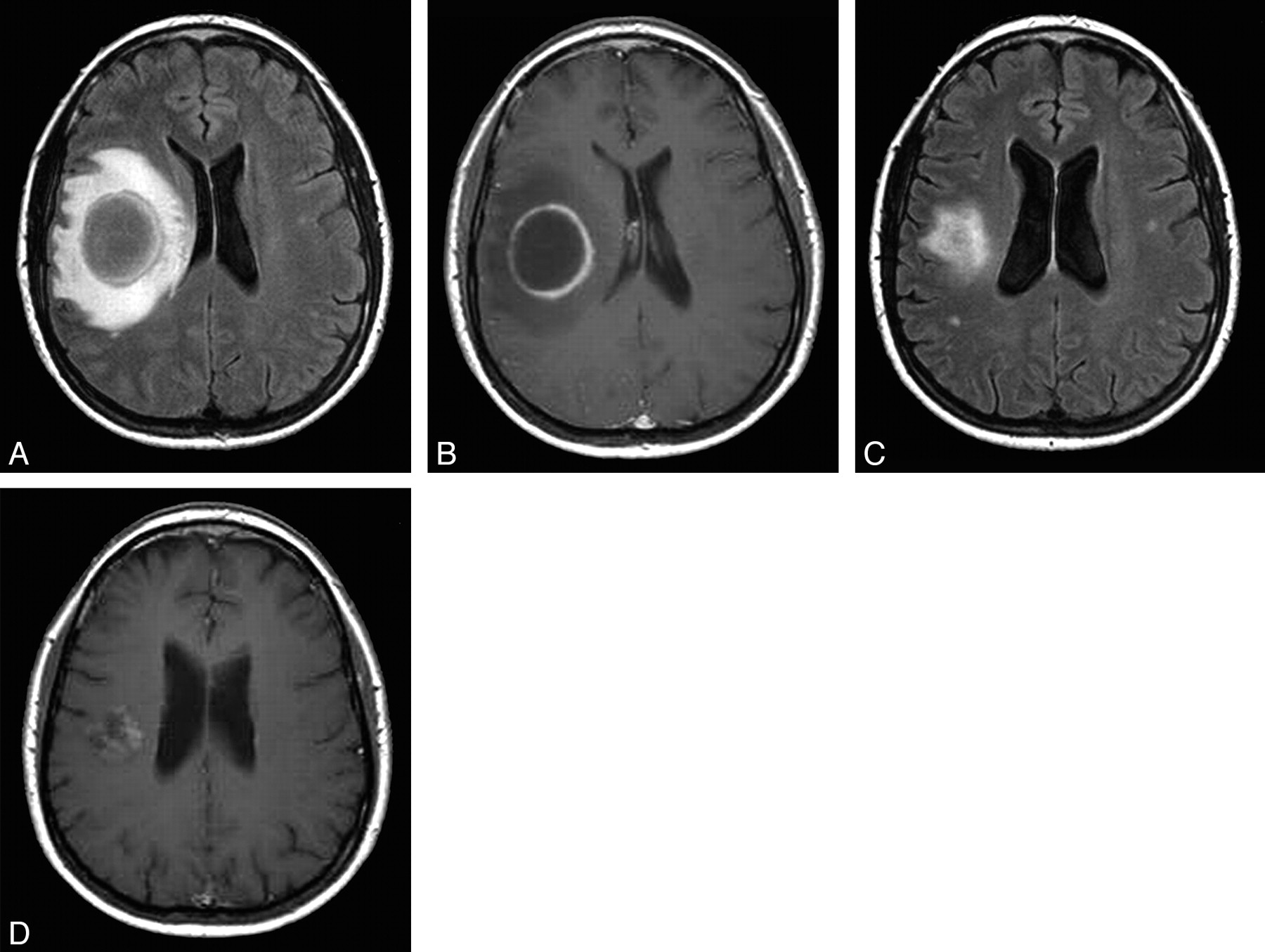
A and B, Axial FLAIR (A) and postgadolinium T1-weighted (B) images show a ring-enhancing tumefactive lesion with edema and mass effect. C and D, Axial FLAIR (C) and postgadolinium T1-weighted (D) images 1 month after treatment. The pattern of contrast enhancement thicker medially suggests demyelination. The lesion does not show restricted diffusion on diffusion-weighted images and is hyperintense on the apparent diffusion coefficient images.

The patient remained stable until 2 years later, when she experienced right hemifacial paresthesias. MR imaging revealed 2 small hyperintense lesions in the bulb and in the cervical spinal cord on T2-weighted images. Her oral prednisone dosage was increased to 1 mg/kg daily again, and hydroxychloroquine was added with good response.

Discussion

The frequency and type of CNS involvement in pSS is a matter of discussion. The reported prevalence of CNS manifestations ranges from 0% to 60%.2–4 In a recent cohort study of 400 patients, only 1% presented with CNS complications.1 Peripheral nervous system (PNS) disease is considered the most common neurologic manifestation in pSS. However, the largest study of patients with pSS with neurologic manifestations described CNS manifestations in 68% of patients, a proportion similar to that of PNS involvement in the same study.6

MR imaging abnormalities are common in pSS. These are usually multiple hyperintense areas in the subcortical and periventricular white matter on T2-weighted and fluid-attenuated inversion recovery (FLAIR) sequences.2,3 However, MR imaging lesions are generally smaller and less pronounced in pSS than in multiple sclerosis (MS) or systemic lupus erythematosus.2 Some authors have reported an increased frequency of pSS in patients with MS.3 Older age, PNS or cranial nerve involvement, spinal cord MR imaging lesions spanning multiple spinal segments, MR imaging cortical brain lesions, absence of lesions in the corpus callosum, or lack of oligoclonal bands are atypical in MS and are characteristic of pSS.7

Our patient had a tumorlike lesion with a notable mass effect. Tumefactive or pseudotumoral lesions have been described in other autoimmune CNS diseases like MS.8–10 Differential diagnosis includes lymphoma, glioma, metastases, abscesses, parasitary cysts, adrenoleukodystrophy, histiocytosis X, progressive multifocal leukoencephalopathy, disseminated encephalomyelitis, and Balò-Marburg disease.8

The pathogenesis of CNS damage in pSS remains unknown. Many authors suggest an underlying mechanism of ischemia secondary to small vessel vasculitis. The clinical course, type of MR imaging lesions, and cerebral angiography results in our patient are not consistent with a vasculitic process. Lymphocytic infiltration can produce extraglandular manifestations in pSS,1 and it has also been described in the CNS. Bakchine et al11 reported a pathologic case with ataxia, oculomotor paralysis, and seizures and a large lymphocytic infiltrate at postmortem examination. Pseudolymphoma or pseudotumoral lesions affecting several organs like the liver, kidney, lung, pancreas, and choroid plexus have been reported in patients with pSS.12–15 In our patient, improvement with corticosteroids, the pattern of contrast enhancement thicker medially, and a relapsing-remitting course of the lesions suggest an inflammatory-demyelinating mechanism as the underlying pathogenic process.

References

  1. ↵
    Garcia-Carrasco M, Ramos-Casals M, Rosas J, et al. Primary Sjogren syndrome: clinical and immunologic disease patterns in a cohort of 400 patients. Medicine (Baltimore)2002;81:270–80
    CrossRefPubMed
  2. ↵
    Morgen K, McFarland HF, Pillemer SR. Central nervous system disease in primary Sjögren syndrome: the role of magnetic resonance imaging. Semin Arthritis Rheum 2004;34:623–30
    CrossRefPubMed
  3. ↵
    Soliotis FC, Mavragani CP, Moutsopoulos HM. Central nervous system involvement in Sjögren's syndrome. Ann Rheum Dis 2004;63:616–20
    FREE Full Text
  4. ↵
    de Seze J, Delalande S, Vermersch P. Neurological manifestations in Sjogren syndrome. Rev Med Interne 2005;26:624–36
    PubMed
  5. ↵
    Vitali C, Bombardieri S, Jonsson R, et al, for the European Study Group on Classification Criteria for Sjögren's Syndrome. Classification criteria for Sjögren's syndrome: a revised version of the European criteria proposed by the American-European Consensus Group. Ann Rheum Dis 2002;61:554–58
    Abstract/FREE Full Text
  6. ↵
    Delalande S, de Seze J, Fauchais AL, et al. Neurologic manifestations in primary Sjögren syndrome: a study of 82 patients. Medicine (Baltimore)2004;83:280–91
    PubMed
  7. ↵
    Rosenbaum R, Barkhuizen A. Primary Sjögren's syndrome. In: Noseworthy JH, ed. Neurological Therapeutics: Principles and Practice. 2nd ed. London, UK: Informa Healthcare;2006 :1587–91
  8. ↵
    Comi G. Multiple sclerosis: pseudotumoral forms. Neurol Sci 2004;25:S374–79
    CrossRefPubMed
  9. Khoshyomn S, Braff SP, Penar PL. Tumefactive multiple sclerosis plaque. J Neurol Neurosurg Psychiatry 2002;73:85
    FREE Full Text
  10. ↵
    Selkirk SM, Shi J. Relapsing-remitting tumefactive multiple sclerosis. Mult Scler 2005;11:731–34
    Abstract/FREE Full Text
  11. ↵
    Bakchine S, Duyckaerts C, Hassine L, et al. Lesions neurologiques centrals et peripheriques au cours d'un syndrome de Gougerot-Sjogren primitif. Rev Neurol 1991;147:368–75
    PubMed
  12. ↵
    Okubo H, Maekawa H, Ogawa K, et al. Pseudolymphoma of the liver associated with Sjögren's syndrome. Scand J Rheumatol 2001;30:117–19
    CrossRefPubMed
  13. Cacoub P, Ginsburg C, Tazi Z, et al. Sjögren's syndrome with acute renal failure caused by renal pseudolymphoma. Am J Kidney Dis 1996;28:762–66
    CrossRefPubMed
  14. Eckstein RP, Hollings RM, Martin PA, et al. Pancreatic pseudotumor arising in association with Sjögren's syndrome. Pathology 1995;27:284–88
    CrossRefPubMed
  15. ↵
    Chang Y, Horoupian DS, Lane B, et al. Inflammatory pseudotumor of the choroid plexus in Sjögren's disease. Neurosurgery 1991;29:287–90
    PubMed
  • Received April 4, 2008.
  • Accepted after revision May 9, 2008.
  • Copyright © American Society of Neuroradiology
View Abstract
PreviousNext
Back to top

In this issue

American Journal of Neuroradiology: 29 (10)
American Journal of Neuroradiology
Vol. 29, Issue 10
November 2008
  • Table of Contents
  • Index by author
Advertisement
Print
Download PDF
Email Article

Thank you for your interest in spreading the word on American Journal of Neuroradiology.

NOTE: We only request your email address so that the person you are recommending the page to knows that you wanted them to see it, and that it is not junk mail. We do not capture any email address.

Enter multiple addresses on separate lines or separate them with commas.
Primary Sjögren Syndrome with Tumefactive Central Nervous System Involvement
(Your Name) has sent you a message from American Journal of Neuroradiology
(Your Name) thought you would like to see the American Journal of Neuroradiology web site.
CAPTCHA
This question is for testing whether or not you are a human visitor and to prevent automated spam submissions.
Citation Tools
Primary Sjögren Syndrome with Tumefactive Central Nervous System Involvement
J. Sanahuja, S. Ordoñez-Palau, R. Begué, L. Brieva, D. Boquet
American Journal of Neuroradiology Nov 2008, 29 (10) 1878-1879; DOI: 10.3174/ajnr.A1204

Citation Manager Formats

  • BibTeX
  • Bookends
  • EasyBib
  • EndNote (tagged)
  • EndNote 8 (xml)
  • Medlars
  • Mendeley
  • Papers
  • RefWorks Tagged
  • Ref Manager
  • RIS
  • Zotero
Share
Primary Sjögren Syndrome with Tumefactive Central Nervous System Involvement
J. Sanahuja, S. Ordoñez-Palau, R. Begué, L. Brieva, D. Boquet
American Journal of Neuroradiology Nov 2008, 29 (10) 1878-1879; DOI: 10.3174/ajnr.A1204
Reddit logo Twitter logo Facebook logo Mendeley logo
  • Tweet Widget
  • Facebook Like
  • Google Plus One
Purchase

Jump to section

  • Article
    • Abstract
    • Case Report
    • Discussion
    • References
  • Figures & Data
  • Info & Metrics
  • References
  • PDF

Related Articles

  • No related articles found.
  • PubMed
  • Google Scholar

Cited By...

  • Tumefactive demyelination: an approach to diagnosis and management
  • Crossref
  • Google Scholar

This article has not yet been cited by articles in journals that are participating in Crossref Cited-by Linking.

More in this TOC Section

  • Predictors of Reperfusion in Patients with Acute Ischemic Stroke
  • Enhanced Axonal Metabolism during Early Natalizumab Treatment in Relapsing-Remitting Multiple Sclerosis
  • Progression of Microstructural Damage in Spinocerebellar Ataxia Type 2: A Longitudinal DTI Study
Show more BRAIN

Similar Articles

Advertisement

News and Updates

  • Lucien Levy Best Research Article Award
  • Thanks to our 2022 Distinguished Reviewers
  • Press Releases

Resources

  • Evidence-Based Medicine Level Guide
  • How to Participate in a Tweet Chat
  • AJNR Podcast Archive
  • Ideas for Publicizing Your Research
  • Librarian Resources
  • Terms and Conditions

Opportunities

  • Share Your Art in Perspectives
  • Get Peer Review Credit from Publons
  • Moderate a Tweet Chat

American Society of Neuroradiology

  • Neurographics
  • ASNR Annual Meeting
  • Fellowship Portal
  • Position Statements

© 2023 by the American Society of Neuroradiology | Print ISSN: 0195-6108 Online ISSN: 1936-959X

Powered by HighWire