
Article Figures & Data
Figures
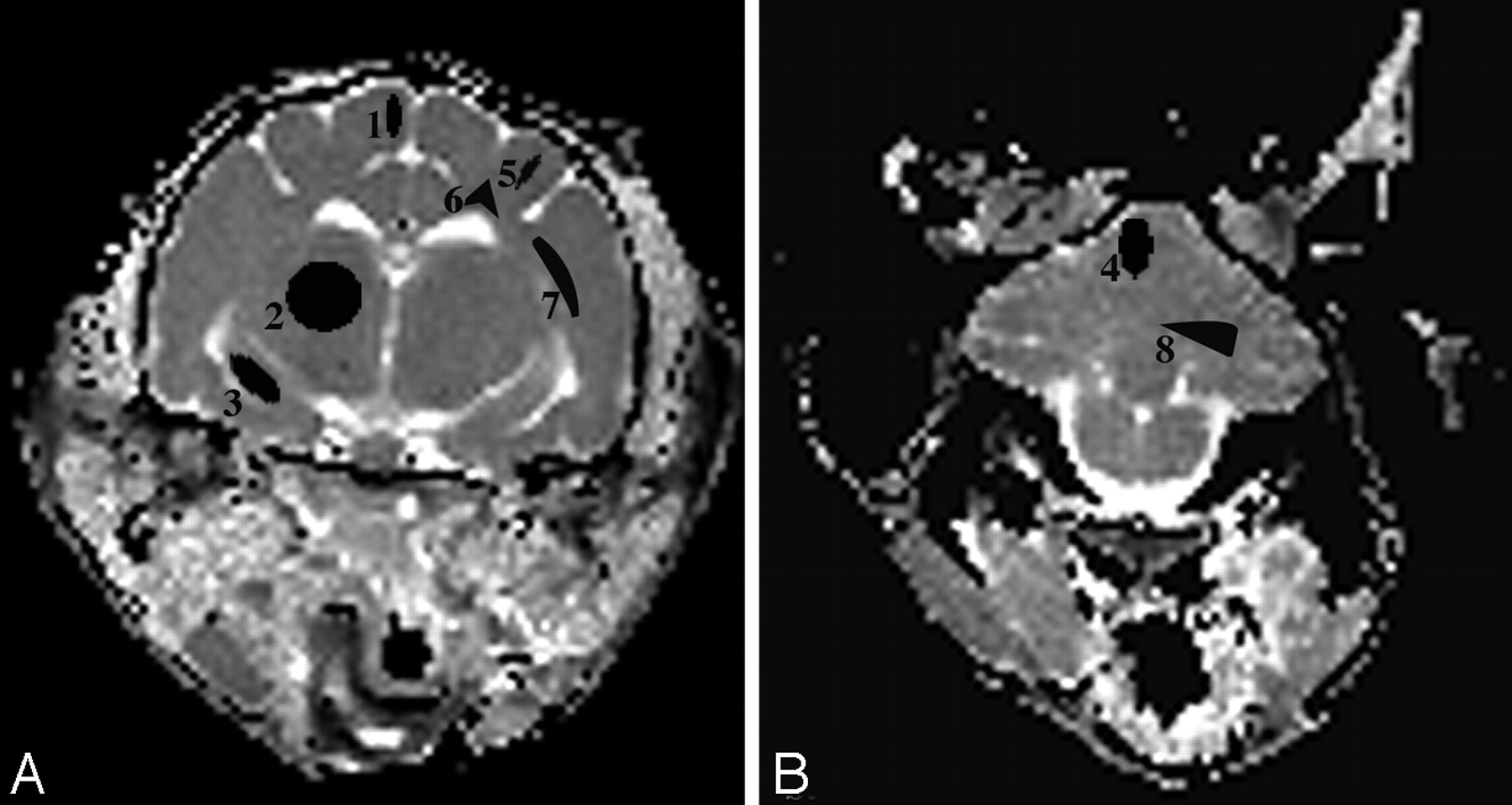
- Fig 1.
A and B, ADCav maps of the brain at the level of the thalamus (A) and the cerebellum (B) are shown. The brain regions for which data were acquired include 4 gray matter regions (1 indicates cerebral cortex; 2, thalamus; 3, hippocampus; 4, cerebellar gray matter) and 4 white matter regions (5 indicates corona radiata; 6, centrum semiovale; 7, internal capsule; 8, cerebellar white matter).
- Fig 2.
T2 relaxation times of gray matter regions (cerebral cortex, thalamus, hippocampus, and cerebellar cortex and white matter regions (corona radiata, centrum semiovale, internal capsule, and cerebellar white matter [bottom]) were determined in unaffected and AMD cats. Significant increases in T2 were found in the white matter of AMD cats compared with unaffected cats (*P < .05). No significant differences in T2 were found between affected and normal cats when gray matter regions were compared.
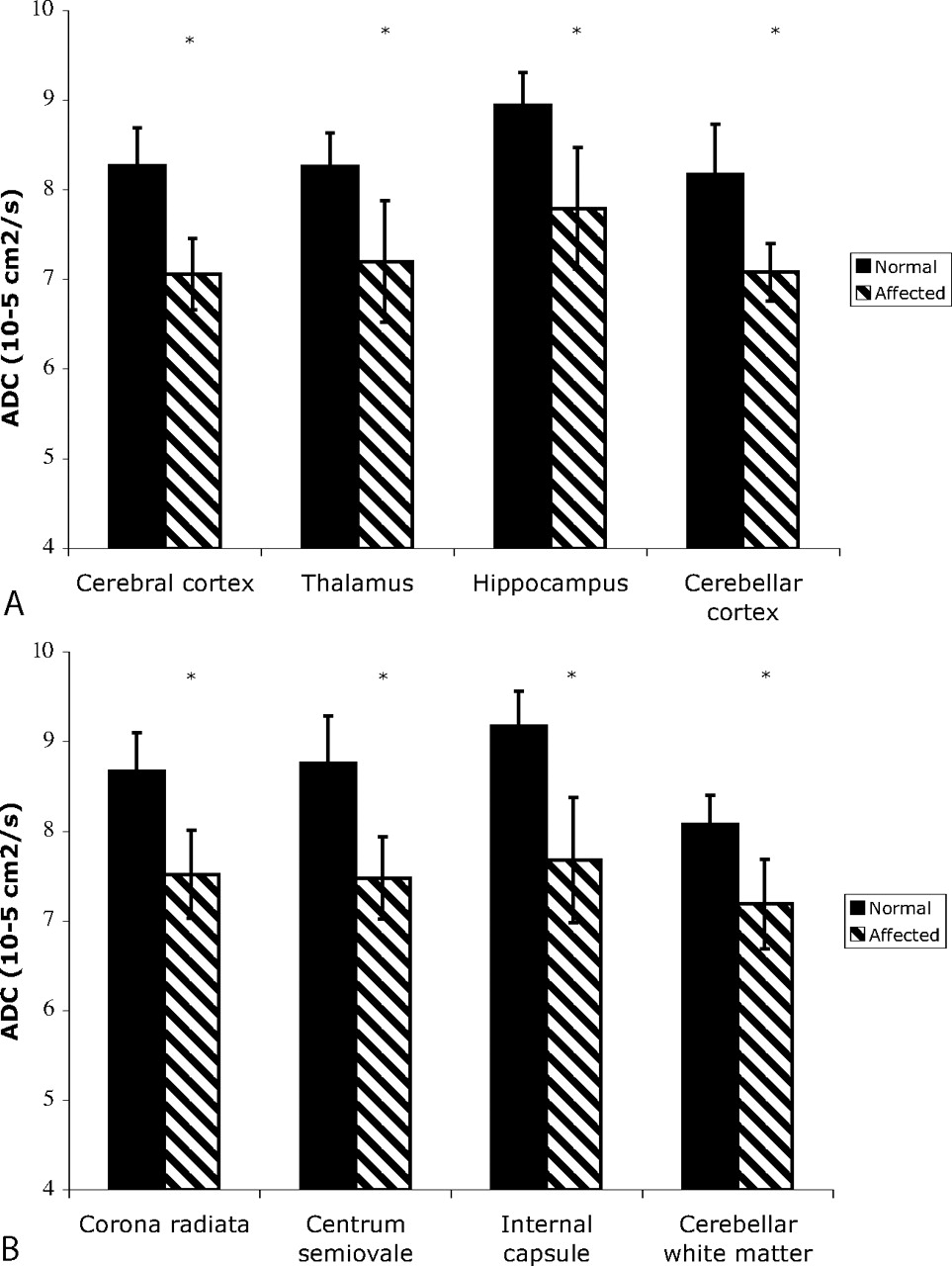
- Fig 3.
ADCav of gray matter regions (cerebral cortex, thalamus, hippocampus, and cerebellar cortex and white matter regions (corona radiata, centrum semiovale, internal capsule, and cerebellar white matter [bottom]) were determined in unaffected and AMD cats. Significant decreases in ADVav were found in AMD cats in all gray and white matter regions examined (*P < .05).
- Fig 4.
A and B, H&E-stained sections of the cerebellar cortex (original magnification ×400) from a normal cat (A) and an AMD cat (B), showing cytoplasmic vacuolation and distention of Purkinje cells (arrowhead), granular cells (small arrow), and glia (large arrow). C and D, Luxol fast blue staining of the cerebellar white matter (original magnification ×50) from a normal cat (C) and an AMD cat (D). A decrease in myelination is seen as a decrease in stain intensity and as an increase in space between myelin sheaths. E and F, GFAP immunohistochemistry of the cerebellum (original magnification ×200) from a normal (E) and an AMD cat (F), showing diffuse astrogliosis (brown staining) present throughout the gray and white matter of affected cats.


In this issue


Jump to section
Related Articles
Cited By...
- No citing articles found.