Advertisement

Main menu

  • Home
  • Content
    • Current Issue
    • Accepted Manuscripts
    • Article Preview
    • Past Issue Archive
    • Video Articles
    • AJNR Case Collection
    • Case of the Week Archive
    • Case of the Month Archive
    • Classic Case Archive
  • Special Collections
    • AJNR Awards
    • ASNR Foundation Special Collection
    • Most Impactful AJNR Articles
    • Photon-Counting CT
    • Spinal CSF Leak Articles (Jan 2020-June 2024)
  • Multimedia
    • AJNR Podcasts
    • AJNR SCANtastic
    • Trainee Corner
    • MRI Safety Corner
    • Imaging Protocols
  • For Authors
    • Submit a Manuscript
    • Submit a Video Article
    • Submit an eLetter to the Editor/Response
    • Manuscript Submission Guidelines
    • Statistical Tips
    • Fast Publishing of Accepted Manuscripts
    • Graphical Abstract Preparation
    • Imaging Protocol Submission
    • Author Policies
  • About Us
    • About AJNR
    • Editorial Board
    • Editorial Board Alumni
  • More
    • Become a Reviewer/Academy of Reviewers
    • Subscribers
    • Permissions
    • Alerts
    • Feedback
    • Advertisers
    • ASNR Home

User menu

  • Alerts
  • Log in

Search

  • Advanced search
American Journal of Neuroradiology

American Journal of Neuroradiology

ASHNR American Society of Functional Neuroradiology ASHNR American Society of Pediatric Neuroradiology ASSR
  • Alerts
  • Log in

Advanced Search

  • Home
  • Content
    • Current Issue
    • Accepted Manuscripts
    • Article Preview
    • Past Issue Archive
    • Video Articles
    • AJNR Case Collection
    • Case of the Week Archive
    • Case of the Month Archive
    • Classic Case Archive
  • Special Collections
    • AJNR Awards
    • ASNR Foundation Special Collection
    • Most Impactful AJNR Articles
    • Photon-Counting CT
    • Spinal CSF Leak Articles (Jan 2020-June 2024)
  • Multimedia
    • AJNR Podcasts
    • AJNR SCANtastic
    • Trainee Corner
    • MRI Safety Corner
    • Imaging Protocols
  • For Authors
    • Submit a Manuscript
    • Submit a Video Article
    • Submit an eLetter to the Editor/Response
    • Manuscript Submission Guidelines
    • Statistical Tips
    • Fast Publishing of Accepted Manuscripts
    • Graphical Abstract Preparation
    • Imaging Protocol Submission
    • Author Policies
  • About Us
    • About AJNR
    • Editorial Board
    • Editorial Board Alumni
  • More
    • Become a Reviewer/Academy of Reviewers
    • Subscribers
    • Permissions
    • Alerts
    • Feedback
    • Advertisers
    • ASNR Home
  • Follow AJNR on Twitter
  • Visit AJNR on Facebook
  • Follow AJNR on Instagram
  • Join AJNR on LinkedIn
  • RSS Feeds

AJNR is seeking candidates for the AJNR Podcast Editor. Read the position description.

OtherPEDIATRICS

Malignant Transformation of a Lipomyelocele into a Rhabdomyosarcoma?

A.M. O'Connell, D. Allcutt, F. Brett and S. Ryan
American Journal of Neuroradiology March 2008, 29 (3) 434-435; DOI: https://doi.org/10.3174/ajnr.A0924
A.M. O'Connell
  • Find this author on Google Scholar
  • Find this author on PubMed
  • Search for this author on this site
D. Allcutt
  • Find this author on Google Scholar
  • Find this author on PubMed
  • Search for this author on this site
F. Brett
  • Find this author on Google Scholar
  • Find this author on PubMed
  • Search for this author on this site
S. Ryan
  • Find this author on Google Scholar
  • Find this author on PubMed
  • Search for this author on this site
  • Article
  • Figures & Data
  • Info & Metrics
  • Responses
  • References
  • PDF
Loading

Abstract

SUMMARY: We report the unusual transformation of a lipomyelocele to a rhabdomyosarcoma in a 3-year-old boy. A lipomyelocele is not considered pre-malignant, but the possibility of developing such a tumor may represent another reason for close neurological follow-up in patients with spinal dysraphism.

Alipomyelocele is an intraspinal lipoma that is tightly attached to the dorsal surface of an open spinal cord.1 The spinal cord remains within the spinal canal unlike a lipomyelomeningocele, in which the thecal sac herniates through the spina bifida. Lipomyeloceles are associated with tethering of the cord but not with malignancy.2

Case Report

A spinal sonography was performed on the first day of life in a full-term neonate with a complex hemangioma over the sacrum (Fig 1). This showed the spinal cord ending in a sacral lipoma. The diagnosis of a lipomyelocele was confirmed by MR imaging at 3 months of age (Fig 2). The baby was managed conservatively with frequent neurosurgical assessment. At 3 years of age, he presented with an unwillingness to walk and general malaise. He had developed a large sacral bulge with a superimposed hematoma. Spinal MR imaging demonstrated a new large mass arising from the lipoma and distal spinal cord within the sacral canal (Fig 3A). There was surface enhancement of the cord (Fig 3B) and brain stem (Fig 3C) consistent with meningeal seeding of the tumor. These findings were confirmed at surgery, and pathologic examination of the partly excised tumor revealed a rhabdomyosarcoma. Histology showed a pleomorphic malignant spindle cell tumor (Fig 4) positive for muscle markers.

Fig 1.
  • Download figure
  • Open in new tab
  • Download powerpoint
Fig 1.

Photograph of a complex ulcerating hemangioma over the sacrum in a full-term neonate on day 1.

Fig 2.
  • Download figure
  • Open in new tab
  • Download powerpoint
Fig 2.

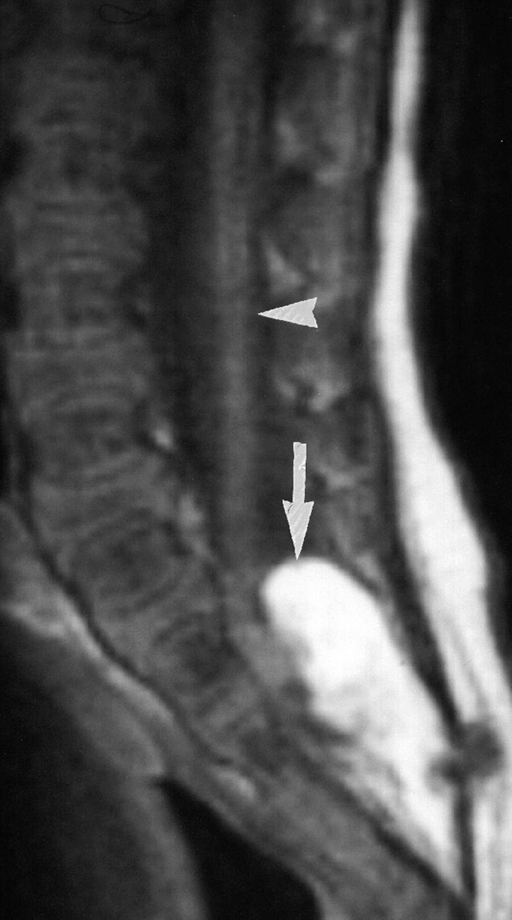
Sagittal T1-weighted MR image obtained at 3 months confirms the sonographic finding of the tethered spinal cord (arrowhead) ending in a lipomyelocele (arrow).

Fig 3.
  • Download figure
  • Open in new tab
  • Download powerpoint
Fig 3.

A, Sagittal T1-weighted MR image at 3 years shows a new large homogeneous mass of low signal intensity (arrow), apparently arising from the lipomyelocele and distal spinal cord within the spinal canal. B, Postcontrast T1-weighted sagittal MR image shows multiple foci (arrowheads) of high signal intensity in the distal thoracic and lumbar spinal cord consistent with metastatic seeding. C, Sagittal postcontrast T1-weighted MR image demonstrates multiple foci (arrowheads) of high signal intensity in the brain stem and cervical and upper thoracic spinal cord, consistent with metastatic seeding.

Fig 4.
  • Download figure
  • Open in new tab
  • Download powerpoint
Fig 4.

Resection of this sacral mass revealed a pleomorphic malignant spindle cell tumor at pathology, consistent with a rhabdomyosarcoma (hematoxylin-eosin).

Discussion

The clinical finding of sacrococcygeal cutaneous stigmata, such as that seen in this baby, has been clearly linked with occult spinal dysraphism. Screening spinal sonography is indicated in these babies, and positive findings can be confirmed by MR imaging. Management is usually conservative. Follow-up assessments are focused on the detection of any possible evidence of neurologic complications of the tethered cord and, in particular, of any sign of neurologic deterioration. Such findings may be indications for surgical untethering of the cord and resection of the lipoma.

The possibility of development of tumors in neural tube defects has been described.3–6 Lipomyeloceles are not, however, considered premalignant conditions. To our knowledge, the development of a rhabdomyosarcoma within a lipomyelocele has not been previously described. It has, however, been described originating from the central nervous system (CNS). There are no skeletal muscle cells in a lipomyelocele, but it is tightly attached to the dorsal aspect of the open spinal cord.

Primary intracranial rhabdomyosarcoma is a rare tumor in infancy and childhood, which is found in various locations in the CNS.7 The clinical course worsens rapidly, and the final outcome is poor, with a median survival time of 8–10 months.7 Invasion of the meninges, spontaneous intratumoral bleeding, spinal leptomeningeal CSF spreading of tumor cells, and early recurrence of the mass are the distinctive features of rhabdomyosarcoma.7

Intracranial rhabdomyosarcoma has also been described in association with subdural hematoma. The authors propose the possibility of sarcoma development secondary to chronic inflammation and proliferation in subdural collection.8 The coexistence in this instance seems coincidental, however, and subdural hematoma was known to be secondary to a fall.

The development of a rhabdomyosarcoma in this case report is likely embryonal. Although originating from a benign lesion such as a lipomyelocele has not been described previously, to our knowledge, unlike CNS origin, it may represent a very rare transformation.

The possibility of development of such a tumor, however rare, presents another reason for close neurologic follow-up of children with spinal dysraphism. It emphasizes the importance of early neuroimaging of the spinal canal when any new neurologic signs or symptoms develop.

References

  1. ↵
    Unsinn KM, Geley T, Freund MC, et al. US of the spinal cord in newborns: spectrum of normal findings, variants, congenital anomalies, and acquired diseases. Radiographics 2000;20:923–38
    PubMed
  2. ↵
    Rossi A. Imaging in spine and spinal cord malformations. Eur J Radiol 2004;50:177–200
    CrossRefPubMed
  3. ↵
    Little J. Childhood cancer and neural tube defects. Am J Hum Genet 1997;61:1204–05
    CrossRefPubMed
  4. Freyer DR, Hutchinson RJ, McKeever PE. Primary primitive neuroectodermal tumor of the spinal cord associated with neural tube defect. Pediatr Neurosci 1989;15:181–87
    PubMed
  5. Thorp RH. Carcinoma associated with myelomeningocele: case report. J Neurosurg 1967;27:446–48
    PubMed
  6. ↵
    Kimura S, Kawaguchi S, Kikuchi K, et al. Rhabdomyosarcoma arising from a dormant dumbbell ganglioneuroma of the lumbar spine: a case report. Spine 2002;27:E513–17
    CrossRefPubMed
  7. ↵
    Tomei G, Grimoldi N, Masini B, et al. Primary intracranial rhabdomyosarcoma: report of two cases. Childs Nerv Syst 2004;5:246–49
    CrossRef
  8. ↵
    Nejat F, Keshavarzi S, Kalaghchi B, et al. Chronic subdural hematoma associated with subdural rhabdomyosarcoma: case report. Neurosurgery 2007;60:E774–75, discussion E775
    PubMed
  • Received October 27, 2007.
  • Accepted after revision October 31, 2007.
  • Copyright © American Society of Neuroradiology
View Abstract
PreviousNext
Back to top

In this issue

American Journal of Neuroradiology: 29 (3)
American Journal of Neuroradiology
Vol. 29, Issue 3
March 2008
  • Table of Contents
  • Index by author
Advertisement
Print
Download PDF
Email Article

Thank you for your interest in spreading the word on American Journal of Neuroradiology.

NOTE: We only request your email address so that the person you are recommending the page to knows that you wanted them to see it, and that it is not junk mail. We do not capture any email address.

Enter multiple addresses on separate lines or separate them with commas.
Malignant Transformation of a Lipomyelocele into a Rhabdomyosarcoma?
(Your Name) has sent you a message from American Journal of Neuroradiology
(Your Name) thought you would like to see the American Journal of Neuroradiology web site.
CAPTCHA
This question is for testing whether or not you are a human visitor and to prevent automated spam submissions.
Cite this article
A.M. O'Connell, D. Allcutt, F. Brett, S. Ryan
Malignant Transformation of a Lipomyelocele into a Rhabdomyosarcoma?
American Journal of Neuroradiology Mar 2008, 29 (3) 434-435; DOI: 10.3174/ajnr.A0924

Citation Manager Formats

  • BibTeX
  • Bookends
  • EasyBib
  • EndNote (tagged)
  • EndNote 8 (xml)
  • Medlars
  • Mendeley
  • Papers
  • RefWorks Tagged
  • Ref Manager
  • RIS
  • Zotero
0 Responses
Respond to this article
Share
Bookmark this article
Malignant Transformation of a Lipomyelocele into a Rhabdomyosarcoma?
A.M. O'Connell, D. Allcutt, F. Brett, S. Ryan
American Journal of Neuroradiology Mar 2008, 29 (3) 434-435; DOI: 10.3174/ajnr.A0924
del.icio.us logo Twitter logo Facebook logo Mendeley logo
  • Tweet Widget
  • Facebook Like
  • Google Plus One
Purchase

Jump to section

  • Article
    • Abstract
    • Case Report
    • Discussion
    • References
  • Figures & Data
  • Info & Metrics
  • Responses
  • References
  • PDF

Related Articles

  • No related articles found.
  • PubMed
  • Google Scholar

Cited By...

  • No citing articles found.
  • Crossref (4)
  • Google Scholar

This article has been cited by the following articles in journals that are participating in Crossref Cited-by Linking.

  • Congenital Spinal Lipomatous Malformations. Part 1. Spinal Lipomas, Lipomyeloceles, and Lipomyelomeningoceles
    Roy H. Rhodes
    Fetal and Pediatric Pathology 2020 39 3
  • Primary spinal epidural rhabdomyosarcoma: a case report and review of the literature
    Mahmoud Reza Khalatbari, Hassan Jalaeikhoo, Mehrdokht Hamidi, Yashar Moharamzad
    Child's Nervous System 2012 28 11
  • SACRAL LIPOMYELOCELE PRESENTING AS A HUMAN TAIL
    Rajesh Raman, Geetha M. J.
    Journal of Evolution of medical and Dental Sciences 2013 2 8
  • Spinal rhabdomyosarcoma in a child at the site of a lumbosacral lipoma: a rare entity
    Francesca Vitulli, Panduranga Seetahal-Maraj, Islam Adbelfattah, Paola Angelini, Julie Chandra, Jean Marie U-King-Im, Andrew King, Difei Wang, Cristina Bleil, Bassel Zebian
    Child's Nervous System 2025 41 1

More in this TOC Section

  • SyMRI & MR Fingerprinting in Brainstem Myelination
  • Comparison of Image Quality and Radiation Dose in Pediatric Temporal Bone CT Using Photon-Counting Detector CT and Energy-Integrating Detector CT
  • Neonatal Hypocalcemia: Cerebral MRI Abnormalities
Show more Pediatrics

Similar Articles

Advertisement

Indexed Content

  • Current Issue
  • Accepted Manuscripts
  • Article Preview
  • Past Issues
  • Editorials
  • Editor's Choice
  • Fellows' Journal Club
  • Letters to the Editor
  • Video Articles

Cases

  • Case Collection
  • Archive - Case of the Week
  • Archive - Case of the Month
  • Archive - Classic Case

Special Collections

  • AJNR Awards
  • ASNR Foundation Special Collection
  • Most Impactful AJNR Articles
  • Photon-Counting CT
  • Spinal CSF Leak Articles (Jan 2020-June 2024)

More from AJNR

  • Trainee Corner
  • Imaging Protocols
  • MRI Safety Corner

Multimedia

  • AJNR Podcasts
  • AJNR Scantastics

Resources

  • Turnaround Time
  • Submit a Manuscript
  • Submit a Video Article
  • Submit an eLetter to the Editor/Response
  • Manuscript Submission Guidelines
  • Statistical Tips
  • Fast Publishing of Accepted Manuscripts
  • Graphical Abstract Preparation
  • Imaging Protocol Submission
  • Evidence-Based Medicine Level Guide
  • Publishing Checklists
  • Author Policies
  • Become a Reviewer/Academy of Reviewers
  • News and Updates

About Us

  • About AJNR
  • Editorial Board
  • Editorial Board Alumni
  • Alerts
  • Permissions
  • Not an AJNR Subscriber? Join Now
  • Advertise with Us
  • Librarian Resources
  • Feedback
  • Terms and Conditions
  • AJNR Editorial Board Alumni

American Society of Neuroradiology

  • Not an ASNR Member? Join Now

© 2025 by the American Society of Neuroradiology All rights, including for text and data mining, AI training, and similar technologies, are reserved.
Print ISSN: 0195-6108 Online ISSN: 1936-959X

Powered by HighWire