Advertisement

Main menu

  • Home
  • Content
    • Current Issue
    • Accepted Manuscripts
    • Article Preview
    • Past Issue Archive
    • Video Articles
    • AJNR Case Collection
    • Case of the Week Archive
    • Case of the Month Archive
    • Classic Case Archive
  • Special Collections
    • AJNR Awards
    • ASNR Foundation Special Collection
    • Most Impactful AJNR Articles
    • Photon-Counting CT
    • Spinal CSF Leak Articles (Jan 2020-June 2024)
  • Multimedia
    • AJNR Podcasts
    • AJNR SCANtastic
    • Trainee Corner
    • MRI Safety Corner
    • Imaging Protocols
  • For Authors
    • Submit a Manuscript
    • Submit a Video Article
    • Submit an eLetter to the Editor/Response
    • Manuscript Submission Guidelines
    • Statistical Tips
    • Fast Publishing of Accepted Manuscripts
    • Graphical Abstract Preparation
    • Imaging Protocol Submission
    • Author Policies
  • About Us
    • About AJNR
    • Editorial Board
    • Editorial Board Alumni
  • More
    • Become a Reviewer/Academy of Reviewers
    • Subscribers
    • Permissions
    • Alerts
    • Feedback
    • Advertisers
    • ASNR Home

User menu

  • Alerts
  • Log in

Search

  • Advanced search
American Journal of Neuroradiology

American Journal of Neuroradiology

ASHNR American Society of Functional Neuroradiology ASHNR American Society of Pediatric Neuroradiology ASSR
  • Alerts
  • Log in

Advanced Search

  • Home
  • Content
    • Current Issue
    • Accepted Manuscripts
    • Article Preview
    • Past Issue Archive
    • Video Articles
    • AJNR Case Collection
    • Case of the Week Archive
    • Case of the Month Archive
    • Classic Case Archive
  • Special Collections
    • AJNR Awards
    • ASNR Foundation Special Collection
    • Most Impactful AJNR Articles
    • Photon-Counting CT
    • Spinal CSF Leak Articles (Jan 2020-June 2024)
  • Multimedia
    • AJNR Podcasts
    • AJNR SCANtastic
    • Trainee Corner
    • MRI Safety Corner
    • Imaging Protocols
  • For Authors
    • Submit a Manuscript
    • Submit a Video Article
    • Submit an eLetter to the Editor/Response
    • Manuscript Submission Guidelines
    • Statistical Tips
    • Fast Publishing of Accepted Manuscripts
    • Graphical Abstract Preparation
    • Imaging Protocol Submission
    • Author Policies
  • About Us
    • About AJNR
    • Editorial Board
    • Editorial Board Alumni
  • More
    • Become a Reviewer/Academy of Reviewers
    • Subscribers
    • Permissions
    • Alerts
    • Feedback
    • Advertisers
    • ASNR Home
  • Follow AJNR on Twitter
  • Visit AJNR on Facebook
  • Follow AJNR on Instagram
  • Join AJNR on LinkedIn
  • RSS Feeds

AJNR is seeking candidates for the AJNR Podcast Editor. Read the position description.

Research ArticleSPINE

A Comparison of Angiographic CT and Multisection CT in Lumbar Myelographic Imaging

J.-H. Buhk, E. Elolf, D. Jacob, H.-H. Rustenbeck, A. Mohr and M. Knauth
American Journal of Neuroradiology March 2008, 29 (3) 442-446; DOI: https://doi.org/10.3174/ajnr.A0853
J.-H. Buhk
  • Find this author on Google Scholar
  • Find this author on PubMed
  • Search for this author on this site
E. Elolf
  • Find this author on Google Scholar
  • Find this author on PubMed
  • Search for this author on this site
D. Jacob
  • Find this author on Google Scholar
  • Find this author on PubMed
  • Search for this author on this site
H.-H. Rustenbeck
  • Find this author on Google Scholar
  • Find this author on PubMed
  • Search for this author on this site
A. Mohr
  • Find this author on Google Scholar
  • Find this author on PubMed
  • Search for this author on this site
M. Knauth
  • Find this author on Google Scholar
  • Find this author on PubMed
  • Search for this author on this site
  • Article
  • Figures & Data
  • Info & Metrics
  • Responses
  • References
  • PDF
Loading

Article Figures & Data

Figures

  • Tables
  • Fig 1.
    • Download figure
    • Open in new tab
    • Download powerpoint
    Fig 1.

    Images from a 58-year-old woman with persistent back pain. The left lateromedial prolapsed intervertebral disk in the segment L2/3 is equally well visible in both ACT (A and C) and MSCT (B and D) images. ACT images in the left posterior oblique position are displayed as 3D-MIP (E) and 3D-VRT (F). G, The myelographic projection was derived by transferring the position of the 3D volume from the workstation to the C-arm. Hence, no additional fluoroscopy was necessary to determine the optimal angles for projection myelographies.

  • Fig 2.
    • Download figure
    • Open in new tab
    • Download powerpoint
    Fig 2.

    Images from a 62-year-old man with symptoms of spinal claudication. Coronal MPR planes of ACT (A) and MSCT (B) are of equal quality. C, A curved coronal reconstruction of the ACT provides an appropriate depiction of the segmental nerve roots.
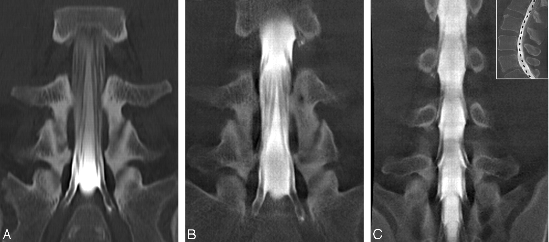
  • Fig 3.
    • Download figure
    • Open in new tab
    • Download powerpoint
    Fig 3.

    MPR images from a 75-year-old woman with persistent back pain after multisegmental laminectomy. In the sagittal sections (A, ACT; B, MSCT), the relevant degenerative changes can be depicted equally well: ligamental hyperplasia in the segment L1/2, ventral osteochondrosis in the segment L2/3, and the slight listhesis in the segment L4/5. A, Note the loss of detail and the enhanced image noise in the ACT section at the level from L5 to S2 due to the high attenuation of contrast media in the thecal sac. The transverse sections through the vertebral body of L4 (C, ACT; D, MSCT) show the hypertrophic facet joints and the thecal sac herniating into the laminectomy site. Both techniques show a well-defined L4 nerve root of the right side and an increase of tissue around the L4 nerve root of the contralateral side.

Tables

  • Figures
    • View popup
    Table 1:

    Cumulative results after all ratings

    VariableVery Good, n (%)Good, n (%)Moderate, n (%)Poor, n (%)Very Poor, n (%)Total, n (%)
    Overall impressionACT46 (28.8)79 (49.4)7 (4.4)27 (16.9)1 (0.6)160 (100)
    MSCT90 (58.1)47 (30.3)4 (2.6)11 (7.1)3 (1.94)155 (100)
    Contrast media distributionACT49 (30.6)80 (50.0)13 (8.1)18 (11.3)0 (0.0)160 (100)
    MSCT90 (58.1)46 (29.7)5 (3.2)11 (7.1)3 (1.9)155 (100)
    Perceptibility of intervertebral disksACT22 (13.8)17 (10.7)9 (5.7)77 (48.4)35 (22.0)160 (100)
    MSCT55 (35.3)60 (38.5)15 (9.6)25 (16.0)0 (0.0)155 (100)
    Perceptibility of bone structureACT26 (16.3)71 (44.4)10 (6.3)50 (31.3)3 (1.9)160 (100)
    MSCT117 (74.5)34 (21.9)2 (1.3)2 (1.3)0 (0.0)155 (100)
    Delineation of the spinal canalACT71 (44.4)76 (47.5)3 (1.9)10 (6.3)0 (0.0)160 (100)
    MSCT128 (82.6)26 (16.8)0 (0.0)1 (0.7)0 (0.0)155 (100)
    Delineation of spinal nerve rootsACT35 (22.0)60 (37.5)13 (8.1)46 (28.8)6 (3.8)160 (100)
    MSCT87 (56.1)50 (32.3)8 (5.2)10 (6.5)0 (0.0)155 (100)
    Diagnostic applicabilityACT30 (37.0)36 (44.4)4 (4.9)10 (12.4)1 (1.2)81 (100)
    MSCT48 (61.6)28 (35.9)1 (1.3)1 (1.3)0 (0.0)78 (100)
    • Note:—ACT indicates angiographic CT; MSCT, multisection CT.

    • View popup
    Table 2:

    Interobserver agreement

    VariableACT, Mean κ Values ± SDMSCT, Mean κ Values ± SDP
    Overall impression0.73 ± 0.060.78 ± 0.04.117
    Contrast media distribution0.74 ± 0.050.65 ± 0.09.059
    Perceptibility of intervertebral disks0.65 ± 0.220.69 ± 0.09.689
    Perceptibility of bone structure0.64 ± 0.120.78 ± 0.05.030
    Delineation of the spinal canal0.75 ± 0.050.78 ± 0.03.172
    Delineation of spinal nerve roots0.66 ± 0.090.72 ± 0.03.193
    Diagnostic applicability0.74 ± 0.060.75 ± 0.03.826
    • Note:—ACT indicates angiographic CT; MSCT, multisection CT. Data are n = 6, except for “diagnostic applicability,” where n = 3 by Student t test.

    • View popup
    Table 3:

    Intraobserver agreement

    VariableACT, Mean κ Values ± SDMSCT, Mean κ Values ± SDP
    Overall impression0.81 ± 0.050.73 ± 0.15.454
    Contrast media distribution0.75 ± 0.020.79 ± 0.08.533
    Perceptibility of intervertebral disks0.65 ± 0.260.68 ± 0.17.875
    Perceptibility of bone structure0.74 ± 0.020.78 ± 0.02.068
    Delineation of the spinal canal0.75 ± 0.080.78 ± 0.02.659
    Delineation of spinal nerve roots0.74 ± 0.040.78 ± 0.03.256
    • Note:—ACT indicates angiographic CT; MSCT, multisection CT. Data are n = 3 by Student t test.

PreviousNext
Back to top

In this issue

American Journal of Neuroradiology: 29 (3)
American Journal of Neuroradiology
Vol. 29, Issue 3
March 2008
  • Table of Contents
  • Index by author
Advertisement
Print
Download PDF
Email Article

Thank you for your interest in spreading the word on American Journal of Neuroradiology.

NOTE: We only request your email address so that the person you are recommending the page to knows that you wanted them to see it, and that it is not junk mail. We do not capture any email address.

Enter multiple addresses on separate lines or separate them with commas.
A Comparison of Angiographic CT and Multisection CT in Lumbar Myelographic Imaging
(Your Name) has sent you a message from American Journal of Neuroradiology
(Your Name) thought you would like to see the American Journal of Neuroradiology web site.
CAPTCHA
This question is for testing whether or not you are a human visitor and to prevent automated spam submissions.
Cite this article
J.-H. Buhk, E. Elolf, D. Jacob, H.-H. Rustenbeck, A. Mohr, M. Knauth
A Comparison of Angiographic CT and Multisection CT in Lumbar Myelographic Imaging
American Journal of Neuroradiology Mar 2008, 29 (3) 442-446; DOI: 10.3174/ajnr.A0853

Citation Manager Formats

  • BibTeX
  • Bookends
  • EasyBib
  • EndNote (tagged)
  • EndNote 8 (xml)
  • Medlars
  • Mendeley
  • Papers
  • RefWorks Tagged
  • Ref Manager
  • RIS
  • Zotero
0 Responses
Respond to this article
Share
Bookmark this article
A Comparison of Angiographic CT and Multisection CT in Lumbar Myelographic Imaging
J.-H. Buhk, E. Elolf, D. Jacob, H.-H. Rustenbeck, A. Mohr, M. Knauth
American Journal of Neuroradiology Mar 2008, 29 (3) 442-446; DOI: 10.3174/ajnr.A0853
del.icio.us logo Twitter logo Facebook logo Mendeley logo
  • Tweet Widget
  • Facebook Like
  • Google Plus One
Purchase

Jump to section

  • Article
    • Abstract
    • Methods
    • Results
    • Discussion
    • Footnotes
    • References
  • Figures & Data
  • Info & Metrics
  • Responses
  • References
  • PDF

Related Articles

  • No related articles found.
  • PubMed
  • Google Scholar

Cited By...

  • Use of Angiographic CT Imaging in the Cardiac Catheterization Laboratory for Congenital Heart Disease
  • Crossref (2)
  • Google Scholar

This article has been cited by the following articles in journals that are participating in Crossref Cited-by Linking.

  • Use of Angiographic CT Imaging in the Cardiac Catheterization Laboratory for Congenital Heart Disease
    Andrew C. Glatz, Xiaowei Zhu, Matthew J. Gillespie, Brian D. Hanna, Jonathan J. Rome
    JACC: Cardiovascular Imaging 2010 3 11
  • Brain imaging with a flat detector C-arm
    M. Söderman, D. Babic, S. Holmin, T. Andersson
    Neuroradiology 2008 50 10

More in this TOC Section

  • Bern Score Validity for SIH
  • MP2RAGE 7T in MS Lesions of the Cervical Spine
  • Deep Learning for STIR Spine MRI Quality
Show more Spine

Similar Articles

Advertisement

Indexed Content

  • Current Issue
  • Accepted Manuscripts
  • Article Preview
  • Past Issues
  • Editorials
  • Editor's Choice
  • Fellows' Journal Club
  • Letters to the Editor
  • Video Articles

Cases

  • Case Collection
  • Archive - Case of the Week
  • Archive - Case of the Month
  • Archive - Classic Case

Special Collections

  • AJNR Awards
  • ASNR Foundation Special Collection
  • Most Impactful AJNR Articles
  • Photon-Counting CT
  • Spinal CSF Leak Articles (Jan 2020-June 2024)

More from AJNR

  • Trainee Corner
  • Imaging Protocols
  • MRI Safety Corner

Multimedia

  • AJNR Podcasts
  • AJNR Scantastics

Resources

  • Turnaround Time
  • Submit a Manuscript
  • Submit a Video Article
  • Submit an eLetter to the Editor/Response
  • Manuscript Submission Guidelines
  • Statistical Tips
  • Fast Publishing of Accepted Manuscripts
  • Graphical Abstract Preparation
  • Imaging Protocol Submission
  • Evidence-Based Medicine Level Guide
  • Publishing Checklists
  • Author Policies
  • Become a Reviewer/Academy of Reviewers
  • News and Updates

About Us

  • About AJNR
  • Editorial Board
  • Editorial Board Alumni
  • Alerts
  • Permissions
  • Not an AJNR Subscriber? Join Now
  • Advertise with Us
  • Librarian Resources
  • Feedback
  • Terms and Conditions
  • AJNR Editorial Board Alumni

American Society of Neuroradiology

  • Not an ASNR Member? Join Now

© 2025 by the American Society of Neuroradiology All rights, including for text and data mining, AI training, and similar technologies, are reserved.
Print ISSN: 0195-6108 Online ISSN: 1936-959X

Powered by HighWire