Advertisement

Main menu

  • Home
  • Content
    • Current Issue
    • Accepted Manuscripts
    • Article Preview
    • Past Issue Archive
    • Video Articles
    • AJNR Case Collection
    • Case of the Week Archive
    • Case of the Month Archive
    • Classic Case Archive
  • Special Collections
    • AJNR Awards
    • ASNR Foundation Special Collection
    • Most Impactful AJNR Articles
    • Photon-Counting CT
    • Spinal CSF Leak Articles (Jan 2020-June 2024)
  • Multimedia
    • AJNR Podcasts
    • AJNR SCANtastic
    • Trainee Corner
    • MRI Safety Corner
    • Imaging Protocols
  • For Authors
    • Submit a Manuscript
    • Submit a Video Article
    • Submit an eLetter to the Editor/Response
    • Manuscript Submission Guidelines
    • Statistical Tips
    • Fast Publishing of Accepted Manuscripts
    • Graphical Abstract Preparation
    • Imaging Protocol Submission
    • Author Policies
  • About Us
    • About AJNR
    • Editorial Board
    • Editorial Board Alumni
  • More
    • Become a Reviewer/Academy of Reviewers
    • Subscribers
    • Permissions
    • Alerts
    • Feedback
    • Advertisers
    • ASNR Home

User menu

  • Alerts
  • Log in

Search

  • Advanced search
American Journal of Neuroradiology

American Journal of Neuroradiology

ASHNR American Society of Functional Neuroradiology ASHNR American Society of Pediatric Neuroradiology ASSR
  • Alerts
  • Log in

Advanced Search

  • Home
  • Content
    • Current Issue
    • Accepted Manuscripts
    • Article Preview
    • Past Issue Archive
    • Video Articles
    • AJNR Case Collection
    • Case of the Week Archive
    • Case of the Month Archive
    • Classic Case Archive
  • Special Collections
    • AJNR Awards
    • ASNR Foundation Special Collection
    • Most Impactful AJNR Articles
    • Photon-Counting CT
    • Spinal CSF Leak Articles (Jan 2020-June 2024)
  • Multimedia
    • AJNR Podcasts
    • AJNR SCANtastic
    • Trainee Corner
    • MRI Safety Corner
    • Imaging Protocols
  • For Authors
    • Submit a Manuscript
    • Submit a Video Article
    • Submit an eLetter to the Editor/Response
    • Manuscript Submission Guidelines
    • Statistical Tips
    • Fast Publishing of Accepted Manuscripts
    • Graphical Abstract Preparation
    • Imaging Protocol Submission
    • Author Policies
  • About Us
    • About AJNR
    • Editorial Board
    • Editorial Board Alumni
  • More
    • Become a Reviewer/Academy of Reviewers
    • Subscribers
    • Permissions
    • Alerts
    • Feedback
    • Advertisers
    • ASNR Home
  • Follow AJNR on Twitter
  • Visit AJNR on Facebook
  • Follow AJNR on Instagram
  • Join AJNR on LinkedIn
  • RSS Feeds

AJNR is seeking candidates for the AJNR Podcast Editor. Read the position description.

Research ArticleBRAIN

Altered Diffusion in the Frontal Lobe in Parkinson Disease

A.T. Karagulle Kendi, S. Lehericy, M. Luciana, K. Ugurbil and P. Tuite
American Journal of Neuroradiology March 2008, 29 (3) 501-505; DOI: https://doi.org/10.3174/ajnr.A0850
A.T. Karagulle Kendi
  • Find this author on Google Scholar
  • Find this author on PubMed
  • Search for this author on this site
S. Lehericy
  • Find this author on Google Scholar
  • Find this author on PubMed
  • Search for this author on this site
M. Luciana
  • Find this author on Google Scholar
  • Find this author on PubMed
  • Search for this author on this site
K. Ugurbil
  • Find this author on Google Scholar
  • Find this author on PubMed
  • Search for this author on this site
P. Tuite
  • Find this author on Google Scholar
  • Find this author on PubMed
  • Search for this author on this site
  • Article
  • Figures & Data
  • Info & Metrics
  • Responses
  • References
  • PDF
Loading

Article Figures & Data

Figures

  • Tables
  • Fig 1.
    • Download figure
    • Open in new tab
    • Download powerpoint
    Fig 1.

    Axial FA (A) and MD (B) images of a control subject at the level of the basal ganglia.
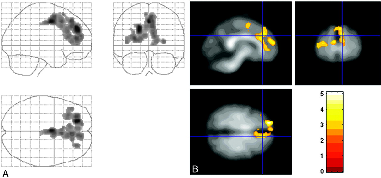
  • Fig 2.
    • Download figure
    • Open in new tab
    • Download powerpoint
    Fig 2.

    A, A glass brain showing significant clusters of reduced FA in patients with PD compared with control subjects as viewed from the right (upper left), front (upper right), and above (lower left). SPM display is according to radiologic convention (right-to-left orientation). B, The statistical map is superimposed on the normalized average FA image. The images are in MNI space. Color bar indicates z-score. Clusters were considered significant at P < .05, corrected for multiple comparisons.

Tables

  • Figures
    • View popup
    Table 1:

    Demographic data and clinical scores of subject population

    Group (No.)Sex (M/F)Age (yr)Disease Duration (yr)HYHYUPDRSUPDRSMMSE
    Mean (SD)Mean (SD)(ON)(OFF)(ON)(OFF)(ON)
    PD (12)5/762.1 (12.7)5.8 (4.5)1.8 (0.4)*1.9 (0.6)43 (13.8)a53.3 (22.1)28.2
    Controls (13)8/558.0 (7.3)N/AN/AN/AN/AN/A28.8
    • Note:—NA indicates data not available; M, male; F, female; HY, Hoehn and Yahr clinical staging; UPDRS, Unified Parkinson Disease Rating Scale; MMSE, Mini-Mental State Examination; ON, taking medication as normal; OFF, cessation of medication.

    • * Levodopa equivalent dosage = 585.2 ± 509.8 mg.

    • View popup
    Table 2:

    Peaks for significant clusters of decreased FA in patients with PD*

    PD Versus ControlsHemisphereBrodmann AreaMNI CoordinatesZ-Score
    xyz
    Medial frontal cortex
        SMAL6−414463.3
        Pre-SMAR8426522.79
    R6618602.83
        Rostral medial frontal gyrusR9646362.97
    Cingulate cortex
        Anterior cingulate gyrus240−6464.12
    L24−634463.41
    32014383.00
    L32−826283.11
        Rostral cingulate gyrusR241030183.00
    L32−84422.82
    L9−846223.18
    Dorsolateral prefrontal cortex
        Superior frontal gyrusR91844324.14
        Middle frontal gyrusR462842143.50
        Inferior frontal gyrusR46423462.93
        Frontal pole
        Middle frontal gyrusR102046103.17
    L10−284283.06
    WM bundles
        Superior longitudinal fasciculusR3026163.09
        Corpus callosumL−1634103.02
    • Note:—SMA indicates supplementary motor area; MNI, Montreal Neurologic Institute.

    • * All significant maxima were part of a single large cluster, including 3927 voxels.

PreviousNext
Back to top

In this issue

American Journal of Neuroradiology: 29 (3)
American Journal of Neuroradiology
Vol. 29, Issue 3
March 2008
  • Table of Contents
  • Index by author
Advertisement
Print
Download PDF
Email Article

Thank you for your interest in spreading the word on American Journal of Neuroradiology.

NOTE: We only request your email address so that the person you are recommending the page to knows that you wanted them to see it, and that it is not junk mail. We do not capture any email address.

Enter multiple addresses on separate lines or separate them with commas.
Altered Diffusion in the Frontal Lobe in Parkinson Disease
(Your Name) has sent you a message from American Journal of Neuroradiology
(Your Name) thought you would like to see the American Journal of Neuroradiology web site.
CAPTCHA
This question is for testing whether or not you are a human visitor and to prevent automated spam submissions.
Cite this article
A.T. Karagulle Kendi, S. Lehericy, M. Luciana, K. Ugurbil, P. Tuite
Altered Diffusion in the Frontal Lobe in Parkinson Disease
American Journal of Neuroradiology Mar 2008, 29 (3) 501-505; DOI: 10.3174/ajnr.A0850

Citation Manager Formats

  • BibTeX
  • Bookends
  • EasyBib
  • EndNote (tagged)
  • EndNote 8 (xml)
  • Medlars
  • Mendeley
  • Papers
  • RefWorks Tagged
  • Ref Manager
  • RIS
  • Zotero
0 Responses
Respond to this article
Share
Bookmark this article
Altered Diffusion in the Frontal Lobe in Parkinson Disease
A.T. Karagulle Kendi, S. Lehericy, M. Luciana, K. Ugurbil, P. Tuite
American Journal of Neuroradiology Mar 2008, 29 (3) 501-505; DOI: 10.3174/ajnr.A0850
del.icio.us logo Twitter logo Facebook logo Mendeley logo
  • Tweet Widget
  • Facebook Like
  • Google Plus One
Purchase

Jump to section

  • Article
    • Abstract
    • Subjects and Methods
    • Results
    • Discussion
    • Conclusion
    • Acknowledgments
    • Footnotes
    • References
  • Figures & Data
  • Info & Metrics
  • Responses
  • References
  • PDF

Related Articles

  • No related articles found.
  • PubMed
  • Google Scholar

Cited By...

  • Magnetoencephalography-based interpretable automated differential diagnosis in neurodegenerative diseases
  • NfL as a biomarker for neurodegeneration and survival in Parkinson disease
  • Modifiable cardiovascular risk factors and axial motor impairments in Parkinson disease
  • White matter microstructure deteriorates across cognitive stages in Parkinson disease
  • Progressive changes in a recognition memory network in Parkinson's disease
  • Diffusion tensor imaging in parkinsonian syndromes: A systematic review and meta-analysis
  • Individual Detection of Patients with Parkinson Disease using Support Vector Machine Analysis of Diffusion Tensor Imaging Data: Initial Results
  • Characterizing dementia with Lewy bodies by means of diffusion tensor imaging
  • White Matter Alteration of the Cingulum in Parkinson Disease with and without Dementia: Evaluation by Diffusion Tensor Tract-Specific Analysis
  • Structural Brain Abnormalities in Patients with Parkinson Disease: A Comparative Voxel-Based Analysis Using T1-Weighted MR Imaging and Magnetization Transfer Imaging
  • Regional Volume Analysis of the Parkinson Disease Brain in Early Disease Stage: Gray Matter, White Matter, Striatum, and Thalamus
  • T2 lesion location really matters: a 10 year follow-up study in primary progressive multiple sclerosis
  • White Matter Microstructure Changes in the Thalamus in Parkinson Disease with Depression: A Diffusion Tensor MR Imaging Study
  • White Matter Involvement in Idiopathic Parkinson Disease: A Diffusion Tensor Imaging Study
  • Crossref (155)
  • Google Scholar

This article has been cited by the following articles in journals that are participating in Crossref Cited-by Linking.

  • Functional network disruption in the degenerative dementias
    Michela Pievani, Willem de Haan, Tao Wu, William W Seeley, Giovanni B Frisoni
    The Lancet Neurology 2011 10 9
  • Parkinson&#039;s disease: The quintessential neuropsychiatric disorder
    Daniel Weintraub, David J. Burn
    Movement Disorders 2011 26 6
  • Problem solving, working memory, and motor correlates of association and commissural fiber bundles in normal aging: A quantitative fiber tracking study
    Natalie M. Zahr, Torsten Rohlfing, Adolf Pfefferbaum, Edith V. Sullivan
    NeuroImage 2009 44 3
  • Diffusion tensor imaging in parkinsonian syndromes
    Claire J. Cochrane, Klaus P. Ebmeier
    Neurology 2013 80 9
  • Magnetic resonance imaging for the diagnosis of Parkinson’s disease
    Beatrice Heim, Florian Krismer, Roberto De Marzi, Klaus Seppi
    Journal of Neural Transmission 2017 124 8
  • White Matter Involvement in Idiopathic Parkinson Disease: A Diffusion Tensor Imaging Study
    G. Gattellaro, L. Minati, M. Grisoli, C. Mariani, F. Carella, M. Osio, E. Ciceri, A. Albanese, M.G. Bruzzone
    American Journal of Neuroradiology 2009 30 6
  • White matter lesions in Parkinson disease
    Nicolaas I. Bohnen, Roger L. Albin
    Nature Reviews Neurology 2011 7 4
  • Mild cognitive impairment is linked with faster rate of cortical thinning in patients with Parkinson’s disease longitudinally
    Alexandru Hanganu, Christophe Bedetti, Clotilde Degroot, Béatriz Mejia-Constain, Anne-Louise Lafontaine, Valérie Soland, Sylvain Chouinard, Marie-Andrée Bruneau, Samira Mellah, Sylvie Belleville, Oury Monchi
    Brain 2014 137 4
  • Free-water imaging in Parkinson’s disease and atypical parkinsonism
    Peggy J. Planetta, Edward Ofori, Ofer Pasternak, Roxana G. Burciu, Priyank Shukla, Jesse C. DeSimone, Michael S. Okun, Nikolaus R. McFarland, David E. Vaillancourt
    Brain 2016 139 2
  • Regional alterations of brain microstructure in Parkinson&#039;s disease using diffusion tensor imaging
    Wang Zhan, Gail A. Kang, Graham A. Glass, Yu Zhang, Cheryl Shirley, Rachel Millin, Katherine L. Possin, Marzieh Nezamzadeh, Michael W. Weiner, William J. Marks, Norbert Schuff
    Movement Disorders 2012 27 1

More in this TOC Section

  • Usefulness of Quantitative Susceptibility Mapping for the Diagnosis of Parkinson Disease
  • White Matter Alterations in the Brains of Patients with Active, Remitted, and Cured Cushing Syndrome: A DTI Study
  • Qualitative and Quantitative Analysis of MR Imaging Findings in Patients with Middle Cerebral Artery Stroke Implanted with Mesenchymal Stem Cells
Show more BRAIN

Similar Articles

Advertisement

Indexed Content

  • Current Issue
  • Accepted Manuscripts
  • Article Preview
  • Past Issues
  • Editorials
  • Editor's Choice
  • Fellows' Journal Club
  • Letters to the Editor
  • Video Articles

Cases

  • Case Collection
  • Archive - Case of the Week
  • Archive - Case of the Month
  • Archive - Classic Case

Special Collections

  • AJNR Awards
  • ASNR Foundation Special Collection
  • Most Impactful AJNR Articles
  • Photon-Counting CT
  • Spinal CSF Leak Articles (Jan 2020-June 2024)

More from AJNR

  • Trainee Corner
  • Imaging Protocols
  • MRI Safety Corner

Multimedia

  • AJNR Podcasts
  • AJNR Scantastics

Resources

  • Turnaround Time
  • Submit a Manuscript
  • Submit a Video Article
  • Submit an eLetter to the Editor/Response
  • Manuscript Submission Guidelines
  • Statistical Tips
  • Fast Publishing of Accepted Manuscripts
  • Graphical Abstract Preparation
  • Imaging Protocol Submission
  • Evidence-Based Medicine Level Guide
  • Publishing Checklists
  • Author Policies
  • Become a Reviewer/Academy of Reviewers
  • News and Updates

About Us

  • About AJNR
  • Editorial Board
  • Editorial Board Alumni
  • Alerts
  • Permissions
  • Not an AJNR Subscriber? Join Now
  • Advertise with Us
  • Librarian Resources
  • Feedback
  • Terms and Conditions
  • AJNR Editorial Board Alumni

American Society of Neuroradiology

  • Not an ASNR Member? Join Now

© 2025 by the American Society of Neuroradiology All rights, including for text and data mining, AI training, and similar technologies, are reserved.
Print ISSN: 0195-6108 Online ISSN: 1936-959X

Powered by HighWire