
Abstract
BACKGROUND AND PURPOSE: Our aim was to investigate the extent and severity of changes in spinal cord diffusion tensor imaging (DTI) parameters in patients with cervical cord injury.
MATERIALS AND METHODS: DTI was performed in 20 symptomatic patients (mean, 45.7 ± 17.7 years of age; 2 women, 18 men) with cervical spine trauma and 8 volunteers (mean, 34.2 ± 10.7 years of age; 6 men, 2 women). The whole cord and regional apparent diffusion coefficient (ADC), fractional anisotropy (FA), relative anisotropy (RA), and volume ratio (VR) of patients and volunteers were compared. DTI parameters were calculated in 16 patients. MR imaging demonstrated hemorrhagic cord contusions (n = 6), nonhemorrhagic cord contusions (n = 4), and soft-tissue injury (n = 6). Medical records were reviewed for extent of neurologic deficit.
RESULTS: Regional ADC values differed significantly between upper and mid and upper and lower (both, P < .004) cervical cord sections. FA was significantly different between upper and lower sections (P < .03). Whole cord ADC values were significantly lower in patients than in volunteers (P < .0001). Whole spine FA was not significantly decreased in patients (P < .06). ADC and FA values were significantly decreased at injury sites when compared with volunteers (P < .031 and .0001, respectively). The greatest differences in whole cord ADC, FA, RA, and VR were in patients with hemorrhagic cord contusions compared with healthy volunteers (P < .0001, .003, .0005, and .008, respectively).
CONCLUSION: DTI parameters are sensitive markers of cervical cord injury, with ADC showing the greatest sensitivity. Changes in DTI parameters are most marked at injury sites and reflect the severity of cord injury.
Traumatic injuries of the cervical spine are potentially catastrophic. When associated with neurologic damage, they can result in devastating medical, social, emotional, and financial consequences. About 11,000 new spinal cord injuries occur in the United States every year.1 Most (78%) involve males between the ages of 16 and 30 years of age.1
Conventional MR imaging is performed routinely to demonstrate soft-tissue and spinal cord injuries in spinal trauma.2–4 Edema and hemorrhage in the spinal cord following trauma are well demonstrated by MR imaging and may help to predict neurologic outcomes.5–7
Diffusion-weighted MR imaging (DWI) assesses in vivo changes in random motion of protons in water.8,9 DWI and tissue perfusion imaging have now become integral parts of routine imaging to evaluate patients for acute stroke.8,9 Recent animal and human studies have attempted to assess changes in the normal and pathologic cervical spinal cord by using diffusion-tensor imaging (DTI).10–13 DTI of the cervical cord is technically challenged by the low signal intensity-to-noise ratio of the small volume of cord tissue, pulsation artifacts arising from the CSF, cardiac and respiratory motion, and magnetic susceptibility artifacts caused by adjacent bone.10–12 The ability of DTI to complement conventional MR imaging by diagnosing subtle injuries to the cord or predict the need for early therapeutic intervention for optimal clinical outcome has yet to be demonstrated. To our knowledge, no previous studies have reported DTI changes following cervical spine injury in humans. We hypothesize that DTI changes are sensitive and will correlate with the severity of spinal cord injury. We attempted to study changes in the apparent diffusion coefficient (ADC), fractional anisotropy (FA), relative anisotropy (RA), and volume ratio (VR) after traumatic cervical cord injury and to compare these with corresponding data from a control group of individuals with no spinal cord injuries.
Materials and Methods
Our institutional review board approved this study. The study complied with the requirements of the Health Insurance Portability and Accountability Act.
Patients
Data from 20 patients (mean, 45.7 ± 17.7 years of age; 2 women, 18 men) who were admitted to our trauma center following blunt force trauma with cervical spine injuries and who were imaged by using MR imaging as part of a standard protocol were retrospectively compared with data from 8 healthy volunteers (mean, 34.2 ± 10.7 years of age; 6 men, 2 women). Both groups were imaged by using conventional MR imaging and DTI. MR imaging studies were performed 2 hours to 15 days (mean, 32 ± 22 hours) following injury. Indications for MR imaging studies in the trauma group included neurologic deficit on clinical examination localized to the cervical spine (n = 16), assessment of the extent of ligamentous injury following cervical spine fracture demonstrated on the admission CT of the cervical spine (n = 2), and neck pain or tenderness unexplained by admission cervical spine radiographs or CT examination (n = 2). Mechanisms of injury included motor vehicle collision (n = 7), motorcycle collision (n = 1), fall (n = 6), diving/surfing accident (n = 3), assault (n = 1), and all-terrain-vehicle crash (n = 1). The medical record was not available to determine the mechanism of injury in 1 patient. DTI data from 4 patients were not used for further analysis because of poor quality, including images with metal artifacts. The 16 remaining patients formed the study group.
MR Imaging Technique
All MR imaging was performed on a 1.5T Avanto scanner (Siemens, Erlangan, Germany) with parallel imaging capability. Conventional MR imaging included sagittal T2 (TE/TR, 109/4000 ms), fluid-attenuated inversion recovery (TE/TR/echo train, 102/8000 ms/13), and axial T2 and T2* images. DTIs were obtained by using an echo-planar imaging (EPI) sequence at a TE/TR of 76/8000 ms and a resolution of 128 × 128 over a 20-cm FOV. Diffusion-weighting was applied in 6 noncollinear directions in the axial plane at an effective b-value of 1000 s/mm2. A total of 67 axial sections at 3-mm thickness were obtained to cover from the medullary cervical junction to the cervical-thoracic junction. Partial Fourier with a kernel size of 32 was used for a total scanning time of 3.66 minutes. A 12-channel head-neck array coil was used on all patients, and parallel imaging was used with the phase-encoding in the anterior-to-posterior direction. Sagittal T2-weighted images and b = 0 s/mm2 images from the DTIs were used for anatomic reference.
Image Analysis
Maps of ADC and FA were generated with background noise suppressed by using the DTI task card that runs on the Siemens MR imaging workstations (courtesy Drs. G. Sorenson and T. Benner, Massachusetts General Hospital, Boston, Mass). RA and VR were calculated for each region of interest by using the eigenvalues of the tensor as described by Le Bihan et al.14 The conventional images were viewed for evidence of signal-intensity abnormality to indicate the presence of a cord injury. The anatomic location of the cord injury and the presence of edema or hemorrhage were noted. Conventional MR images demonstrated hemorrhagic cord contusion in 6 patients, nonhemorrhagic cord contusions in 4 patients, and soft-tissue-only injury in 6 patients. The cord contusions involved the upper cervical cord level in 1 patient, the midcervical cord level in 9 patients, and the lower cervical cord level in 8 patients. The cervical cord was divided into 3 anatomic regions. The upper cervical cord included the region from the lower brain stem to the lower C2 vertebral body, the mid cervical cord included the region from the upper C3 vertebral body to the lower C5 vertebral body, and the lower cervical cord included the region from upper C6 vertebral body to the lower T1 vertebral body. Regions of interest were drawn (Fig. 1A, -B) in each of the 3 regions by using the T2-weighted and/or the b = 0 portion of the DTI images for both controls and patients; and the ADC, FA, RA, and VR were measured. The regions of interest were carefully placed so that they included both the central gray and white matter. Region-of-interest locations were confirmed by using the optimal conventional MR imaging sequence, and care was taken to avoid inclusion of CSF.
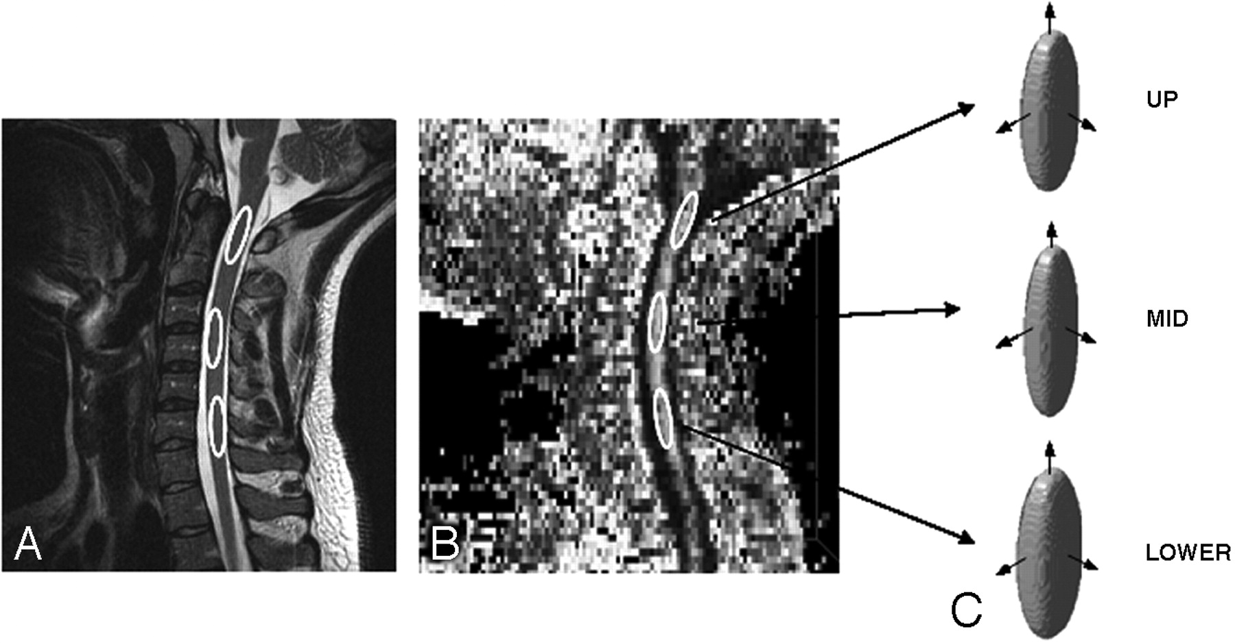
Images showing the placement of regions of interest in the sagittal T2-weighted image (A), FA image (B), and the corresponding ellipsoid representation of the tensor at each level of the cord (C).
Whole cord ADC, FA, RA, and VR measures were obtained by averaging the 3 regions of interest per subject. To assess normal regional variation of the DTI parameters, we made comparisons between the 3 regions among the healthy subjects. Regions from the patients were compared with corresponding regions from the healthy volunteers to assess differences in DTI parameters. The same parameters were also compared at the whole spine level. Regions of interest were also placed within the area of the spinal cord injury identified by using conventional MR imaging, from which the DTI parameters were measured. These parameters were compared with the average whole cord data from the control subjects. To determine whether patients presenting with hemorrhagic contusions demonstrated more severe changes in DTI parameters, we compared data from the 6 patients with hemorrhagic cord contusions with those from the healthy controls. All comparisons were made by using a 1-sided t test and corrected for multiple comparisons by using the Bonferroni method. Significance was defined as P < .05.
Medical records (available for 15 of the 16 patients) were reviewed to determine the extent of neurologic deficit. Neurologic deficits were seen in 11 patients, including quadriplegia in 8 patients, diplegia in 1, monoplegia in 1, and a radiculopathy in 1 patient. Conventional MR imaging demonstrated hemorrhagic cord contusion in 5 of the 8 patients with quadriplegia.
Results
The Table summarizes the ADC, FA, RA, and VR values for the 3 cervical cord regions among the control subjects and patients.
Regional DTI parameters of the normal cord in control individuals and patients
Control Group.
Significant differences were observed in the ADC between the upper and mid cord (P < .004) and also between the upper and lower cord levels (P < .004) for the controls. However, significant FA differences were seen only between the mid and lower cord levels (P < .03). The principal eigenvalue E1 was significantly different between the upper and mid (P < .001) and upper and lower (P < .007) cord levels and mirrored the behavior of ADCs. The eigenvalues E2 and E3 were found to be significantly different only between the upper and lower cord levels (P < .02 and P < .035, respectively). Figure 1C shows the ellipsoid representation of the diffusion tensor for the control subjects at each of the 3 levels at which it was measured.
Patient Group.
ADC values for patients were significantly lower than those in the control group for all 3 cord levels (P < .012, .0005, and .0001 for the upper, mid, and lower sections, respectively) with the lower cervical level (C6-T1 region) showing the largest decrease in ADC values. Corresponding E1 (P < .0018, .0001, and .0001, respectively) and E3 (P < .09, .039, and .01, respectively) values also exhibited significant reductions in these areas (Fig 2). Although FA showed a trend toward lower values in all 3 regions, none was significantly (at the P < .01 level) reduced from the corresponding values in controls. The largest reduction was seen in the mid cervical cord (C3-C5, P < .07).

A 56-year-old man admitted following a fall, with central cord syndrome. A, T2-weighted image shows a cord contusion (arrowhead) posterior to the C4-5 disk space. Ellipsoid representation of the tensor of the axial cervical cord of the patient and healthy volunteer at corresponding anatomic levels of the upper cord (B), adjacent to the contusion (C), cord contusion (D), and the lower cord (E) shows abnormal ellipsoid representation of the cord at and away from the cord contusion, where the conventional MR imaging shows no signal-intensity abnormality
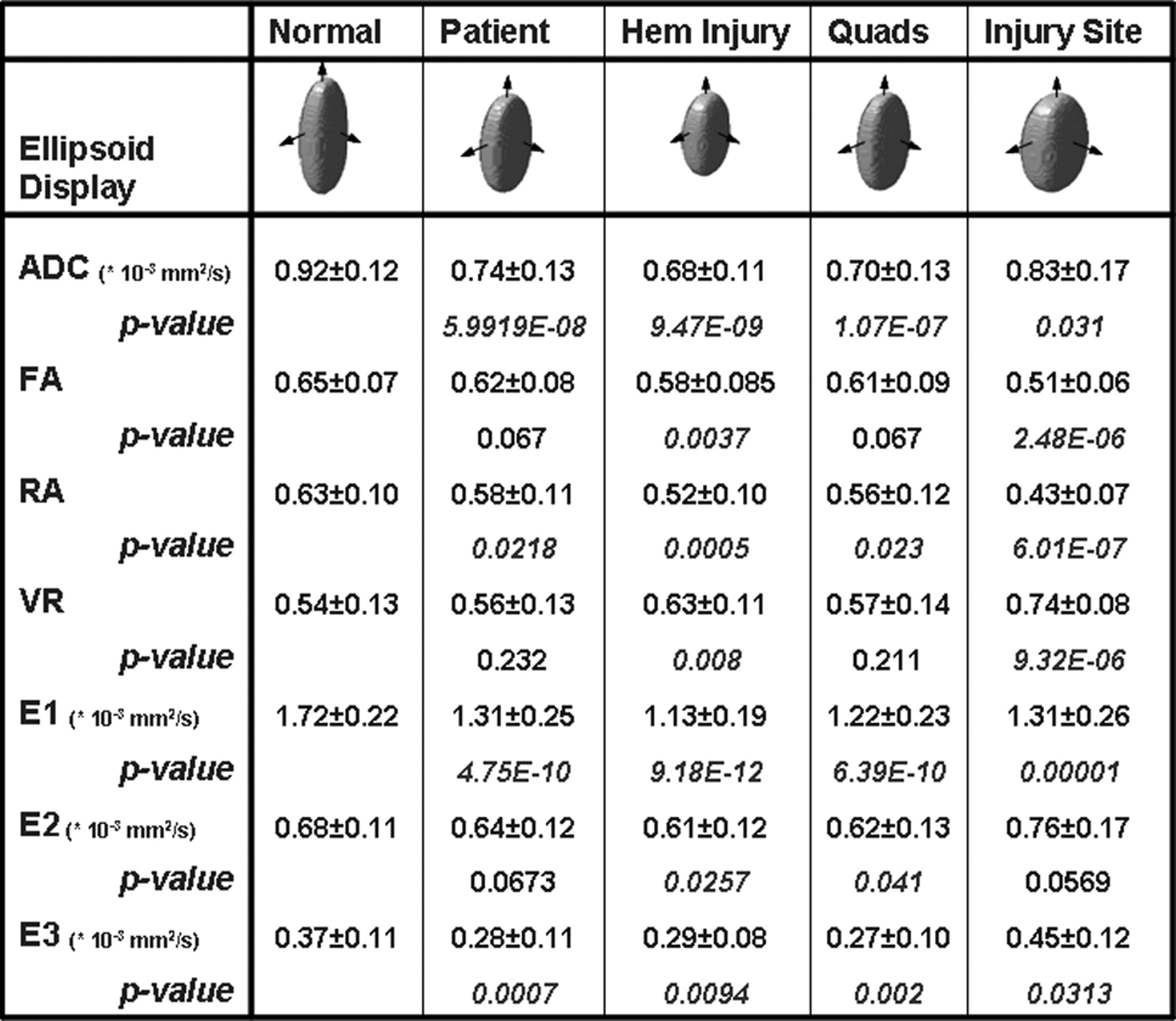
Figure 3 summarizes the DTI parameters for the whole cervical cord for all 16 patients, for the 6 patients exhibiting hemorrhagic cord contusions, and for the 8 patients with quadriplegia and compares these with whole cord DTI values obtained from controls. The Table also provides results of the comparison between the DTI parameters at the cord injury site (n = 10) and whole cord DTI values from controls. The data clearly indicate that the ADC is significantly decreased among all patient groups, with patients with hemorrhage exhibiting the greatest decrease, followed by patients with quadriplegia. The DTI parameters of the quadriplegic patient group are similar to those in the entire patient group, and both are significantly different from those of the hemorrhagic group (Fig 3). Although FA decreased among all patient groups, this decrease was significant only for patients with hemorrhage and when FA was measured at the injury site. However, RA, which typically tends to follow the trend of FA, was significantly decreased among all groups of patients. The greatest decreases in the FA and RA were observed at the cord injury site. VRs in the hemorrhagic patient group and at the injury site were significantly increased compared with those in control subjects, suggesting an increased isotropic behavior among patients with cord injury.

Comparison of whole spine control (n = 8) DTI parameters with the patient population (n = 16), patients exhibiting hemorrhagic (Hem) injury (n = 6), patients with quadriplegia (Quads, n = 7), and site of injury (n = 11).
Discussion
Conventional MR imaging is routinely used to image the spinal cord to demonstrate the location of cord injury and the amount of cord compression from extramedullary bleeding or posterior disk herniation. The amount of cord compression and the presence of hemorrhage or edema at the injury site may help in treatment planning and in predicting neurologic outcome. T2-weighted and fat-suppressed inversion recovery images are the most useful sequences to demonstrate these findings.
A few studies have reported DTI changes in cervical spine in animal models.9,10,15,16 These studies have investigated the utility of various novel MR images in demonstrating DTI differences seen in white and gray matter and morphologic differences in white matter tracts. DTI changes in the cervical spine have also been reported in patients with cervical spondylosis and primary progressive multiple sclerosis.11–13 The DWI parameters were not only abnormal in patients with clinical symptoms of cervical myelopathy but also showed increased sensitivity in demonstrating early changes not visible on conventional MR imaging.12,13 DTI imaging of the cervical cord in patients with primary progressive multiple sclerosis was able to evaluate and quantify the extent of axonal injury and reactive gliosis.11
The cylindrical anatomy and symmetry of white matter tracts that run in the craniocaudal direction within the spinal cord make it possible to obtain reasonably accurate measurements of water diffusion longitudinal and transverse to the white matter tracts.15 Previous studies have reported a variation in the mean ADC and FA values between the upper and lower cervical cord. Mamata et al13 reported higher mean values for ADC and FA in the upper cervical spine (C2-C3) than in the lower levels (C4-C7) in 11 healthy volunteers. Wheeler-Kingshott et al,17 however, reported no significant differences in mean diffusivity along the length of the normal cord but observed a significant increase in the FA in the mid and lower section of the cord compared with the upper cord. Our study demonstrated significantly higher mean ADC values in the mid (C3-C5) and lower (C6-T1) cervical cord when compared with the upper (cervicomedullary junction to C2) cervical cord among the control subjects. FA values were significantly lower in the mid and lower cervical cord.
The differences in these findings may be attributed to measurements made at different anatomic locations as reported by Mamata et al13 and variations in the choice of pulse sequences. We used the most popular commercially available single-shot echo-planar imaging (EPI) technique, whereas Mamata et al used a line-scan diffusion technique and Wheeler-Kingshott et al17 used a zonally magnified oblique multi-slice (ZOOM)-EPI technique. The line-scan technique and the ZOOM-EPI techniques are less prone to susceptibility artifacts than the standard EPI technique. However, the scanning times from these techniques can be quite long, making them impractical for evaluation of patients with cervical trauma. The fast spin-echo-based line-scanning technique is also prone to specific absorption ratio limitations at higher field strengths that may further increase scanning time.
Although some findings in controls differed in our study and that of Wheeler-Kingshott et al,17 other findings were similar. Trends in FA measurements were similar in both studies, though values in our study did not reach significance between the upper and mid and upper and lower cord segments. FA and ADC values were similar to those observed by Wheeler-Kingshott et al, and the behaviors of eigenvalues were also analogous, suggesting that similar results can be obtained by using the standard EPI technique.
This study demonstrates that whole cervical spine DTI parameters, including ADC and RA values for patients with cervical spine contusions, were significantly lower and the VR was significantly higher than comparative values in healthy individuals. Regional measurements of ADC values at all 3 cord levels were decreased among patients, with the greatest decrease at these site of cord injury (mid and lower cervical levels). These results indicate that DTI parameters are sensitive markers of cord injury compared with conventional MR imaging. Such remote changes from the injury site are not seen on conventional MR imaging and are evident only by measuring DTI parameters (Fig 2). Among the 4 DTI parameters, ADC was the most sensitive marker of cord injury. To the authors' knowledge, these results have not been reported in previous cervical spine trauma studies in humans. Demonstration of abnormalities in the DTI parameters in areas of the spinal cord that appear normal on conventional MR imaging could be used to document the true extent of injury and help to correlate better the neurologic deficit with MR imaging. Future studies are required to see whether ADC values may be used as markers of cord injury in patients with significant neurologic deficit who have normal findings on conventional MR imaging studies of the cervical spine.
Patients with hemorrhagic cord contusions on conventional MR imaging have more severe injuries with poorer long-term clinical outcomes than patients with nonhemorragic cord contusions.2–7 In our study, whole spine ADC, FA, and RA values were most significantly reduced and VR was increased significantly for patients with cord hemorrhage. These changes were noted on a regional basis when compared with the control group. The extent of decrease was more marked when compared with that of patients with nonhemorrhagic cord contusions, indicating that changes in DTI parameters reflected the severity of injury. VR is indicative of the ellipsoid volume in comparison with a sphere with radius equal to the mean diffusivity of the water protons. Consequently, a high VR is indicative of isotropic diffusion. An increase in VR for the hemorrhagic patient groups and the measurements made at the site of injury is consistent with corresponding decreases seen in FA and RA for both these groups compared with control subjects. Similar findings have not been reported in previous studies. These results indicate that DTI parameters, in addition to the clinical examination, could be an independent predictor of the severity of spinal cord injury and may be used to monitor recovery following treatment.
The ADC, FA, RA, and VR values for patients with quadriplegia were also compared with those from the control group. Although FA showed only a near-significant decrease (P < .067), other DTI values were significantly decreased (increased in the case of VR) in the quadriplegic group compared with volunteers. The increase in VR and the decrease in the ADC, FA, and RA values were less marked than those noted among patients with hemorrhagic cord contusions but greater than the changes noted in the entire group of patients with spinal cord injury. These results also indicate that DTI parameters in addition to the clinical examination could be an independent predictor of the severity of spinal cord injury. In this study, 5 of the 6 hemorrhagic cord contusions were seen in patients with quadriplegia. These findings are consistent with the observation that cord contusions in patients with quadriplegia as seen on conventional MR imaging usually extend over multiple levels and involve a large area of the transverse diameter of the cord.2–7
Previous animal studies have noted significant differences in transverse and longitudinal diffusivity between white matter tracts within the spinal cord.16 These differences may be the result of changes in axonal diameters seen within the various white matter tracts. The highest transverse and longitudinal diffusivity is usually seen in axons with the largest diameter.16,18 A higher transverse diffusivity also may be seen when there is an increase in the space between axons. In this study, both the transverse and longitudinal diffusivity decreased significantly at the injury site in patients with hemorrhagic cord contusions. The authors postulate that these values may reflect a decrease in axonal diameter at the injury level from external compression by hemorrhage within the extracellular space (Fig 2). The presence of hemorrhage at the injury site may also prevent free movement of water in the extracellular space. This concept may help explain the decrease in FA, ADC, and RA. Unfortunately, the histology of the cord injuries was not available for this patient group, and future studies with histologic correlation are necessary to confirm these findings.
Several limitations were inherent in this study. In addition to its retrospective design and small sample size, the study did not correlate DTI parameters with clinical outcomes, nor as noted, were histologic observations and correlations included. Future studies with a large sample size are necessary to validate our results.
Conclusion
This preliminary study demonstrates that DTI parameter abnormalities are seen throughout the cervical spinal cord following injury. These changes are not seen on conventional MR imaging. The abnormalities are greatest at the cord injury site and reflect injury severity.
Acknowledgments
We acknowledge the assistance of Nancy Knight, PhD, in the preparation of this manuscript for publication.
References
- Received April 9, 2007.
- Accepted after revision November 4, 2007.
- Copyright © American Society of Neuroradiology
In this issue


Jump to section
Related Articles
Cited By...
- Cervical Cordotomy for Intractable Pain: Do Postoperative Imaging Features Correlate with Pain Outcomes and Mirror Pain?
- Local BDNF Delivery to the Injured Cervical Spinal Cord using an Engineered Hydrogel Enhances Diaphragmatic Respiratory Function
- Cervical Spinal Cord DTI Is Improved by Reduced FOV with Specific Balance between the Number of Diffusion Gradient Directions and Averages
- Pulse-Triggered DTI Sequence with Reduced FOV and Coronal Acquisition at 3T for the Assessment of the Cervical Spinal Cord in Patients with Myelitis
- Diffusion tensor imaging of the spinal cord and its clinical applications
- Diffusion Tensor Imaging of the Pediatric Spinal Cord at 1.5T: Preliminary Results