Advertisement

Main menu

  • Home
  • Content
    • Current Issue
    • Accepted Manuscripts
    • Article Preview
    • Past Issue Archive
    • Video Articles
    • AJNR Case Collection
    • Case of the Week Archive
    • Case of the Month Archive
    • Classic Case Archive
  • Special Collections
    • AJNR Awards
    • ASNR Foundation Special Collection
    • Most Impactful AJNR Articles
    • Photon-Counting CT
    • Spinal CSF Leak Articles (Jan 2020-June 2024)
  • Multimedia
    • AJNR Podcasts
    • AJNR SCANtastic
    • Trainee Corner
    • MRI Safety Corner
    • Imaging Protocols
  • For Authors
    • Submit a Manuscript
    • Submit a Video Article
    • Submit an eLetter to the Editor/Response
    • Manuscript Submission Guidelines
    • Statistical Tips
    • Fast Publishing of Accepted Manuscripts
    • Graphical Abstract Preparation
    • Imaging Protocol Submission
    • Author Policies
  • About Us
    • About AJNR
    • Editorial Board
    • Editorial Board Alumni
  • More
    • Become a Reviewer/Academy of Reviewers
    • Subscribers
    • Permissions
    • Alerts
    • Feedback
    • Advertisers
    • ASNR Home

User menu

  • Alerts
  • Log in

Search

  • Advanced search
American Journal of Neuroradiology

American Journal of Neuroradiology

ASHNR American Society of Functional Neuroradiology ASHNR American Society of Pediatric Neuroradiology ASSR
  • Alerts
  • Log in

Advanced Search

  • Home
  • Content
    • Current Issue
    • Accepted Manuscripts
    • Article Preview
    • Past Issue Archive
    • Video Articles
    • AJNR Case Collection
    • Case of the Week Archive
    • Case of the Month Archive
    • Classic Case Archive
  • Special Collections
    • AJNR Awards
    • ASNR Foundation Special Collection
    • Most Impactful AJNR Articles
    • Photon-Counting CT
    • Spinal CSF Leak Articles (Jan 2020-June 2024)
  • Multimedia
    • AJNR Podcasts
    • AJNR SCANtastic
    • Trainee Corner
    • MRI Safety Corner
    • Imaging Protocols
  • For Authors
    • Submit a Manuscript
    • Submit a Video Article
    • Submit an eLetter to the Editor/Response
    • Manuscript Submission Guidelines
    • Statistical Tips
    • Fast Publishing of Accepted Manuscripts
    • Graphical Abstract Preparation
    • Imaging Protocol Submission
    • Author Policies
  • About Us
    • About AJNR
    • Editorial Board
    • Editorial Board Alumni
  • More
    • Become a Reviewer/Academy of Reviewers
    • Subscribers
    • Permissions
    • Alerts
    • Feedback
    • Advertisers
    • ASNR Home
  • Follow AJNR on Twitter
  • Visit AJNR on Facebook
  • Follow AJNR on Instagram
  • Join AJNR on LinkedIn
  • RSS Feeds

AJNR is seeking candidates for the AJNR Podcast Editor. Read the position description.

Research ArticlePEDIATRICS

Aberrant Midsagittal Fiber Tracts in Patients with Hemimegalencephaly

N. Sato, M. Ota, A. Yagishita, Y. Miki, T. Takahashi, Y. Adachi, Y. Nakata, K. Sugai and M. Sasaki
American Journal of Neuroradiology April 2008, 29 (4) 823-827; DOI: https://doi.org/10.3174/ajnr.A0919
N. Sato
  • Find this author on Google Scholar
  • Find this author on PubMed
  • Search for this author on this site
M. Ota
  • Find this author on Google Scholar
  • Find this author on PubMed
  • Search for this author on this site
A. Yagishita
  • Find this author on Google Scholar
  • Find this author on PubMed
  • Search for this author on this site
Y. Miki
  • Find this author on Google Scholar
  • Find this author on PubMed
  • Search for this author on this site
T. Takahashi
  • Find this author on Google Scholar
  • Find this author on PubMed
  • Search for this author on this site
Y. Adachi
  • Find this author on Google Scholar
  • Find this author on PubMed
  • Search for this author on this site
Y. Nakata
  • Find this author on Google Scholar
  • Find this author on PubMed
  • Search for this author on this site
K. Sugai
  • Find this author on Google Scholar
  • Find this author on PubMed
  • Search for this author on this site
M. Sasaki
  • Find this author on Google Scholar
  • Find this author on PubMed
  • Search for this author on this site
  • Article
  • Figures & Data
  • Info & Metrics
  • Responses
  • References
  • PDF
Loading

Article Figures & Data

Figures

  • Fig 1.
    • Download figure
    • Open in new tab
    • Download powerpoint
    Fig 1.

    A 19-year-old female patient with left hemimegalencephaly evaluated in the retrospective analysis. A, T2-weighted image shows increased width (arrows) between the 2 anterior horns due to a white matter–intensity structure (arrowhead). B and C, The white matter–intensity structure appears to extend posteriorly and to pass through the bodies of the lateral ventricles beneath the corpus callosum (arrow).

  • Fig 2.
    • Download figure
    • Open in new tab
    • Download powerpoint
    Fig 2.

    An 8-year-old boy with left hemimegalencephaly (case 1). A, Coronal T2-weighted inversion recovery image shows a white matter–intensity structure between the 2 anterior horns (arrow). B, Axial T2-weighted image shows a midsagittal bandlike structure between the bodies of the 2 lateral ventricles (arrow). It appears to cross from anterior-left to posterior-right. C, Coronal T2-weighted inversion recovery image. The left fornix (arrowhead) is normal, while the right one (arrow) is thick. D–F, 2D coronal (D), 2D axial (E), and 2D coronal (F) views on fiber tract (FT) reconstruction. The images correspond to A, B, and C, respectively. Aberrant midsagittal fibers penetrate the midsagittal bandlike structures shown in A–C. G and H, 3D superior (G) and 3D right lateral (H) views on FT reconstruction. The fibers run between the 2 frontal lobes and the contralateral occipital and parietal lobes. The main fibers pass through the right crus of the fornix.
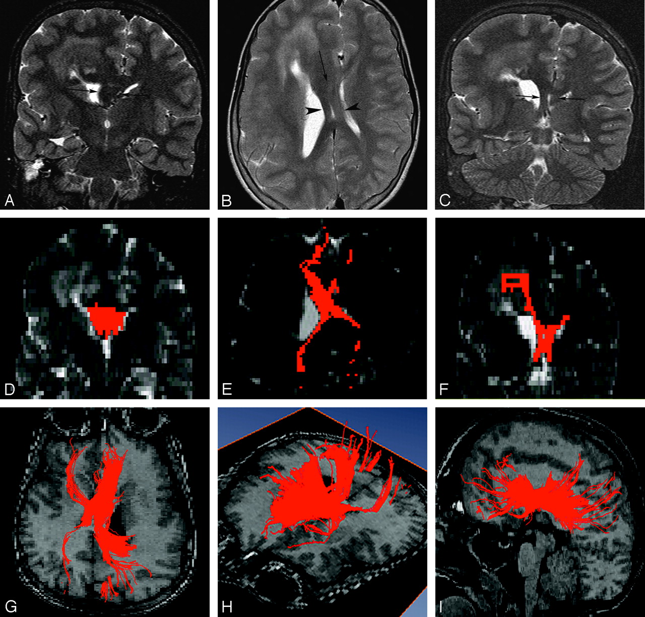
  • Fig 3.
    • Download figure
    • Open in new tab
    • Download powerpoint
    Fig 3.

    An 11-year-old boy with right hemimegalencephaly (case 2). A, Coronal T2-weighted inversion recovery image shows markedly increased width between the 2 anterior horns occupied by a white matter–intensity structure (arrows). B, Axial T2-weighted image demonstrates an abnormal white matter–intensity bandlike structure between the 2 lateral ventricles (arrow), which separates in the posterior portion and runs a course similar to that of the cruces of the fornices (arrowheads). C, Coronal T2-weighted inversion recovery image posterior to that in A also detects bilateral marked thickening of the cruces of the fornices (arrows). D–F, 2D coronal (D), 2D axial (E), and 2D coronal (F) views on FT reconstruction. These images correspond to A, B, and C, respectively. Aberrant midsagittal fibers penetrate the midsagittal abnormal white matter–intensity structures shown in A–C. G–I, 3D superior (G), 3D left lateral superior (H), and 3D left lateral (I) views on FT reconstruction. Aberrant massive fibers connect the bilateral frontal and occipital and parietal lobes passing through abnormal midsagittal structures. They are dominant ipsilaterally.

PreviousNext
Back to top

In this issue

American Journal of Neuroradiology: 29 (4)
American Journal of Neuroradiology
Vol. 29, Issue 4
April 2008
  • Table of Contents
  • Index by author
Advertisement
Print
Download PDF
Email Article

Thank you for your interest in spreading the word on American Journal of Neuroradiology.

NOTE: We only request your email address so that the person you are recommending the page to knows that you wanted them to see it, and that it is not junk mail. We do not capture any email address.

Enter multiple addresses on separate lines or separate them with commas.
Aberrant Midsagittal Fiber Tracts in Patients with Hemimegalencephaly
(Your Name) has sent you a message from American Journal of Neuroradiology
(Your Name) thought you would like to see the American Journal of Neuroradiology web site.
CAPTCHA
This question is for testing whether or not you are a human visitor and to prevent automated spam submissions.
Cite this article
N. Sato, M. Ota, A. Yagishita, Y. Miki, T. Takahashi, Y. Adachi, Y. Nakata, K. Sugai, M. Sasaki
Aberrant Midsagittal Fiber Tracts in Patients with Hemimegalencephaly
American Journal of Neuroradiology Apr 2008, 29 (4) 823-827; DOI: 10.3174/ajnr.A0919

Citation Manager Formats

  • BibTeX
  • Bookends
  • EasyBib
  • EndNote (tagged)
  • EndNote 8 (xml)
  • Medlars
  • Mendeley
  • Papers
  • RefWorks Tagged
  • Ref Manager
  • RIS
  • Zotero
0 Responses
Respond to this article
Share
Bookmark this article
Aberrant Midsagittal Fiber Tracts in Patients with Hemimegalencephaly
N. Sato, M. Ota, A. Yagishita, Y. Miki, T. Takahashi, Y. Adachi, Y. Nakata, K. Sugai, M. Sasaki
American Journal of Neuroradiology Apr 2008, 29 (4) 823-827; DOI: 10.3174/ajnr.A0919
del.icio.us logo Twitter logo Facebook logo Mendeley logo
  • Tweet Widget
  • Facebook Like
  • Google Plus One
Purchase

Jump to section

  • Article
    • Abstract
    • Materials and Methods
    • Results
    • Discussion
    • Conclusions
    • References
  • Figures & Data
  • Info & Metrics
  • Responses
  • References
  • PDF

Related Articles

  • No related articles found.
  • PubMed
  • Google Scholar

Cited By...

  • Variability of Homotopic and Heterotopic Callosal Connectivity in Partial Agenesis of the Corpus Callosum: A 3T Diffusion Tensor Imaging and Q-Ball Tractography Study
  • Crossref (30)
  • Google Scholar

This article has been cited by the following articles in journals that are participating in Crossref Cited-by Linking.

  • The corpus callosum, the other great forebrain commissures, and the septum pellucidum: anatomy, development, and malformation
    Charles Raybaud
    Neuroradiology 2010 52 6
  • Definitions and classification of malformations of cortical development: practical guidelines
    Mariasavina Severino, Ana Filipa Geraldo, Norbert Utz, Domenico Tortora, Ivana Pogledic, Wlodzimierz Klonowski, Fabio Triulzi, Filippo Arrigoni, Kshitij Mankad, Richard J Leventer, Grazia M S Mancini, James A Barkovich, Maarten H Lequin, Andrea Rossi
    Brain 2020 143 10
  • Variability of Homotopic and Heterotopic Callosal Connectivity in Partial Agenesis of the Corpus Callosum: A 3T Diffusion Tensor Imaging and Q-Ball Tractography Study
    M. Wahl, Z. Strominger, R.J. Jeremy, A.J. Barkovich, M. Wakahiro, E.H. Sherr, P. Mukherjee
    American Journal of Neuroradiology 2009 30 2
  • Diffusion tensor imaging and fiber tractography in brain malformations
    Andrea Poretti, Avner Meoded, Andrea Rossi, Charles Raybaud, Thierry A. G. M. Huisman
    Pediatric Radiology 2013 43 1
  • Development and Dysgenesis of the Cerebral Cortex: Malformations of Cortical Development
    Charles Raybaud, Elysa Widjaja
    Neuroimaging Clinics of North America 2011 21 3
  • Update on neuroimaging phenotypes of mid-hindbrain malformations
    Patrice Jissendi-Tchofo, Mariasavina Severino, Béatrice Nguema-Edzang, Cissé Toure, Gustavo Soto Ares, Anthony James Barkovich
    Neuroradiology 2015 57 2
  • Magnetic Resonance Imaging of Malformations of Midbrain-Hindbrain
    Ahmed Abdel Khalek Abdel Razek, Mauricio Castillo
    Journal of Computer Assisted Tomography 2016 40 1
  • Diagnostic Approach to Macrocephaly in Children
    Andrea Accogli, Ana Filipa Geraldo, Gianluca Piccolo, Antonella Riva, Marcello Scala, Ganna Balagura, Vincenzo Salpietro, Francesca Madia, Mohamad Maghnie, Federico Zara, Pasquale Striano, Domenico Tortora, Mariasavina Severino, Valeria Capra
    Frontiers in Pediatrics 2022 9
  • Structural brain anomalies in patients with FOXG1 syndrome and in Foxg1+/− mice
    Milka Pringsheim, Diana Mitter, Simone Schröder, Rita Warthemann, Kim Plümacher, Gerhard Kluger, Martina Baethmann, Thomas Bast, Sarah Braun, Hans‐Martin Büttel, Elizabeth Conover, Carolina Courage, Alexandre N. Datta, Angelika Eger, Theresa A. Grebe, Annette Hasse‐Wittmer, Marion Heruth, Karen Höft, Angela M. Kaindl, Stephanie Karch, Torsten Kautzky, Georg C. Korenke, Bernd Kruse, Richard E. Lutz, Heymut Omran, Steffi Patzer, Heike Philippi, Keri Ramsey, Tina Rating, Angelika Rieß, Mareike Schimmel, Rachel Westman, Frank‐Martin Zech, Birgit Zirn, Pauline A. Ulmke, Godwin Sokpor, Tran Tuoc, Andreas Leha, Martin Staudt, Knut Brockmann
    Annals of Clinical and Translational Neurology 2019 6 4
  • Diffusion Tensor Imaging of Brain Malformations
    Avner Meoded, Thierry A.G.M. Huisman
    Neuroimaging Clinics of North America 2019 29 3

More in this TOC Section

  • SyMRI & MR Fingerprinting in Brainstem Myelination
  • Comparison of Image Quality and Radiation Dose in Pediatric Temporal Bone CT Using Photon-Counting Detector CT and Energy-Integrating Detector CT
  • Venous Sinus Stenosis in Mucopolysaccharidosis I
Show more Pediatrics

Similar Articles

Advertisement

Indexed Content

  • Current Issue
  • Accepted Manuscripts
  • Article Preview
  • Past Issues
  • Editorials
  • Editor's Choice
  • Fellows' Journal Club
  • Letters to the Editor
  • Video Articles

Cases

  • Case Collection
  • Archive - Case of the Week
  • Archive - Case of the Month
  • Archive - Classic Case

Special Collections

  • AJNR Awards
  • ASNR Foundation Special Collection
  • Most Impactful AJNR Articles
  • Photon-Counting CT
  • Spinal CSF Leak Articles (Jan 2020-June 2024)

More from AJNR

  • Trainee Corner
  • Imaging Protocols
  • MRI Safety Corner

Multimedia

  • AJNR Podcasts
  • AJNR Scantastics

Resources

  • Turnaround Time
  • Submit a Manuscript
  • Submit a Video Article
  • Submit an eLetter to the Editor/Response
  • Manuscript Submission Guidelines
  • Statistical Tips
  • Fast Publishing of Accepted Manuscripts
  • Graphical Abstract Preparation
  • Imaging Protocol Submission
  • Evidence-Based Medicine Level Guide
  • Publishing Checklists
  • Author Policies
  • Become a Reviewer/Academy of Reviewers
  • News and Updates

About Us

  • About AJNR
  • Editorial Board
  • Editorial Board Alumni
  • Alerts
  • Permissions
  • Not an AJNR Subscriber? Join Now
  • Advertise with Us
  • Librarian Resources
  • Feedback
  • Terms and Conditions
  • AJNR Editorial Board Alumni

American Society of Neuroradiology

  • Not an ASNR Member? Join Now

© 2025 by the American Society of Neuroradiology All rights, including for text and data mining, AI training, and similar technologies, are reserved.
Print ISSN: 0195-6108 Online ISSN: 1936-959X

Powered by HighWire