Advertisement

Main menu

  • Home
  • Content
    • Current Issue
    • Accepted Manuscripts
    • Article Preview
    • Past Issue Archive
    • Video Articles
    • AJNR Case Collection
    • Case of the Week Archive
    • Case of the Month Archive
    • Classic Case Archive
  • Special Collections
    • AJNR Awards
    • ASNR Foundation Special Collection
    • Most Impactful AJNR Articles
    • Photon-Counting CT
    • Spinal CSF Leak Articles (Jan 2020-June 2024)
  • Multimedia
    • AJNR Podcasts
    • AJNR SCANtastic
    • Trainee Corner
    • MRI Safety Corner
    • Imaging Protocols
  • For Authors
    • Submit a Manuscript
    • Submit a Video Article
    • Submit an eLetter to the Editor/Response
    • Manuscript Submission Guidelines
    • Statistical Tips
    • Fast Publishing of Accepted Manuscripts
    • Graphical Abstract Preparation
    • Imaging Protocol Submission
    • Author Policies
  • About Us
    • About AJNR
    • Editorial Board
    • Editorial Board Alumni
  • More
    • Become a Reviewer/Academy of Reviewers
    • Subscribers
    • Permissions
    • Alerts
    • Feedback
    • Advertisers
    • ASNR Home

User menu

  • Alerts
  • Log in

Search

  • Advanced search
American Journal of Neuroradiology

American Journal of Neuroradiology

ASHNR American Society of Functional Neuroradiology ASHNR American Society of Pediatric Neuroradiology ASSR
  • Alerts
  • Log in

Advanced Search

  • Home
  • Content
    • Current Issue
    • Accepted Manuscripts
    • Article Preview
    • Past Issue Archive
    • Video Articles
    • AJNR Case Collection
    • Case of the Week Archive
    • Case of the Month Archive
    • Classic Case Archive
  • Special Collections
    • AJNR Awards
    • ASNR Foundation Special Collection
    • Most Impactful AJNR Articles
    • Photon-Counting CT
    • Spinal CSF Leak Articles (Jan 2020-June 2024)
  • Multimedia
    • AJNR Podcasts
    • AJNR SCANtastic
    • Trainee Corner
    • MRI Safety Corner
    • Imaging Protocols
  • For Authors
    • Submit a Manuscript
    • Submit a Video Article
    • Submit an eLetter to the Editor/Response
    • Manuscript Submission Guidelines
    • Statistical Tips
    • Fast Publishing of Accepted Manuscripts
    • Graphical Abstract Preparation
    • Imaging Protocol Submission
    • Author Policies
  • About Us
    • About AJNR
    • Editorial Board
    • Editorial Board Alumni
  • More
    • Become a Reviewer/Academy of Reviewers
    • Subscribers
    • Permissions
    • Alerts
    • Feedback
    • Advertisers
    • ASNR Home
  • Follow AJNR on Twitter
  • Visit AJNR on Facebook
  • Follow AJNR on Instagram
  • Join AJNR on LinkedIn
  • RSS Feeds

AJNR is seeking candidates for the AJNR Podcast Editor. Read the position description.

Research ArticleINTERVENTIONAL

3D Rotational Angiography: The New Gold Standard in the Detection of Additional Intracranial Aneurysms

W.J. van Rooij, M.E. Sprengers, A.N. de Gast, J.P.P. Peluso and M. Sluzewski
American Journal of Neuroradiology May 2008, 29 (5) 976-979; DOI: https://doi.org/10.3174/ajnr.A0964
W.J. van Rooij
  • Find this author on Google Scholar
  • Find this author on PubMed
  • Search for this author on this site
M.E. Sprengers
  • Find this author on Google Scholar
  • Find this author on PubMed
  • Search for this author on this site
A.N. de Gast
  • Find this author on Google Scholar
  • Find this author on PubMed
  • Search for this author on this site
J.P.P. Peluso
  • Find this author on Google Scholar
  • Find this author on PubMed
  • Search for this author on this site
M. Sluzewski
  • Find this author on Google Scholar
  • Find this author on PubMed
  • Search for this author on this site
  • Article
  • Figures & Data
  • Info & Metrics
  • Responses
  • References
  • PDF
Loading

Article Figures & Data

Figures

  • Fig 1.
    • Download figure
    • Open in new tab
    • Download powerpoint
    Fig 1.

    A 2-mm middle cerebral artery aneurysm missed on DSA in 45-year-old man. A–D, DSA in 4 projections fails to depict an aneurysm. E, Demonstration of an aneurysm on 3DRA (arrow).
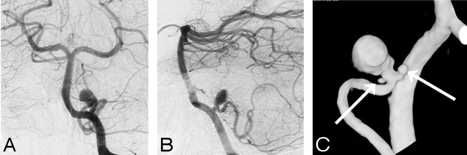
  • Fig 2.
    • Download figure
    • Open in new tab
    • Download powerpoint
    Fig 2.

    Two angiographically occult additional microaneurysms adjacent to a ruptured posterior inferior cerebellar artery aneurysm in a 53-year-old woman. A and B, DSA in 2 projections demonstrates a posterior inferior cerebellar artery aneurysm. C, 3DRA detects 2 additional microaneurysms (arrows).
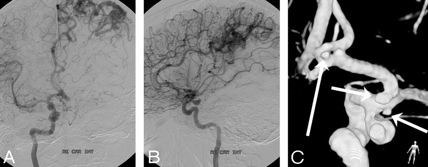
  • Fig 3.
    • Download figure
    • Open in new tab
    • Download powerpoint
    Fig 3.

    A 44-year-old woman with subarachnoid hemorrhage. A and B, DSA in 2 projections reveals a posterior communicating artery aneurysm and an asymptomatic left parietal arteriovenous malformation. C, 3DRA shows, besides the posterior communicating artery aneurysm, 3 additional small aneurysms on the supraclinoidal carotid and proximal A1 arteries (short arrows) and on a fenestrated anterior communicating artery (long arrow).
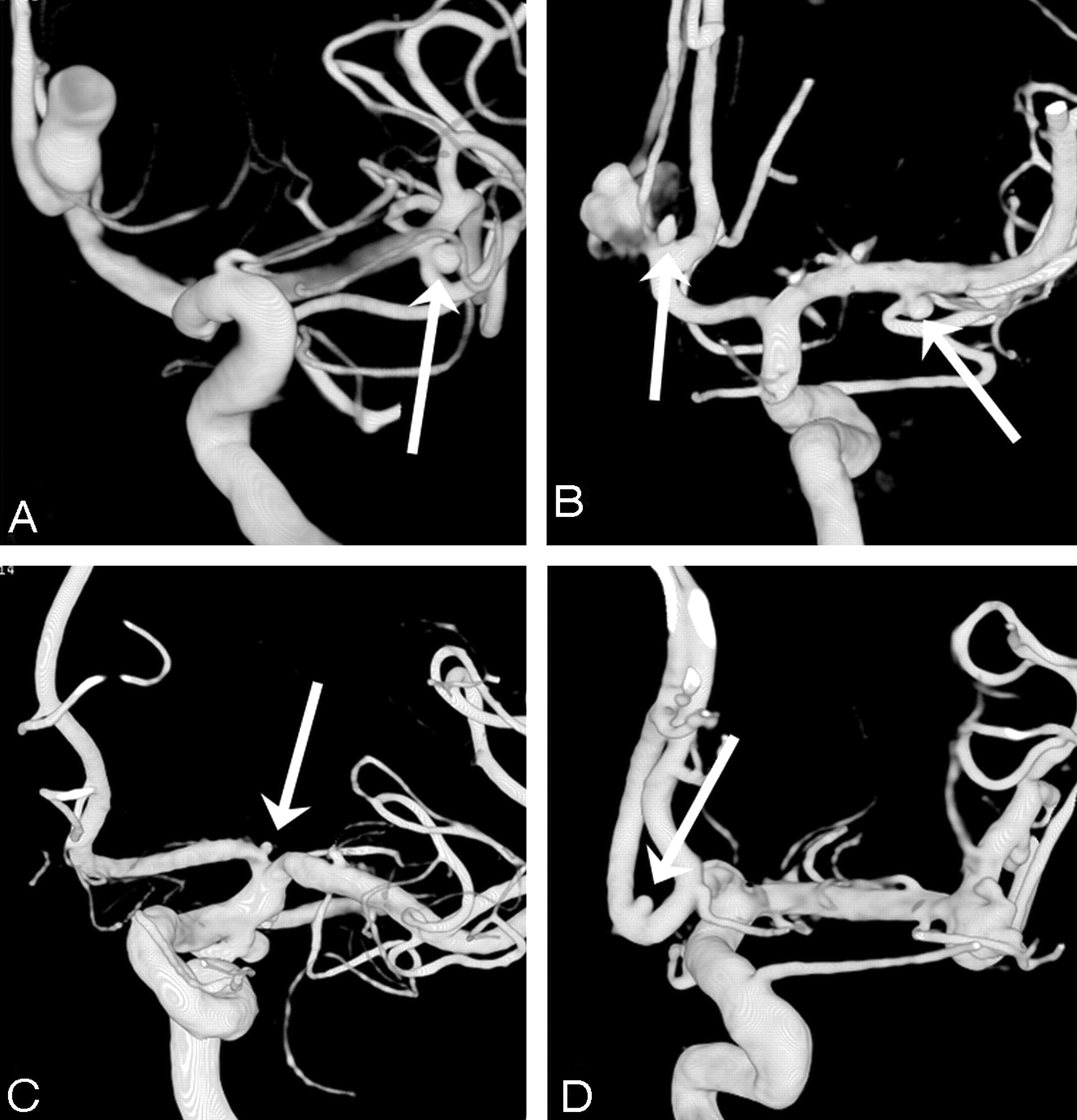
  • Fig 4.
    • Download figure
    • Open in new tab
    • Download powerpoint
    Fig 4.

    Four examples of small additional aneurysms missed on DSA. A, A very small (0.5 mm) A1 aneurysm (arrow) in a patient with a ruptured middle cerebral artery aneurysm. B, Two supraclinoidal aneurysms (arrows) in a patient with a ruptured posterior communicating artery aneurysm. C, Two middle cerebral artery aneurysms (arrows) in a patient with a ruptured pericallosal artery aneurysm (not shown). D, An intracavernous carotid artery aneurysm (arrow) in a patient with a large ophthalmic artery aneurysm, symptomatic by mass effect.

  • Fig 5.
    • Download figure
    • Open in new tab
    • Download powerpoint
    Fig 5.

    Four more examples of missed additional aneurysms on DSA. A, A small middle cerebral artery aneurysm (arrow) in a patient with a ruptured anterior communicating artery aneurysm. B, Two small additional aneurysms on the anterior communicating and middle cerebral arteries (arrows) in a patient with a ruptured anterior communicating artery aneurysm. C, A very small (0.5 mm) A1 aneurysm (arrow) in a patient with a ruptured posterior communicating artery aneurysm. D, A small additional anterior communicating artery aneurysm (arrow) in a patient with a ruptured middle cerebral artery aneurysm.

PreviousNext
Back to top

In this issue

American Journal of Neuroradiology: 29 (5)
American Journal of Neuroradiology
Vol. 29, Issue 5
May 2008
  • Table of Contents
  • Index by author
Advertisement
Print
Download PDF
Email Article

Thank you for your interest in spreading the word on American Journal of Neuroradiology.

NOTE: We only request your email address so that the person you are recommending the page to knows that you wanted them to see it, and that it is not junk mail. We do not capture any email address.

Enter multiple addresses on separate lines or separate them with commas.
3D Rotational Angiography: The New Gold Standard in the Detection of Additional Intracranial Aneurysms
(Your Name) has sent you a message from American Journal of Neuroradiology
(Your Name) thought you would like to see the American Journal of Neuroradiology web site.
CAPTCHA
This question is for testing whether or not you are a human visitor and to prevent automated spam submissions.
Cite this article
W.J. van Rooij, M.E. Sprengers, A.N. de Gast, J.P.P. Peluso, M. Sluzewski
3D Rotational Angiography: The New Gold Standard in the Detection of Additional Intracranial Aneurysms
American Journal of Neuroradiology May 2008, 29 (5) 976-979; DOI: 10.3174/ajnr.A0964

Citation Manager Formats

  • BibTeX
  • Bookends
  • EasyBib
  • EndNote (tagged)
  • EndNote 8 (xml)
  • Medlars
  • Mendeley
  • Papers
  • RefWorks Tagged
  • Ref Manager
  • RIS
  • Zotero
0 Responses
Respond to this article
Share
Bookmark this article
3D Rotational Angiography: The New Gold Standard in the Detection of Additional Intracranial Aneurysms
W.J. van Rooij, M.E. Sprengers, A.N. de Gast, J.P.P. Peluso, M. Sluzewski
American Journal of Neuroradiology May 2008, 29 (5) 976-979; DOI: 10.3174/ajnr.A0964
del.icio.us logo Twitter logo Facebook logo Mendeley logo
  • Tweet Widget
  • Facebook Like
  • Google Plus One
Purchase

Jump to section

  • Article
    • Abstract
    • Patients and Methods
    • Results
    • Discussion
    • Conclusion
    • References
  • Figures & Data
  • Info & Metrics
  • Responses
  • References
  • PDF

Related Articles

  • No related articles found.
  • PubMed
  • Google Scholar

Cited By...

  • Acceleration of Brain TOF-MRA with Compressed Sensitivity Encoding: A Multicenter Clinical Study
  • Plaque morphology in acute symptomatic intracranial atherosclerotic disease
  • Current Hospital Demographics of Subarachnoid Hemorrhage Based on CT Angiography and 3D Rotational Angiography in a Neurosurgical Center
  • Surveillance of Unruptured Intracranial Saccular Aneurysms Using Noncontrast 3D-Black-Blood MRI: Comparison of 3D-TOF and Contrast-Enhanced MRA with 3D-DSA
  • Morphologic Change of Flow-Related Aneurysms in Brain Arteriovenous Malformations after Stereotactic Radiosurgery
  • Morphologic Change of Flow-Related Aneurysms in Brain Arteriovenous Malformations after Stereotactic Radiosurgery
  • Retrograde 3D rotational venography (3DRV) for venous sinus stent placement in idiopathic intracranial hypertension
  • Yield of Repeat 3D Angiography in Patients with Aneurysmal-Type Subarachnoid Hemorrhage
  • Two-color 3D-3D fusion of selective rotational cerebral angiograms: a novel approach to imaging in cerebrovascular neurosurgery
  • Hemodynamic Differences in Intracranial Aneurysms before and after Rupture
  • Guidelines for the Management of Patients With Unruptured Intracranial Aneurysms: A Guideline for Healthcare Professionals From the American Heart Association/American Stroke Association
  • Diagnostic quality and accuracy of low dose 3D-DSA protocols in the evaluation of intracranial aneurysms
  • Use of Follow-Up Imaging in Isolated Perimesencephalic Subarachnoid Hemorrhage: A Meta-Analysis
  • Cost-Effectiveness of Angiographic Imaging in Isolated Perimesencephalic Subarachnoid Hemorrhage
  • Reducing radiation dose while maintaining diagnostic image quality of cerebral three-dimensional digital subtraction angiography: an in vivo study in swine
  • Diagnosing Intracranial Aneurysms With MR Angiography: Systematic Review and Meta-Analysis
  • Flat Panel Catheter Angiotomography of the Spinal Venous System: An Enhanced Venous Phase for Spinal Digital Subtraction Angiography
  • Contrast-free MRA at 3.0 T for the detection of intracranial aneurysms
  • Patient-Specific Computational Hemodynamics of Intracranial Aneurysms from 3D Rotational Angiography and CT Angiography: An In Vivo Reproducibility Study
  • Utility of CT Angiography in the Identification and Characterization of Supraclinoid Internal Carotid Artery Blister Aneurysms
  • Opinion: Imaging Follow-Up after Coiling of Intracranial Aneurysms
  • Large-Cohort Comparison Between Three-Dimensional Time-of-Flight Magnetic Resonance and Rotational Digital Subtraction Angiographies in Intracranial Aneurysm Detection
  • Comparison of 2D Digital Subtraction Angiography and 3D Rotational Angiography in the Evaluation of Dome-to-Neck Ratio
  • MR Angiography Follow-Up 5 Years after Coiling: Frequency of New Aneurysms and Enlargement of Untreated Aneurysms
  • Coil Embolization of Very Small (2 mm or Smaller) Berry Aneurysms: Feasibility and Technical Issues
  • Crossref (222)
  • Google Scholar

This article has been cited by the following articles in journals that are participating in Crossref Cited-by Linking.

  • Guidelines for the Management of Patients With Unruptured Intracranial Aneurysms
    B. Gregory Thompson, Robert D. Brown, Sepideh Amin-Hanjani, Joseph P. Broderick, Kevin M. Cockroft, E. Sander Connolly, Gary R. Duckwiler, Catherine C. Harris, Virginia J. Howard, S. Claiborne (Clay) Johnston, Philip M. Meyers, Andrew Molyneux, Christopher S. Ogilvy, Andrew J. Ringer, James Torner
    Stroke 2015 46 8
  • Enhancement of Vascular Structures in3D and 2D Angiographic Images
    Tim Jerman, Franjo Pernus, Bostjan Likar, Ziga Spiclin
    IEEE Transactions on Medical Imaging 2016 35 9
  • Diagnosing Intracranial Aneurysms With MR Angiography
    Anna M.H. Sailer, Bart A.J.M. Wagemans, Patricia J. Nelemans, Rick de Graaf, Willem H. van Zwam
    Stroke 2014 45 1
  • Diagnosis and Treatment of Unruptured Intracranial Aneurysms and Aneurysmal Subarachnoid Hemorrhage
    Rabih G. Tawk, Tasneem F. Hasan, Caitlin E. D’Souza, Jeffrey B. Peel, William D. Freeman
    Mayo Clinic Proceedings 2021 96 7
  • Diagnosing cerebral aneurysms by computed tomographic angiography: Meta‐analysis
    Jan Menke, Jörg Larsen, Kai Kallenberg
    Annals of Neurology 2011 69 4
  • Current Imaging Assessment and Treatment of Intracranial Aneurysms
    Lotfi Hacein-Bey, James M. Provenzale
    American Journal of Roentgenology 2011 196 1
  • Intracranial aneurysms: Review of current science and management
    Gabor Toth, Russell Cerejo
    Vascular Medicine 2018 23 3
  • Automated segmentation of cerebral vasculature with aneurysms in 3DRA and TOF‐MRA using geodesic active regions: An evaluation study
    Hrvoje Bogunović, José María Pozo, María Cruz Villa‐Uriol, Charles B. L. M. Majoie, Rene van den Berg, Hugo A. F. Gratama van Andel, Juan M. Macho, Jordi Blasco, Luis San Román, Alejandro F. Frangi
    Medical Physics 2011 38 1
  • Benefits of 3D Rotational DSA Compared with 2D DSA in the Evaluation of Intracranial Aneurysm
    Siong Chuong Wong, Ouzreiah Nawawi, Norlisah Ramli, Khairul Azmi Abd Kadir
    Academic Radiology 2012 19 6
  • Coil Embolization of Very Small (2 mm or Smaller) Berry Aneurysms: Feasibility and Technical Issues
    V. Gupta, M. Chugh, A.N. Jha, B.S. Walia, S. Vaishya
    American Journal of Neuroradiology 2009 30 2

More in this TOC Section

  • SAVE vs. Solumbra Techniques for Thrombectomy
  • Contrast-Induced Encephalopathy after NeuroIR
  • CT Perfusion&Reperfusion in Acute Ischemic Stroke
Show more Interventional

Similar Articles

Advertisement

Indexed Content

  • Current Issue
  • Accepted Manuscripts
  • Article Preview
  • Past Issues
  • Editorials
  • Editor's Choice
  • Fellows' Journal Club
  • Letters to the Editor
  • Video Articles

Cases

  • Case Collection
  • Archive - Case of the Week
  • Archive - Case of the Month
  • Archive - Classic Case

Special Collections

  • AJNR Awards
  • ASNR Foundation Special Collection
  • Most Impactful AJNR Articles
  • Photon-Counting CT
  • Spinal CSF Leak Articles (Jan 2020-June 2024)

More from AJNR

  • Trainee Corner
  • Imaging Protocols
  • MRI Safety Corner

Multimedia

  • AJNR Podcasts
  • AJNR Scantastics

Resources

  • Turnaround Time
  • Submit a Manuscript
  • Submit a Video Article
  • Submit an eLetter to the Editor/Response
  • Manuscript Submission Guidelines
  • Statistical Tips
  • Fast Publishing of Accepted Manuscripts
  • Graphical Abstract Preparation
  • Imaging Protocol Submission
  • Evidence-Based Medicine Level Guide
  • Publishing Checklists
  • Author Policies
  • Become a Reviewer/Academy of Reviewers
  • News and Updates

About Us

  • About AJNR
  • Editorial Board
  • Editorial Board Alumni
  • Alerts
  • Permissions
  • Not an AJNR Subscriber? Join Now
  • Advertise with Us
  • Librarian Resources
  • Feedback
  • Terms and Conditions
  • AJNR Editorial Board Alumni

American Society of Neuroradiology

  • Not an ASNR Member? Join Now

© 2025 by the American Society of Neuroradiology All rights, including for text and data mining, AI training, and similar technologies, are reserved.
Print ISSN: 0195-6108 Online ISSN: 1936-959X

Powered by HighWire