
Article Figures & Data
Figures
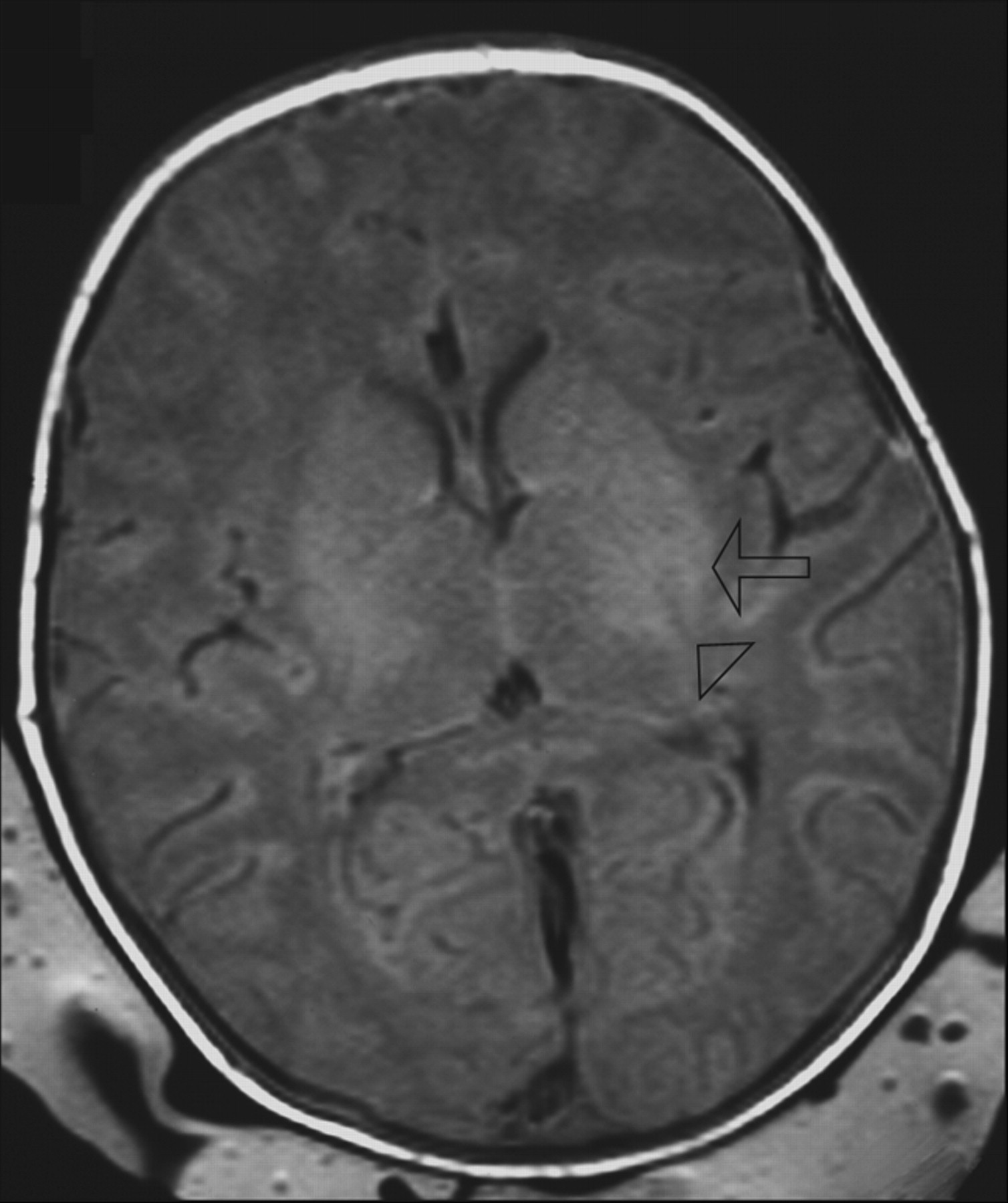
- Fig 1.
T1-weighted image (TR/TE, 550/14; signals acquired, 2; matrix, 205/256; section thickness, 5 mm; section gap, 0.5 mm; FOV, 16 cm) of a neonate from the HIE group (HIE grade 2) imaged at 3 days of age. The image shows higher SI in the posterolateral putamen (arrow) than in the posterior limb of the internal capsule (arrowhead) and abnormal SI in the lateral thalami. The neonate was born at a gestational age of 40+4 weeks, the mother underwent an emergency cesarean delivery for fetal distress, and the neonate was resuscitated. The Apgar score was 0–4–5 (at 1, 5, and 10 minutes). At 5 years of age, this child has an abnormal development (outcome group 3).
- Fig 2.
T1-weighted image (TR/TE, 550/14; signals acquired, 2; matrix, 205/256; section thickness, 5 mm; section gap, 0.5 mm; FOV, 16 cm) of a neonate from the control group imaged at 2 days of age. The image shows higher SI in the posterior limb of the internal capsule (arrowhead) than in the posterolateral putamen (arrow). The child was a term-born neonate with a simple lumbar meningocele. At the age of 5 years, this child has a normal development (outcome group 1).


Tables
- Table 1:
Sensitivity, specificity, and predictive value of the neonatal clinical classification for outcome
Outcome Sensitivity Specificity PPV NPV Abnormal 85% 97% 96% 88% Adverse 100% 76% 52% 100% Note:—PPV indicates positive predictive value; NPV, negative predictive value.
- Table 2:
Sensitivity, specificity, and predictive value of the brain structure T1-weighted comparison of the SI of the posterolateral putamen equal to or greater than that of the posterior limb of the internal capsule for outcome
Outcome Sensitivity Specificity PPV NPV Abnormal 54% 94% 88% 71% Adverse 92% 89% 69% 98% Note:—PPV indicates positive predictive value; NPV, negative predictive value. SI, signal intensity.
- Table 3:
Sensitivity, specificity, and predictive value of the brain structure T1-weighted comparison of the SI of the posterolateral putamen less than that of the posterior limb of the internal capsule for outcome
Outcome Sensitivity Specificity PPV NPV Abnormal 46% 6% 29% 13% Adverse 8% 11% 2% 31% Note:—PPV indicates positive predictive value; NPV, negative predictive value; SI, signal intensity.
- Table 4:
Predictive value of the neonatal clinical classification and the brain structure T1-weighted SI comparisons for adverse outcome in HIE grade 2 infants only
PPV NPV Neonatal classification 45% 0% SI PP ≥ PLIC 67% 88% SI PP < PLIC 88% 67% Note:—PPV indicates positive predictive value; NPV, negative predictive value; PP, posterolateral putamen; PLIC, posterior limb of the internal capsule; SI, signal intensity.


