
Article Figures & Data
Figures
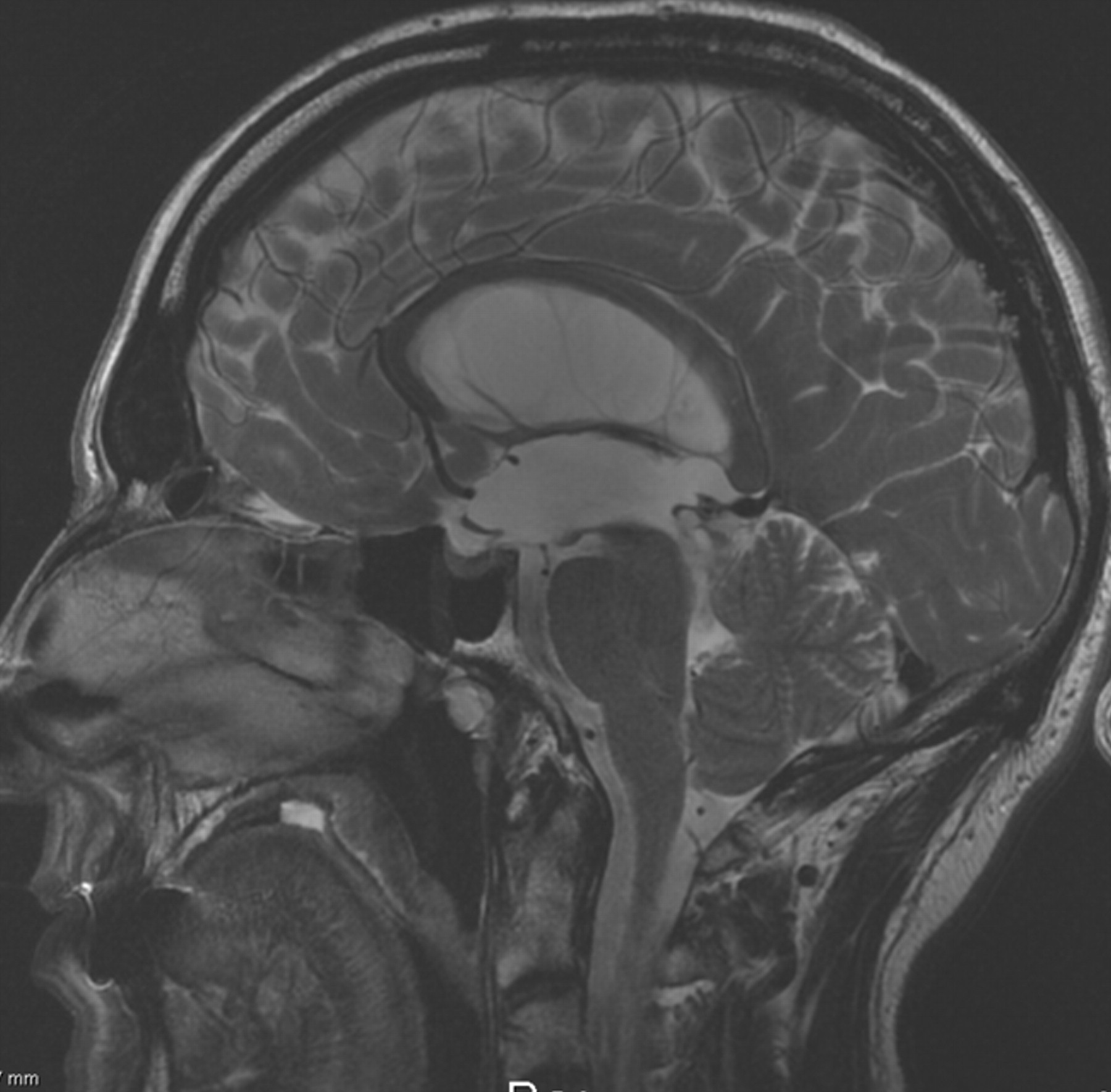
- Fig 1.
Sagittal scout view sequences are used as localizers to select the anatomic levels for flow quantification. The acquisition planes are selected perpendicular to the presumed direction of the flow. Sections through the C2–C3 subarachnoid space level (a), fourth ventricle (b), and Sylvian aqueduct (c) are used for CSF. By varying the velocity encoding, the same cervical section level (a) is used to measure vascular flows in the left and right internal carotid arteries, vertebral arteries, and internal jugular veins.
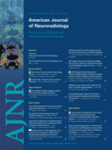
- Fig 2.
Sagittal T2 conventional MR image in a 50-year-old patient admitted for recent gait and urinary dysfunction, with a medical history of chronic headaches. Note the dilated lateral and 3rd Vs associated with a comparatively small 4th V. There is a slight downward bulging of the floor of the 3rd V, but no direct signs of obstruction at the aqueductal level. PC-MR imaging (not shown) showed a total absence of CSF flow at the aqueductal level and helped the neurosurgeon with the diagnosis of aqueductal stenosis.

Tables
Symptoms Acute Onset* Subacute Onset† Chronic Onset‡ Headache 5 4 1 Gait disturbance 0 1 6 Memory disturbance 0 1 6 Incontinence 0 1 5 Seizure 0 2 0 Note:—AS indicates aqueductal stenosis.
* Duration between symptoms and radiologic examination <1 month.
† Duration between 1 and 6 months.
‡ Chronic onset: duration >6 months. Numbers refer to patients in each category.
Present Absent Unknown Triventricular dilation 17 0 0 Small/normal 4th V 17 0 0 Direct CSF pathway obstruction (sagittal T2) 10 4 3 Downward bulging of the floor of the 3rd V 10 7 0 Space-occupying lesion 3 14 0 Note:—4th V indicates fourth ventricle; 3rd V, third ventricle.
* Numbers refer to the patients in each category.
- Table 3:
Amplitude and temporal parameters of vascular blood flow and CSF (cervical and ventricular) flows in patients with AS and controls*
AS Control P No. of patients 17 20 NS Age (yr) 39 44 NS Vascular flows Arterial mean flow (mL/min) 589 ± 115 607 ± 158 NS Venous mean flow (mL/min) 379 ± 157 417 ± 158 NS Arteriovenous delay (% of CC) 16 ± 12 19 ± 12 NS Arteriovenous stroke volume (mL) 0.8 ± 0.2 0.8 ± 0.3 NS Cervical flows C2–C3 peak flush flow (mL/min) 157 ± 58 160 ± 54 NS C2–C3 peak flush latency (% of CC) 6 ± 3 5 ± 4 NS C2–C3 stroke volume (μL/CC) 499 ± 251 508 ± 166 NS 4th V flows 4th V peak flush latency (% CC) 4 ± 7 13 ± 8 .05 4th V stroke volume (μL/CC) 24 ± 18 24 ± 13 NS Aqueductal level Stroke volume (μL/CC) ∅ 44 ± 25 Note:—NS indicates no significance;∅, absence of flow.
* Results are represented by mean ± SD values.


