
Abstract
SUMMARY: Radiation therapy, a mainstay in the treatment of many brain tumors, results in a variety of well-documented acute and chronic complications. Isolated cortical damage following irradiation represents an extremely rare delayed therapeutic complication, described only twice in the medical literature. We report this rare delayed complication in a patient following treatment of a right frontal anaplastic oligodendroglioma.
Radiation therapy, a mainstay in the treatment of many brain tumors, results in a variety of well-documented acute and chronic complications, ranging from edema1 to radionecrosis2 to vasculopathy3 to vascular malformations4,5 to secondary neoplasms.6 This list is augmented by numerous reports of more uncommon complications. Chronic complications may occur months or years following initial treatment and can be difficult to differentiate from progressive or recurrent tumor.
Isolated cortical damage in the form of neuronal gigantism and cerebral cortical thickening following irradiation represents an extremely rare delayed therapeutic complication, described only twice in the medical literature. We describe this rare delayed complication in a patient following treatment of a right frontal anaplastic oligodendroglioma.
Case Report
A 54-year-old woman with a history of a recurrent extensively treated right frontal anaplastic oligodendroglioma followed in our neuro-oncology clinic was found to have abnormalities on MR imaging, which were interpreted by the radiologist as tumor recurrence. Thus, neurosurgery was consulted for further evaluation.
The patient initially presented 9 years previously with neuroimaging studies showing a solid and cystic heterogeneously enhancing right frontal mass, diagnosed as an anaplastic oligodendroglioma, following gross total resection. Over the course of the next 7 years, she received multiple treatments with fractionated radiation therapy and multiple chemotherapeutic agents for the initial disease and subsequent recurrences along the resection margins. Chemotherapeutic agents included procarbazine, lomustine, vincristine, temozolomide, carboplatin, etoposide, paclitaxel, and imatinib.
MR imaging performed at the completion of her final chemotherapy treatment (Fig 1) demonstrated near-complete resolution of the enhancement along the resection margins. However, at this time, MR imaging showed faint gyriform contrast enhancement over the right frontal lobe surrounding the macrocystic encephalomalacic changes of the resection cavity.

Comparison study. A and B, Axial postcontrast T1-weighted (A) and T2-weighted (B) images obtained at the completion of final chemotherapy treatment demonstrate no enhancement along the resection margins but faint gyriform contrast enhancement (white arrows) and underlying gliosis (black arrows) in the adjacent frontal lobe.
MR imaging performed at the time of presentation (2 years later) (Fig 2) demonstrated macrocystic encephalomalacia of the resection cavity. Postcontrast T1-weighted imaging showed marked enlargement and nodular enhancement of the right frontal lobe cortex and juxtacortical white matter surrounding the resection cavity, with T2-weighted imaging showing extensive edema-like signal intensity throughout the right frontal white matter. The degree of cortical thickening and nodular enhancement had progressed dramatically from the previous MR imaging study, though the encephalomalacic changes were relatively the same. There was no significant enhancement along the resection margins and no masslike area of enhancement.
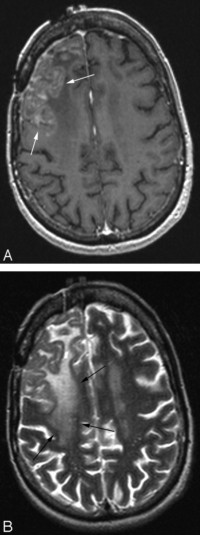
At presentation. A and B, Axial postcontrast T1-weighted (A) and T2-weighted (B) images show marked enlargement and nodular enhancement of the right frontal lobe cortex and juxtacortical white matter (white arrows) adjacent to the resection cavity (not shown), with extensive white matter T2 hyperintensity throughout the right frontal white matter (black arrows). The degree of cortical thickening and nodular enhancement have progressed dramatically from that of the previous study.
Due to the concern for disease recurrence, the patient eventually underwent surgical debulking of portions of the radiographically abnormal tissue.
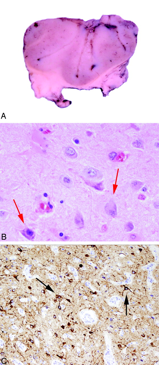
Pathologic examination of the original tumor showed the classic features of an anaplastic oligodendroglioma (not shown). Gross examination of the most recent resection specimen showed thickening of the cortical ribbon with blurring of the gray-white matter junction (Fig 3). Microscopic examination showed diffuse thickening and gliosis of the cortical ribbon and adjacent white matter, with enlarged irregularly arranged neurons and focal areas of perivascular demyelination (Fig 3). Numerous arterioles had thickened and hyalinized vessel walls. There was no evidence of oligodendroglioma.

Gross and microscopic photographs of the recent resection specimen. A, Gross specimen shows subtle cortical thickening and focal blurring of the gray-white matter interface. B, Enlarged cortical neurons show haphazard polarity (red arrows) (hematoxylin-eosin, original magnification ×400). C, Abnormal giant neurons show loss of normal cortical lamination (black arrows) (neurofilament protein [SMI-33] immunostain, original magnification ×200).
The patient's cognition improved following the resection.
Discussion
Isolated cortical damage following irradiation represents an extremely rare delayed therapy complication. In the neurology and pathology literature, there are 2 prior reports describing the pathologic findings in 2 separate cases of presumed postradiation cortical thickening and neuronal gigantism.7,8 No such reports currently exist in the radiology literature.
Lampert and Davis (1964)7 first described the finding of cortical neuronal hypertrophy following high-dose radiation for a glioma. Microscopic examination of brain tissue showed no evidence of the glial tumor. Instead, there were severe white matter gliosis, foci of demyelination, cyst formation, hypertrophic neurons, perivascular macrophage infiltrates, and vascular hyalinization.
Caccamo et al (1989)8 described a similar form of cortical dysplasia, which they dubbed “focal neuronal gigantism,” found in a 32-year-old woman 6 years after resection and irradiation of a pituitary adenoma.
At the time of image interpretation in our patient, the MR imaging characteristics of this lesion were perplexing. Due to the patient's history and lack of any reasonable differential diagnostic considerations, the concern was raised for tumor recurrence. However, the imaging appearance and disease distribution would be extremely atypical for a tumor. The thickening and nodular enhancement were isolated to the cerebral cortex, showing a geographic distribution, with no significant enhancement of the resection margins and no mass. In addition, the signal-intensity changes in the underlying white matter appeared stable from prior MR imaging studies, mitigating against a progressive infiltrating neoplasm.
In summary, the imaging characteristics and pathology in this report support an atypical presentation of radiation changes as the etiology of our findings.
References
- Received March 10, 2009.
- Accepted after revision April 13, 2009.
- Copyright © American Society of Neuroradiology
In this issue


Jump to section
Related Articles
Cited By...
- No citing articles found.