Advertisement

Main menu

  • Home
  • Content
    • Current Issue
    • Accepted Manuscripts
    • Article Preview
    • Past Issue Archive
    • Video Articles
    • AJNR Case Collection
    • Case of the Week Archive
    • Case of the Month Archive
    • Classic Case Archive
  • Special Collections
    • AJNR Awards
    • ASNR Foundation Special Collection
    • Most Impactful AJNR Articles
    • Photon-Counting CT
    • Spinal CSF Leak Articles (Jan 2020-June 2024)
  • Multimedia
    • AJNR Podcasts
    • AJNR SCANtastic
    • Trainee Corner
    • MRI Safety Corner
    • Imaging Protocols
  • For Authors
    • Submit a Manuscript
    • Submit a Video Article
    • Submit an eLetter to the Editor/Response
    • Manuscript Submission Guidelines
    • Statistical Tips
    • Fast Publishing of Accepted Manuscripts
    • Graphical Abstract Preparation
    • Imaging Protocol Submission
    • Author Policies
  • About Us
    • About AJNR
    • Editorial Board
    • Editorial Board Alumni
  • More
    • Become a Reviewer/Academy of Reviewers
    • Subscribers
    • Permissions
    • Alerts
    • Feedback
    • Advertisers
    • ASNR Home

User menu

  • Alerts
  • Log in

Search

  • Advanced search
American Journal of Neuroradiology

American Journal of Neuroradiology

ASHNR American Society of Functional Neuroradiology ASHNR American Society of Pediatric Neuroradiology ASSR
  • Alerts
  • Log in

Advanced Search

  • Home
  • Content
    • Current Issue
    • Accepted Manuscripts
    • Article Preview
    • Past Issue Archive
    • Video Articles
    • AJNR Case Collection
    • Case of the Week Archive
    • Case of the Month Archive
    • Classic Case Archive
  • Special Collections
    • AJNR Awards
    • ASNR Foundation Special Collection
    • Most Impactful AJNR Articles
    • Photon-Counting CT
    • Spinal CSF Leak Articles (Jan 2020-June 2024)
  • Multimedia
    • AJNR Podcasts
    • AJNR SCANtastic
    • Trainee Corner
    • MRI Safety Corner
    • Imaging Protocols
  • For Authors
    • Submit a Manuscript
    • Submit a Video Article
    • Submit an eLetter to the Editor/Response
    • Manuscript Submission Guidelines
    • Statistical Tips
    • Fast Publishing of Accepted Manuscripts
    • Graphical Abstract Preparation
    • Imaging Protocol Submission
    • Author Policies
  • About Us
    • About AJNR
    • Editorial Board
    • Editorial Board Alumni
  • More
    • Become a Reviewer/Academy of Reviewers
    • Subscribers
    • Permissions
    • Alerts
    • Feedback
    • Advertisers
    • ASNR Home
  • Follow AJNR on Twitter
  • Visit AJNR on Facebook
  • Follow AJNR on Instagram
  • Join AJNR on LinkedIn
  • RSS Feeds

AJNR is seeking candidates for the AJNR Podcast Editor. Read the position description.

Research ArticleHEAD & NECK

Extraosseous Chordoma of the Nasopharynx

R.P. Nguyen, K.L. Salzman, H.E. Stambuk, A.T. Ahuja and H.R. Harnsberger
American Journal of Neuroradiology April 2009, 30 (4) 803-807; DOI: https://doi.org/10.3174/ajnr.A1446
R.P. Nguyen
  • Find this author on Google Scholar
  • Find this author on PubMed
  • Search for this author on this site
K.L. Salzman
  • Find this author on Google Scholar
  • Find this author on PubMed
  • Search for this author on this site
H.E. Stambuk
  • Find this author on Google Scholar
  • Find this author on PubMed
  • Search for this author on this site
A.T. Ahuja
  • Find this author on Google Scholar
  • Find this author on PubMed
  • Search for this author on this site
H.R. Harnsberger
  • Find this author on Google Scholar
  • Find this author on PubMed
  • Search for this author on this site
  • Article
  • Figures & Data
  • Info & Metrics
  • Responses
  • References
  • PDF
Loading

Article Figures & Data

Figures

  • Fig 1.
    • Download figure
    • Open in new tab
    • Download powerpoint
    Fig 1.

    A, A 65-year-old woman presented with nasal obstruction. Findings show atypical CT appearance of a pathology-proved extraosseous nasopharyngeal chordoma. Sagittal noncontrast CT scan shows a lobulated mass centered in the nasopharynx with superior extension into the sphenoid sinus (curved arrow) and focal erosion of the anterior clivus (black arrow). B, Axial bone CT scan shows a lobulated soft-tissue mass centered in the nasopharynx, with scalloping of the anterior margin of the clivus. Note the lytic appearance with a sclerotic margin (black arrow). C, Sagittal T1-weighted MR image shows a heterogeneous nasopharyngeal mass with a focal area of T1 hyperintensity (arrow) thought to represent blood products or proteinaceous debris. D, Sagittal T1-weighted MR image with contrast shows heterogeneous enhancement of the mass, which is an enhancement pattern seen in both extraosseous and typical skull base chordomas.

  • Fig 2.
    • Download figure
    • Open in new tab
    • Download powerpoint
    Fig 2.

    A 56-year-old man presented with a 2-year history of nasal obstruction. Axial noncontrast CT scan shows dystrophic calcification and a midline sinus tract (arrow), which can help with the preoperative diagnosis of extraosseous nasopharyngeal chordoma. This sinus tract is thought to represent tumor extending into the medial basal canal.

  • Fig 3.
    • Download figure
    • Open in new tab
    • Download powerpoint
    Fig 3.

    A 53-year-old man presented with dryness of mouth and difficulty breathing. Axial T2-weighted MR image with fat saturation shows a locally invasive heterogeneously hyperintense mass with internal septations (black arrow) centered in the nasopharynx, with invasion of the right masticator space, nasal cavity, and maxillary sinus. Note involvement of the anterior clivus (curved arrow), which may help preoperatively to diagnose this lesion as an extraosseous nasopharyngeal chordoma.

  • Fig 4.
    • Download figure
    • Open in new tab
    • Download powerpoint
    Fig 4.

    A, A 32-year-old woman presented with a mass in the nasopharynx. MR imaging shows a midline sinus tract extending into the clivus of a pathologically proved extraosseous nasopharyngeal chordoma. Axial bone CT scan shows a midline tract (curved arrow) representing the extension of the extraosseous chordoma into the medial basal canal. B, Axial T2-weighted MR image shows the hyperintense midline sinus tract (curved arrow) extending posteriorly from the nasopharyngeal mass. Note fluid in the left mastoid air cells secondary to obstruction of the eustachian tube due to a nasopharyngeal chordoma.

  • Fig 5.
    • Download figure
    • Open in new tab
    • Download powerpoint
    Fig 5.

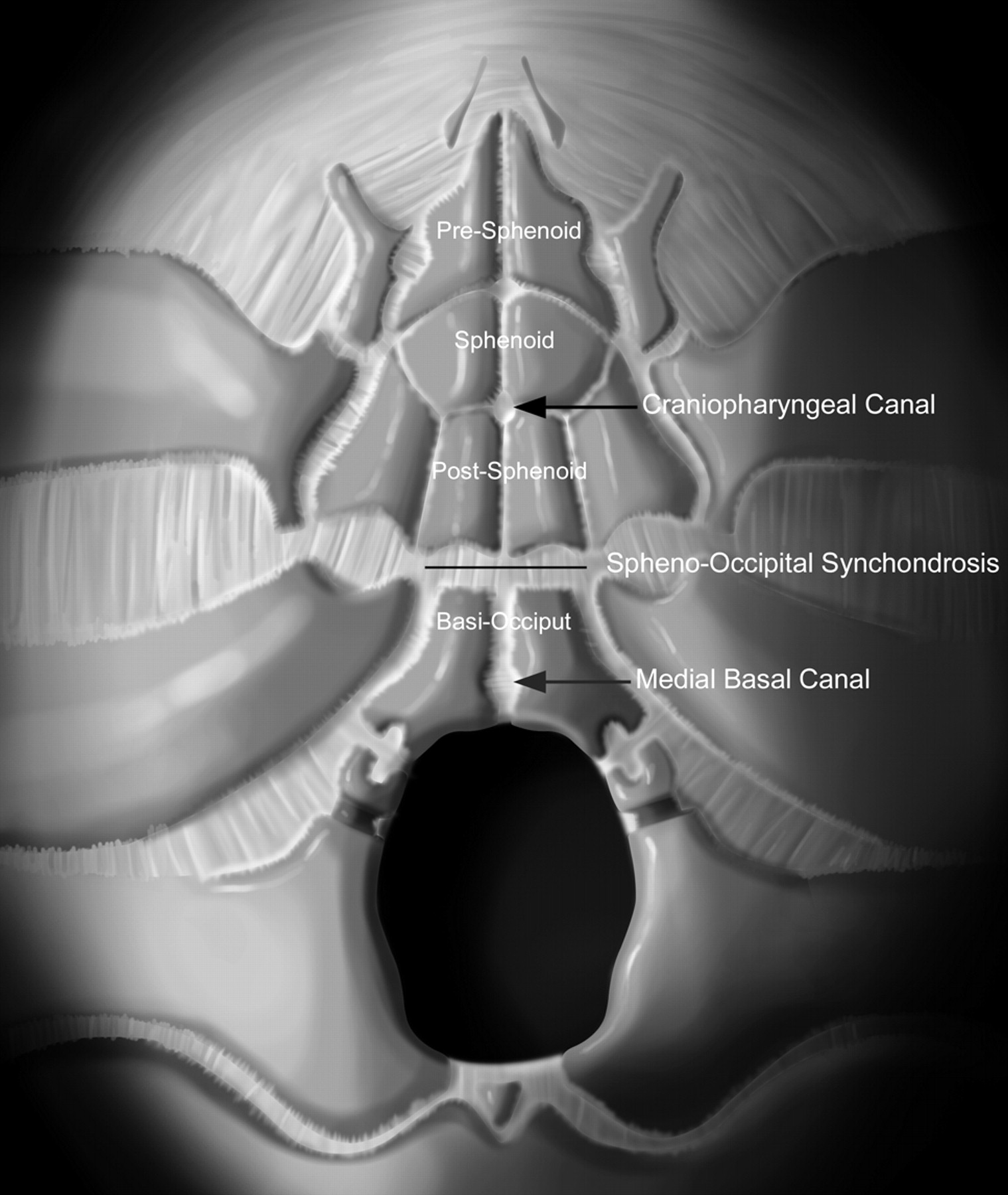
    Graphic drawing of developing skull base ossification centers. Note that the craniopharyngeal canal is anterior to the spheno-occipital synchondrosis, whereas the medial basal canal (canalis basilaris medianus) is posterior to the synchondrosis. Reproduced with permission from Amirsys.

  • Fig 6.
    • Download figure
    • Open in new tab
    • Download powerpoint
    Fig 6.

    Sagittal graphic drawing shows the expected location of the notochordal remnant (blue line). Note the relationship of the spheno-occipital synchondrosis between the craniopharyngeal canal and the medial basal canal (short arrow). Extraosseous chordomas appear to arise from the extraosseous notochordal remnant. If these chordomas arise inferiorly, they may extend into the medial basal canal. Reproduced with permission from Amirsys.

PreviousNext
Back to top

In this issue

American Journal of Neuroradiology: 30 (4)
American Journal of Neuroradiology
Vol. 30, Issue 4
April 2009
  • Table of Contents
  • Index by author
Advertisement
Print
Download PDF
Email Article

Thank you for your interest in spreading the word on American Journal of Neuroradiology.

NOTE: We only request your email address so that the person you are recommending the page to knows that you wanted them to see it, and that it is not junk mail. We do not capture any email address.

Enter multiple addresses on separate lines or separate them with commas.
Extraosseous Chordoma of the Nasopharynx
(Your Name) has sent you a message from American Journal of Neuroradiology
(Your Name) thought you would like to see the American Journal of Neuroradiology web site.
CAPTCHA
This question is for testing whether or not you are a human visitor and to prevent automated spam submissions.
Cite this article
R.P. Nguyen, K.L. Salzman, H.E. Stambuk, A.T. Ahuja, H.R. Harnsberger
Extraosseous Chordoma of the Nasopharynx
American Journal of Neuroradiology Apr 2009, 30 (4) 803-807; DOI: 10.3174/ajnr.A1446

Citation Manager Formats

  • BibTeX
  • Bookends
  • EasyBib
  • EndNote (tagged)
  • EndNote 8 (xml)
  • Medlars
  • Mendeley
  • Papers
  • RefWorks Tagged
  • Ref Manager
  • RIS
  • Zotero
0 Responses
Respond to this article
Share
Bookmark this article
Extraosseous Chordoma of the Nasopharynx
R.P. Nguyen, K.L. Salzman, H.E. Stambuk, A.T. Ahuja, H.R. Harnsberger
American Journal of Neuroradiology Apr 2009, 30 (4) 803-807; DOI: 10.3174/ajnr.A1446
del.icio.us logo Twitter logo Facebook logo Mendeley logo
  • Tweet Widget
  • Facebook Like
  • Google Plus One
Purchase

Jump to section

  • Article
    • Abstract
    • Materials and Methods
    • Results
    • Discussion
    • Conclusions
    • References
  • Figures & Data
  • Info & Metrics
  • Responses
  • References
  • PDF

Related Articles

  • No related articles found.
  • PubMed
  • Google Scholar

Cited By...

  • No citing articles found.
  • Crossref (27)
  • Google Scholar

This article has been cited by the following articles in journals that are participating in Crossref Cited-by Linking.

  • Chordoma of the Head and Neck: A Review
    Jason K. Wasserman, Denis Gravel, Bibianna Purgina
    Head and Neck Pathology 2018 12 2
  • Les chordomes
    B. George, D. Bresson, S. Bouazza, S. Froelich, E. Mandonnet, S. Hamdi, M. Orabi, M. Polivka, A. Cazorla, H. Adle-Biassette, J.-P. Guichard, M. Duet, E. Gayat, F. Vallée, C.-H. Canova, F. Riet, S. Bolle, V. Calugaru, R. Dendale, J.-J. Mazeron, L. Feuvret, E. Boissier, S. Vignot, S. Puget, C. Sainte-Rose, K. Beccaria
    Neurochirurgie 2014 60 3
  • Characterization of Microstructural Injury: A Novel Approach in Infant Abusive Head Trauma—Initial Experience
    Karen Kay Imagawa, Anita Hamilton, Rafeal Ceschin, Elenora Tokar, Phung Pham, Stefan Bluml, Jessica Wisnowski, Ashok Panigrahy
    Journal of Neurotrauma 2014 31 19
  • Imaging of the Central Skull Base
    Lindsey M. Conley, C. Douglas Phillips
    Radiologic Clinics of North America 2017 55 1
  • A Parapharyngeal Soft Tissue Chordoma Presenting with Synchronous Cervical Lymph Node Metastasis: An Unusual Presentation
    S. A. Khurram, D. Biswas, M. Fernando
    Head and Neck Pathology 2016 10 3
  • Pediatric extraosseous sacral chordoma: case report and literature review of embryonic derivation and clinical implications
    Kimberly Hamilton, Susan Rebsamen, Shahriar Salamat, Raheel Ahmed
    Journal of Neurosurgery: Pediatrics 2019 23 5
  • Soft Tissue Special Issue: Chondroid Neoplasms of the Skull
    A. N. Flaman, J. K. Wasserman, D. H. Gravel, B. M. Purgina
    Head and Neck Pathology 2020 14 1
  • Primary Chordoma of the Nasopharynx: A Rare Case Report and Review of the Literatures
    Ery Kus Dwianingsih, Yosinta Snak, Hanggoro Tri Rinonce, Brian Wasita, Ester Lianawati Antoro, Samir S. Amr
    Case Reports in Pathology 2019 2019
  • Primäres extraaxiales chondroides Chordom des anterioren Nasenseptums: Fallbericht eines seltenen Chordoms mit Literaturübersicht
    C. Holley, T. Breining, M. Scheithauer, P. Möller, T. F. E. Barth
    HNO 2021 69 3
  • Skull Base Tumor Mimics
    Jeffrey H. Huang, Mari Hagiwara
    Neuroimaging Clinics of North America 2022 32 2

More in this TOC Section

  • Chondrosarcoma vs Synovial Chondromatosis: Imaging
  • WHO Classification Update: Nasal&Skull Base Tumors
  • Peritumoral Signal in Vestibular Schwannomas
Show more HEAD & NECK

Similar Articles

Advertisement

Indexed Content

  • Current Issue
  • Accepted Manuscripts
  • Article Preview
  • Past Issues
  • Editorials
  • Editor's Choice
  • Fellows' Journal Club
  • Letters to the Editor
  • Video Articles

Cases

  • Case Collection
  • Archive - Case of the Week
  • Archive - Case of the Month
  • Archive - Classic Case

Special Collections

  • AJNR Awards
  • ASNR Foundation Special Collection
  • Most Impactful AJNR Articles
  • Photon-Counting CT
  • Spinal CSF Leak Articles (Jan 2020-June 2024)

More from AJNR

  • Trainee Corner
  • Imaging Protocols
  • MRI Safety Corner

Multimedia

  • AJNR Podcasts
  • AJNR Scantastics

Resources

  • Turnaround Time
  • Submit a Manuscript
  • Submit a Video Article
  • Submit an eLetter to the Editor/Response
  • Manuscript Submission Guidelines
  • Statistical Tips
  • Fast Publishing of Accepted Manuscripts
  • Graphical Abstract Preparation
  • Imaging Protocol Submission
  • Evidence-Based Medicine Level Guide
  • Publishing Checklists
  • Author Policies
  • Become a Reviewer/Academy of Reviewers
  • News and Updates

About Us

  • About AJNR
  • Editorial Board
  • Editorial Board Alumni
  • Alerts
  • Permissions
  • Not an AJNR Subscriber? Join Now
  • Advertise with Us
  • Librarian Resources
  • Feedback
  • Terms and Conditions
  • AJNR Editorial Board Alumni

American Society of Neuroradiology

  • Not an ASNR Member? Join Now

© 2025 by the American Society of Neuroradiology All rights, including for text and data mining, AI training, and similar technologies, are reserved.
Print ISSN: 0195-6108 Online ISSN: 1936-959X

Powered by HighWire