Advertisement

Main menu

  • Home
  • Content
    • Current Issue
    • Accepted Manuscripts
    • Article Preview
    • Past Issue Archive
    • Video Articles
    • AJNR Case Collection
    • Case of the Week Archive
    • Case of the Month Archive
    • Classic Case Archive
  • Special Collections
    • AJNR Awards
    • ASNR Foundation Special Collection
    • Most Impactful AJNR Articles
    • Photon-Counting CT
    • Spinal CSF Leak Articles (Jan 2020-June 2024)
  • Multimedia
    • AJNR Podcasts
    • AJNR SCANtastic
    • Trainee Corner
    • MRI Safety Corner
    • Imaging Protocols
  • For Authors
    • Submit a Manuscript
    • Submit a Video Article
    • Submit an eLetter to the Editor/Response
    • Manuscript Submission Guidelines
    • Statistical Tips
    • Fast Publishing of Accepted Manuscripts
    • Graphical Abstract Preparation
    • Imaging Protocol Submission
    • Author Policies
  • About Us
    • About AJNR
    • Editorial Board
    • Editorial Board Alumni
  • More
    • Become a Reviewer/Academy of Reviewers
    • Subscribers
    • Permissions
    • Alerts
    • Feedback
    • Advertisers
    • ASNR Home

User menu

  • Alerts
  • Log in

Search

  • Advanced search
American Journal of Neuroradiology

American Journal of Neuroradiology

ASHNR American Society of Functional Neuroradiology ASHNR American Society of Pediatric Neuroradiology ASSR
  • Alerts
  • Log in

Advanced Search

  • Home
  • Content
    • Current Issue
    • Accepted Manuscripts
    • Article Preview
    • Past Issue Archive
    • Video Articles
    • AJNR Case Collection
    • Case of the Week Archive
    • Case of the Month Archive
    • Classic Case Archive
  • Special Collections
    • AJNR Awards
    • ASNR Foundation Special Collection
    • Most Impactful AJNR Articles
    • Photon-Counting CT
    • Spinal CSF Leak Articles (Jan 2020-June 2024)
  • Multimedia
    • AJNR Podcasts
    • AJNR SCANtastic
    • Trainee Corner
    • MRI Safety Corner
    • Imaging Protocols
  • For Authors
    • Submit a Manuscript
    • Submit a Video Article
    • Submit an eLetter to the Editor/Response
    • Manuscript Submission Guidelines
    • Statistical Tips
    • Fast Publishing of Accepted Manuscripts
    • Graphical Abstract Preparation
    • Imaging Protocol Submission
    • Author Policies
  • About Us
    • About AJNR
    • Editorial Board
    • Editorial Board Alumni
  • More
    • Become a Reviewer/Academy of Reviewers
    • Subscribers
    • Permissions
    • Alerts
    • Feedback
    • Advertisers
    • ASNR Home
  • Follow AJNR on Twitter
  • Visit AJNR on Facebook
  • Follow AJNR on Instagram
  • Join AJNR on LinkedIn
  • RSS Feeds

AJNR is seeking candidates for the AJNR Podcast Editor. Read the position description.

Research ArticleBRAIN

Hemorrhage in Posterior Reversible Encephalopathy Syndrome: Imaging and Clinical Features

H.M. Hefzy, W.S. Bartynski, J.F. Boardman and D. Lacomis
American Journal of Neuroradiology August 2009, 30 (7) 1371-1379; DOI: https://doi.org/10.3174/ajnr.A1588
H.M. Hefzy
  • Find this author on Google Scholar
  • Find this author on PubMed
  • Search for this author on this site
W.S. Bartynski
  • Find this author on Google Scholar
  • Find this author on PubMed
  • Search for this author on this site
J.F. Boardman
  • Find this author on Google Scholar
  • Find this author on PubMed
  • Search for this author on this site
D. Lacomis
  • Find this author on Google Scholar
  • Find this author on PubMed
  • Search for this author on this site
  • Article
  • Figures & Data
  • Info & Metrics
  • Responses
  • References
  • PDF
Loading

Article Figures & Data

Figures

  • Tables
  • Fig 1.
    • Download figure
    • Open in new tab
    • Download powerpoint
    Fig 1.

    A 27-year-old man with necrotic pneumonia and lung abscess. A, MR FLAIR image demonstrates PRES vasogenic edema in the parietal and frontal lobes (arrows). B, Gradient image demonstrates minute hemorrhages in the left frontal lobe (arrows).
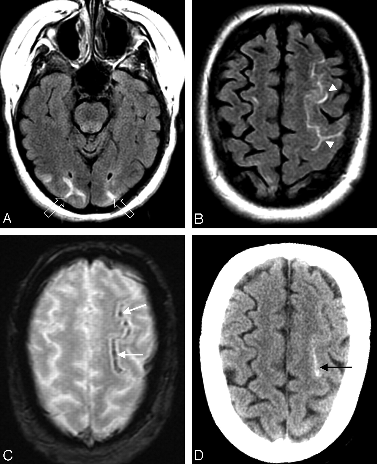
  • Fig 2.
    • Download figure
    • Open in new tab
    • Download powerpoint
    Fig 2.

    A 50-year-old woman with fever and severe hypertension. A and B, FLAIR MR image demonstrates sulcal signal abnormality and PRES vasogenic edema in the left frontal lobe (arrowheads) and edema in the occipital lobes bilaterally (open arrows). C, Gradient MR image demonstrates linear low signal intensity consistent with sulcal subarachnoid hemorrhage (arrows). D, CT image demonstrates high attenuation consistent with the MR imaging appearance, further confirming the sulcal subarachnoid hemorrhage (arrow).
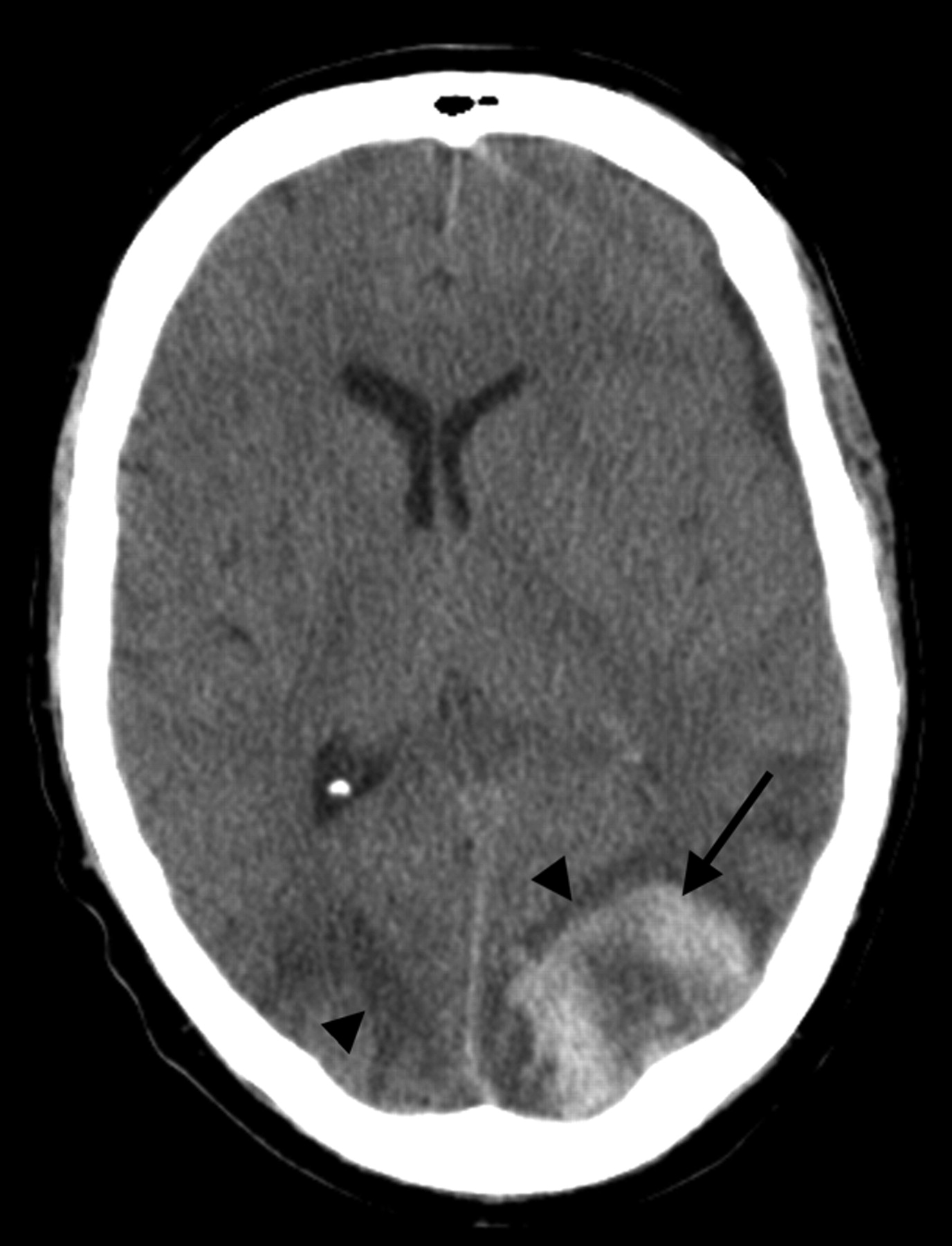
  • Fig 3.
    • Download figure
    • Open in new tab
    • Download powerpoint
    Fig 3.

    A 50-year-old man status post allo-BMT for acute myelogenous leukemia. CT scan demonstrates PRES vasogenic edema in the parietal region bilaterally (arrowheads), along with an acute hematoma in the left parietal lobe (arrow).

Tables

  • Figures
    • View popup
    Table 1:

    Hemorrhage in PRES: relationship to clinical associations and blood pressure at toxicity

    Toxicity AssociationTotal No. Pts with PRESPatients with PRES and Hemorrhage
    No. Hemorrhage (%)Blood Pressure in Pts with PRES Hemorrhage
    Normotensive (MAP ≤ 105)Mild HTN (MAP, 106–115)Severe HTN (MAP ≥ 116)
    Immunosuppression‡5011 (22.0)317
        Solid-organ transplant344 (11.8)211
        Allo-BMT157 (46.7)106
        Other*10 (0.0)–––
    Infection/sepsis/shock526 (11.5)411
    Autoimmune122 (16.7)002
    Chemotherapy101 (10.0)010
    Eclampsia†181 (5.6)001
    Unknown92 (22.2)002
    Total15123 (15.2)7313
    • Note:—Pts indicates patients; CsA, cyclosporine; HTN, hypertension; –, none; PRES, posterior reversible encephalopathy syndrome; MAP, mean arterial pressure; FK-506, tacrolimus; Allo-BMT, allogeneic bone marrow transplantation.

    • * A single patient with psoriasis on cyclosporine.

    • † Patients with eclampsia and delayed eclampsia.

    • ‡ Transplant, on cyclosporine, or tacrolimus (FK-506).

    • View popup
    Table 2:

    Hemorrhage subtypes in PRES: relationship to clinical associations and adverse clinical outcomes

    Toxicity AssociationTotal No. Pts with PRESPatients with PRES and Hemorrhage
    No. Pts with HemorrhageHemorrhage Subtypes (No. Pts)
    MinuteSulcalHematoma
    Immunosuppression‡5011543
        Solid-organ transplant3444 (1D)10
        Allo-BMT1571 (1D)3 (1D)3 (2D)
        Other†10000
    Infection/sepsis/shock526414 (1D)
    Autoimmune1222*02*
    Chemotherapy101100
    Eclampsia181011
    Unknown92011
    Total1512312711
    • Note:—D indicates that the patient died.

    • * A single patient with autoimmune disease and both minute hemorrhage and focal hematoma who sustained a permanent deficit (homonymous hemianopsia, hemiparesis, and facial droop).

    • † A single patient with psoriasis on cyclosporine.

    • ‡ Transplant, on cyclosporine, or tacrolimus (FK-506).

    • View popup
    Table 3:

    Hemorrhage subtype in PRES: relationship to blood pressure at toxicity

    Blood Pressure at ToxicityTotal No. Pts with PRESPatients with PRES and Hemorrhage
    No. (%)Hemorrhage Subtype
    MinuteSulcalFocalHematoma + SulcalHematoma + MinuteSulcal + Minute
    Normotensive (MAP ≤ 105)447 (15.9)220030
    Mildly hypertensive (MAP = 106–115)203 (15)201000
    Severe hypertension (MAP ≥ 116)8713 (14.9)234121
    Total15123 (15.2)655151
    • View popup
    Table 4:

    Coagulation state and blood pressures at toxicity in 150 patients with PRES: 23 with hemorrhage, 127 without hemorrhage

    Blood Pressure at ToxicityNo. PtsNormal Coagulation Platelets, Coagulation Parameters, and No Medications (No. Pts with PRES)Abnormal Coagulation
    Intrinsic Coagulopathy Thrombocytopenia or Abnormal PT, PTT, INR (No. Pts with PRES)Subtherapeutic, on Medications Affecting Platelet Function or Coagulation (No. Pts with PRES)Therapeutic, on Medication Affecting Platelet Function or Coagulation (No. Pts with PRES)
    W/O HemWith HemW/O HemWith HemW/O HemWith HemW/O HemWith Hem
    Normotensive (MAP ≤ 105)441531713122
    Mild HTN (MAP, 106–115)2082513001
    Severe HTN (MAP ≥ 116)8645515610041
    Total150681037816164
    • Note:—W/O indicates without; Hem, hemorrhage; PT, prothrombin time; PTT, partial thromboplastin time; INR, international normalized ratio.

PreviousNext
Back to top

In this issue

American Journal of Neuroradiology: 30 (7)
American Journal of Neuroradiology
Vol. 30, Issue 7
August 2009
  • Table of Contents
  • Index by author
Advertisement
Print
Download PDF
Email Article

Thank you for your interest in spreading the word on American Journal of Neuroradiology.

NOTE: We only request your email address so that the person you are recommending the page to knows that you wanted them to see it, and that it is not junk mail. We do not capture any email address.

Enter multiple addresses on separate lines or separate them with commas.
Hemorrhage in Posterior Reversible Encephalopathy Syndrome: Imaging and Clinical Features
(Your Name) has sent you a message from American Journal of Neuroradiology
(Your Name) thought you would like to see the American Journal of Neuroradiology web site.
CAPTCHA
This question is for testing whether or not you are a human visitor and to prevent automated spam submissions.
Cite this article
H.M. Hefzy, W.S. Bartynski, J.F. Boardman, D. Lacomis
Hemorrhage in Posterior Reversible Encephalopathy Syndrome: Imaging and Clinical Features
American Journal of Neuroradiology Aug 2009, 30 (7) 1371-1379; DOI: 10.3174/ajnr.A1588

Citation Manager Formats

  • BibTeX
  • Bookends
  • EasyBib
  • EndNote (tagged)
  • EndNote 8 (xml)
  • Medlars
  • Mendeley
  • Papers
  • RefWorks Tagged
  • Ref Manager
  • RIS
  • Zotero
0 Responses
Respond to this article
Share
Bookmark this article
Hemorrhage in Posterior Reversible Encephalopathy Syndrome: Imaging and Clinical Features
H.M. Hefzy, W.S. Bartynski, J.F. Boardman, D. Lacomis
American Journal of Neuroradiology Aug 2009, 30 (7) 1371-1379; DOI: 10.3174/ajnr.A1588
del.icio.us logo Twitter logo Facebook logo Mendeley logo
  • Tweet Widget
  • Facebook Like
  • Google Plus One
Purchase

Jump to section

  • Article
    • Abstract
    • Materials and Methods
    • Results
    • Discussion
    • Conclusions
    • References
  • Figures & Data
  • Info & Metrics
  • Responses
  • References
  • PDF

Related Articles

  • No related articles found.
  • PubMed
  • Google Scholar

Cited By...

  • Early Readmissions After Hospitalization for Posterior Reversible Encephalopathy Syndrome
  • Posterior reversible encephalopathy syndrome (PRES): diagnosis and management
  • Antithymocyte globulin-induced atypical haemorrhagic posterior reversible encephalopathy syndrome in severe aplastic anaemia
  • Posterior reversible encephalopathy syndrome associated with focal segmental glomerulosclerosis in a child
  • Serial Imaging of Virus-Associated Necrotizing Disseminated Acute Leukoencephalopathy (VANDAL) in COVID-19
  • Hemorrhagic Posterior Reversible Encephalopathy Syndrome as a Manifestation of COVID-19 Infection
  • Perimesencephalic and sulcal subarachnoid haemorrhage: an interesting presentation of posterior reversible encephalopathy syndrome
  • Controversy of posterior reversible encephalopathy syndrome: what have we learnt in the last 20 years?
  • Interrupted aortic arch complicated with takotsubo cardiomyopathy mimicking aortic dissection
  • Pazopanib-associated posterior reversible encephalopathy syndrome with intracerebral haemorrhage
  • Emergency management of autonomic dysreflexia with neurologic complications
  • Risk of Stroke After the International Classification of Diseases-Ninth Revision Discharge Code Diagnosis of Hypertensive Encephalopathy
  • Neuroimaging Features and Predictors of Outcome in Eclamptic Encephalopathy: A Prospective Observational Study
  • Resolution of neurological deficits secondary to spontaneous intracranial haemorrhage and posterior reversible encephalopathy syndrome (PRES) in a patient with hepatitis C-associated cryoglobulinaemia: a role for plasmapheresis
  • Clinicopathologic and MRI Characteristics of Presumptive Hypertensive Encephalopathy in Two Cats and Two Dogs
  • The many faces of posterior reversible encephalopathy syndrome
  • Posterior reversible encephalopathy syndrome postautologous peripheral stem cell transplantation for multiple myeloma
  • Detection of Microhemorrhage in Posterior Reversible Encephalopathy Syndrome Using Susceptibility-Weighted Imaging
  • Amyloid-{beta} Contributes to Blood-Brain Barrier Leakage in Transgenic Human Amyloid Precursor Protein Mice and in Humans With Cerebral Amyloid Angiopathy
  • The posterior reversible encephalopathy syndrome: what's certain, what's new?
  • Suspicious Neuroimaging Pattern of Thrombotic Microangiopathy
  • Isolated Acute Nontraumatic Cortical Subarachnoid Hemorrhage
  • Posterior reversible encephalopathy syndrome: long-term follow-up
  • TEACHING NEUROIMAGES: HEMORRHAGE ASSOCIATED WITH REVERSIBLE POSTERIOR LEUKOENCEPHALOPATHY SYNDROME
  • Crossref (267)
  • Google Scholar

This article has been cited by the following articles in journals that are participating in Crossref Cited-by Linking.

  • Posterior reversible encephalopathy syndrome: clinical and radiological manifestations, pathophysiology, and outstanding questions
    Jennifer E Fugate, Alejandro A Rabinstein
    The Lancet Neurology 2015 14 9
  • Cerebrovascular disease in patients with COVID-19: neuroimaging, histological and clinical description
    Francisco Hernández-Fernández, Hernán Sandoval Valencia, Rosa Angélica Barbella-Aponte, Rosa Collado-Jiménez, Óscar Ayo-Martín, Cristina Barrena, Juan David Molina-Nuevo, Jorge García-García, Elena Lozano-Setién, Cristian Alcahut-Rodriguez, Álvaro Martínez-Martín, Antonio Sánchez-López, Tomás Segura
    Brain 2020 143 10
  • Amyloid-β Contributes to Blood–Brain Barrier Leakage in Transgenic Human Amyloid Precursor Protein Mice and in Humans With Cerebral Amyloid Angiopathy
    Anika M.S. Hartz, Björn Bauer, Emma L.B. Soldner, Andrea Wolf, Sandra Boy, Roland Backhaus, Ivan Mihaljevic, Ulrich Bogdahn, Hans H. Klünemann, Gerhard Schuierer, Felix Schlachetzki
    Stroke 2012 43 2
  • The clinical and radiological spectrum of posterior reversible encephalopathy syndrome: the retrospective Berlin PRES study
    T. G. Liman, G. Bohner, P. U. Heuschmann, M. Endres, E. Siebert
    Journal of Neurology 2012 259 1
  • Hemorrhagic Posterior Reversible Encephalopathy Syndrome as a Manifestation of COVID-19 Infection
    A.M. Franceschi, O. Ahmed, L. Giliberto, M. Castillo
    American Journal of Neuroradiology 2020 41 7
  • Posterior Reversible Encephalopathy Syndrome: Clinical Features and Outcome
    Archana Hinduja
    Frontiers in Neurology 2020 11
  • Posterior Reversible Encephalopathy Syndrome
    Dimitre Staykov, Stefan Schwab
    Journal of Intensive Care Medicine 2012 27 1
  • Detection of Microhemorrhage in Posterior Reversible Encephalopathy Syndrome Using Susceptibility-Weighted Imaging
    A.M. McKinney, B. Sarikaya, C. Gustafson, C.L. Truwit
    American Journal of Neuroradiology 2012 33 5
  • Isolated Acute Nontraumatic Cortical Subarachnoid Hemorrhage
    V. Cuvinciuc, A. Viguier, L. Calviere, N. Raposo, V. Larrue, C. Cognard, F. Bonneville
    American Journal of Neuroradiology 2010 31 8
  • The many faces of posterior reversible encephalopathy syndrome
    C J Stevens, M K S Heran
    The British Journal of Radiology 2012 85 1020

More in this TOC Section

  • Usefulness of Quantitative Susceptibility Mapping for the Diagnosis of Parkinson Disease
  • White Matter Alterations in the Brains of Patients with Active, Remitted, and Cured Cushing Syndrome: A DTI Study
  • Qualitative and Quantitative Analysis of MR Imaging Findings in Patients with Middle Cerebral Artery Stroke Implanted with Mesenchymal Stem Cells
Show more BRAIN

Similar Articles

Advertisement

Indexed Content

  • Current Issue
  • Accepted Manuscripts
  • Article Preview
  • Past Issues
  • Editorials
  • Editor's Choice
  • Fellows' Journal Club
  • Letters to the Editor
  • Video Articles

Cases

  • Case Collection
  • Archive - Case of the Week
  • Archive - Case of the Month
  • Archive - Classic Case

Special Collections

  • AJNR Awards
  • ASNR Foundation Special Collection
  • Most Impactful AJNR Articles
  • Photon-Counting CT
  • Spinal CSF Leak Articles (Jan 2020-June 2024)

More from AJNR

  • Trainee Corner
  • Imaging Protocols
  • MRI Safety Corner

Multimedia

  • AJNR Podcasts
  • AJNR Scantastics

Resources

  • Turnaround Time
  • Submit a Manuscript
  • Submit a Video Article
  • Submit an eLetter to the Editor/Response
  • Manuscript Submission Guidelines
  • Statistical Tips
  • Fast Publishing of Accepted Manuscripts
  • Graphical Abstract Preparation
  • Imaging Protocol Submission
  • Evidence-Based Medicine Level Guide
  • Publishing Checklists
  • Author Policies
  • Become a Reviewer/Academy of Reviewers
  • News and Updates

About Us

  • About AJNR
  • Editorial Board
  • Editorial Board Alumni
  • Alerts
  • Permissions
  • Not an AJNR Subscriber? Join Now
  • Advertise with Us
  • Librarian Resources
  • Feedback
  • Terms and Conditions
  • AJNR Editorial Board Alumni

American Society of Neuroradiology

  • Not an ASNR Member? Join Now

© 2025 by the American Society of Neuroradiology All rights, including for text and data mining, AI training, and similar technologies, are reserved.
Print ISSN: 0195-6108 Online ISSN: 1936-959X

Powered by HighWire