
Article Figures & Data
Figures
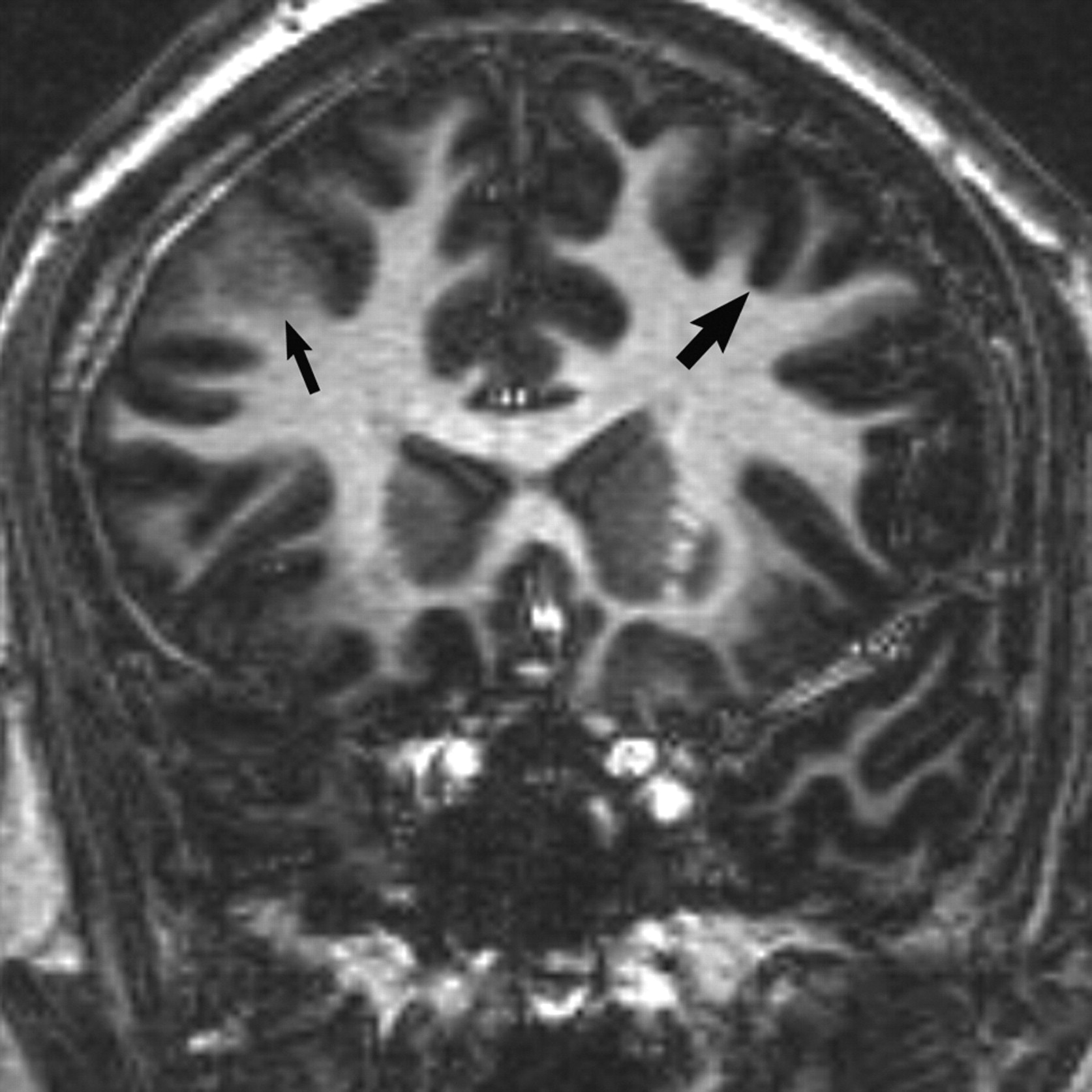
- Fig 1.
Coronal 3D T1 echo-spoiled gradient-echo image demonstrates an area of cortical thickening and indistinctness involving the right frontal lobe (thin arrow). Compare with the normal left frontal cortex (wide arrow).
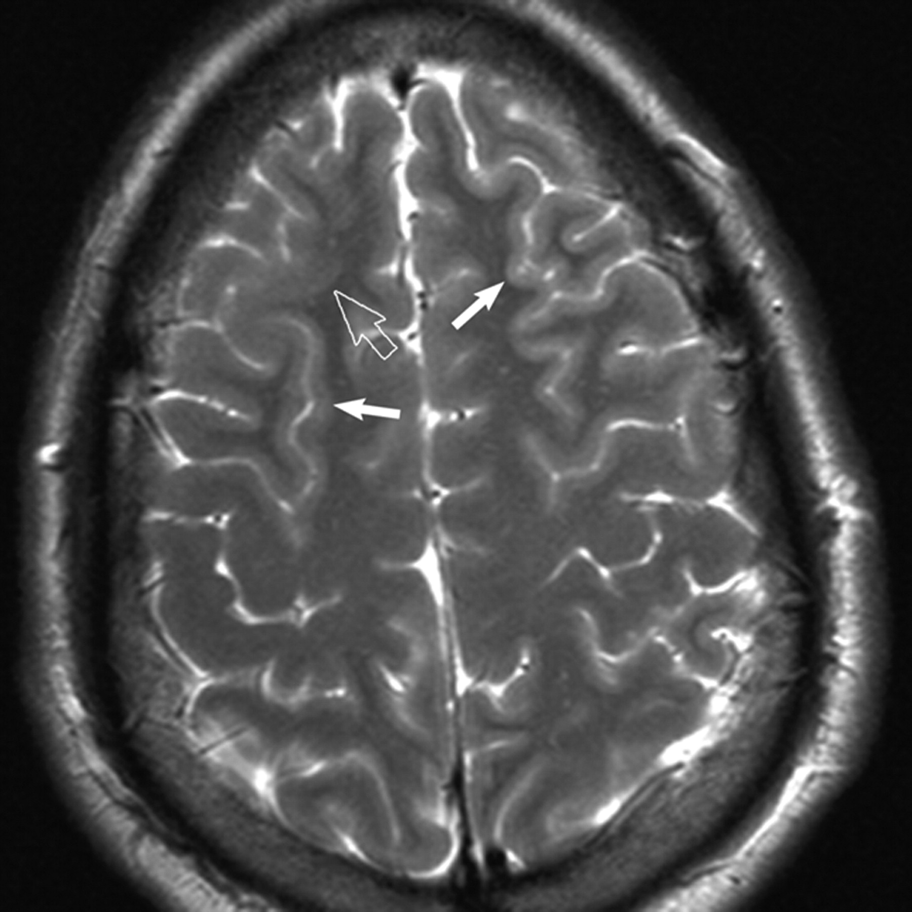
- Fig 2.
Axial T2-weighted MR image reveals subtle cortical thickening and blurring of the gray-white matter junction (open arrow) in the right frontal lobe. Subtle gyral expansion is questioned. Note areas of normal thin-appearing cortex in the ipsilateral and contralateral hemisphere for comparison (closed arrows).


