
Abstract
SUMMARY: Intracranial stent placement is emerging as an effective treatment for acute stroke. As a means to avoid stent-associated complications and capitalize on stent-placement-related advantages, the concept of a “temporary endovascular bypass” (TEB) for stroke therapy was recently reported. In this technique, a stent is temporarily deployed for instant recanalization. Once sufficient flow is established to maintain vessel patency, the stent is recaptured and withdrawn. We report a second case to further characterize the merits of TEB.
Stenting is emerging as a promising treatment option for acute cerebral ischemia.1–4 The main advantage of stenting for acute stroke is rapid recanalization with high technical success rates.3,4 To capitalize on the advantages of stenting for acute stroke but avoid long-term complications, the “temporary endovascular bypass” (TEB) technique, recently described by Kelly et al,2 represents an interesting alternative to permanent device implantation. A stent is deployed and then recaptured after successful vessel recanalization has been achieved. To further characterize the merits of TEB, a second case is reported here.
Case Report
A 41-year-old man presented with a National Institutes of Health Stroke Scale (NIHSS) score of 19 nine hours after stroke onset. Diagnostic arteriography confirmed vertebrobasilar occlusion (Fig 1A). After access was obtained and intravenous heparin given (activated clotting time, 268 seconds), we prepared for TEB as the primary recanalization strategy.2 The lesion was crossed with a Synchro 0.014-inch microguidewire (Boston Scientific, Natick, Mass) and a Prowler Select Plus microcatheter (Cordis, Miami Lakes, Fla). A long 4.5 × 37 mm Enterprise stent (Cordis) was partially deployed, spanning the entire occluded segment in the vertebrobasilar system. Partial unsheathing of the stent “propped” open the distal vertebral artery (VA) and the proximal basilar artery. Using the stent as a buttress device, we reconstituted and maintained sufficient flow (Fig 1B). The stent was recaptured and withdrawn 18 minutes later. Poststent arteriography (Fig 1C) demonstrated moderate residual stenosis. The patient then improved immediately (NIHSS score, 8). However, on postoperative day 2, the patient developed lethargy secondary to a cerebellar infarct. He then underwent ventriculostomy and surgical posterior fossa decompression. The patient continued to recover following surgery (NIHSS score of 2, thirty days postoperatively). Conventional angiography demonstrated patency of the vertebrobasilar system without change 8 weeks post-TEB.
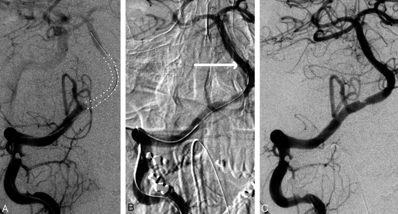
A, Right vertebrobasilar arteriogram, dual-injection technique. Right VA occlusion distal to the posterior inferior cerebellar artery origin. A large vertebrobasilar thrombus is outlined by the dotted lines. B, Right VA angiogram, close-up view. The 4.5 × 37 mm Enterprise stent (Cordis) is partially unsheathed. Close inspection of the image reveals 3 small dots marking the distal end of the stent (arrow). The proximal end is still within its sheath, allowing recapture. Recanalization is already successful but has not yet reached its full extent. C, Right VA angiogram, 10 minutes after B was obtained. The stent is still partially deployed. Recanalization continues to improve (relative to A). After this run, the stent was recaptured and withdrawn.
Discussion
TEB appears to be a promising new technique to treat acute stroke. Technically, TEB is straightforward and restores flow immediately. The bypass can be left in place as long as needed or converted to a permanently implanted stent by completing the unsheathing maneuver. The stent recapture eliminates long-term stent-associated complications, such as in-stent stenosis and the need for aggressive antiplatelet therapy.
Intracranial Stent Placement for Acute Stroke
Recently, a few centers have begun to explore intracranial stent placement for acute stroke treatment.1–4 Zaidat et al4 reported their experience with acute stroke by using Neuroform and Wingspan stents (Boston Scientific). Stroke-related mortality occurred in 3 patients (33%). Survivors had modified Rankin Scale scores ≤2. Follow-up angiography showed no in-stent stenosis. Levy et al3 reported their initial series of 19 lesions treated mainly with the Neuroform stent. Stent deployment at the target occlusion site (technical success) was achieved in all cases. Recanalization was successful in 79%. According to these first reports3,4 and our current experience, the main advantage of intracranial stent placement for acute stroke is the immediate recanalization with a high technical success rate. The TEB technique builds on this success.
In-Stent Stenosis after Target Vessel Revascularization
Initial results for stent placement of chronic intracranial stenosis were promising.5,6 However, disappointingly high rates of recurrent stenosis have been described for midterm results.7,8 On the basis of the Wingspan experience, in-stent stenosis rates are fairly significant (25%–29.7%).9,10 Although most patients with in-stent stenosis remain asymptomatic, it can cause neurologic symptoms and may require target-vessel recanalization.9,11 In the setting of acute stroke, Zaidat et al4 reported 1 case (11%) of immediate in-stent restenosis. The risk of in-stent stenosis may be higher in the setting of symptomatic intracranial stenosis or acute stroke compared with the natural history of intracranial stents placed for aneurysm treatment.12 As a practical concept, TEB avoids stent-associated risks altogether because the device is removed.
Risk of Hemorrhagic Complications in the Setting of Acute Stroke
The need for aggressive antiplatelet and/or anticoagulant therapy associated with intracranial stent placement1–4,10,13–16 is a second major disadvantage if stent placement is used as a treatment technique in the setting of acute stroke. Patients treated for the prevention of recurrent stroke with either warfarin or aspirin face a hemorrhagic complication rate of 2.22 per 100 patient-years versus 1.49 per 100 patient-years.17 With dual antiplatelet therapy or antiplatelets plus anticoagulation, the risk is increased.18–21 Zaidat et al4 reported an 11% hemorrhage rate associated with stent placement for acute stroke. Levy et al3 also reported lethal hemorrhage as a complication in 11% of patients treated with stent placement for acute stroke. With TEB, the need for aggressive (dual) antiplatelet therapy is eliminated with recapture of the stent. Therefore, the risk of bleeding postoperatively might be decreased and emergent neurosurgical procedures, if indicated, might be safer.
Conclusions
TEB represents a novel interventional treatment option for acute stroke. The temporary use is technically feasible and takes advantage of the high success rate of intracranial stent placement for recanalization. Dual antiplatelet therapy postintervention is unnecessary, and the problem of in-stent stenosis is eliminated.
Acknowledgment
We thank Paul H. Dressel for assistance with preparation of the illustrations and Debra J. Zimmer for editorial assistance.
Footnotes
-
Dr. Levy receives research grant support, other research support (devices), and honoraria from Boston Scientific; has an ownership interest in Intratech Medical and Micrus Endovascular; serves as a consultant to Cordis Neurovascular, Micrus Endovascular, ev3, and TheraSyn Sensors; and receives fees for carotid stent training from Abbott Vascular and ev3. Dr. Mocco has received a research grant from the Brain Aneurysm Foundation and a previous, one-time honorarium from Cordis.
References
- Received December 18, 2008.
- Accepted after revision December 31, 2008.
- Copyright © American Society of Neuroradiology
In this issue


Jump to section
Related Articles
Cited By...
- Brief History of Endovascular Acute Ischemic Stroke Treatment
- Primary stenting for acute ischemic stroke using the Enterprise vascular reconstruction device: early results
- Stenting in acute stroke: point
- Impact of intracranial self-expanding stents in the treatment of acute ischemic stroke: efficacy and limitations
- Impact of Retrievable Stents on Acute Ischemic Stroke Treatment
- Stent-assisted basilar reconstruction for a traumatic vertebral dissection with a large basilar artery thrombosis
- In Vivo Evaluation of the First Dedicated Combined Flow-Restoration and Mechanical Thrombectomy Device in a Swine Model of Acute Vessel Occlusion