Advertisement

Main menu

  • Home
  • Content
    • Current Issue
    • Accepted Manuscripts
    • Article Preview
    • Past Issue Archive
    • Video Articles
    • AJNR Case Collection
    • Case of the Week Archive
    • Case of the Month Archive
    • Classic Case Archive
  • Special Collections
    • AJNR Awards
    • ASNR Foundation Special Collection
    • Most Impactful AJNR Articles
    • Photon-Counting CT
    • Spinal CSF Leak Articles (Jan 2020-June 2024)
  • Multimedia
    • AJNR Podcasts
    • AJNR SCANtastic
    • Trainee Corner
    • MRI Safety Corner
    • Imaging Protocols
  • For Authors
    • Submit a Manuscript
    • Submit a Video Article
    • Submit an eLetter to the Editor/Response
    • Manuscript Submission Guidelines
    • Statistical Tips
    • Fast Publishing of Accepted Manuscripts
    • Graphical Abstract Preparation
    • Imaging Protocol Submission
    • Author Policies
  • About Us
    • About AJNR
    • Editorial Board
    • Editorial Board Alumni
  • More
    • Become a Reviewer/Academy of Reviewers
    • Subscribers
    • Permissions
    • Alerts
    • Feedback
    • Advertisers
    • ASNR Home

User menu

  • Alerts
  • Log in

Search

  • Advanced search
American Journal of Neuroradiology

American Journal of Neuroradiology

ASHNR American Society of Functional Neuroradiology ASHNR American Society of Pediatric Neuroradiology ASSR
  • Alerts
  • Log in

Advanced Search

  • Home
  • Content
    • Current Issue
    • Accepted Manuscripts
    • Article Preview
    • Past Issue Archive
    • Video Articles
    • AJNR Case Collection
    • Case of the Week Archive
    • Case of the Month Archive
    • Classic Case Archive
  • Special Collections
    • AJNR Awards
    • ASNR Foundation Special Collection
    • Most Impactful AJNR Articles
    • Photon-Counting CT
    • Spinal CSF Leak Articles (Jan 2020-June 2024)
  • Multimedia
    • AJNR Podcasts
    • AJNR SCANtastic
    • Trainee Corner
    • MRI Safety Corner
    • Imaging Protocols
  • For Authors
    • Submit a Manuscript
    • Submit a Video Article
    • Submit an eLetter to the Editor/Response
    • Manuscript Submission Guidelines
    • Statistical Tips
    • Fast Publishing of Accepted Manuscripts
    • Graphical Abstract Preparation
    • Imaging Protocol Submission
    • Author Policies
  • About Us
    • About AJNR
    • Editorial Board
    • Editorial Board Alumni
  • More
    • Become a Reviewer/Academy of Reviewers
    • Subscribers
    • Permissions
    • Alerts
    • Feedback
    • Advertisers
    • ASNR Home
  • Follow AJNR on Twitter
  • Visit AJNR on Facebook
  • Follow AJNR on Instagram
  • Join AJNR on LinkedIn
  • RSS Feeds

AJNR is seeking candidates for the AJNR Podcast Editor. Read the position description.

Review ArticleReview Articles
Open Access

Neuroimaging and Deep Brain Stimulation

D. Dormont, D. Seidenwurm, D. Galanaud, P. Cornu, J. Yelnik and E. Bardinet
American Journal of Neuroradiology January 2010, 31 (1) 15-23; DOI: https://doi.org/10.3174/ajnr.A1644
D. Dormont
  • Find this author on Google Scholar
  • Find this author on PubMed
  • Search for this author on this site
D. Seidenwurm
  • Find this author on Google Scholar
  • Find this author on PubMed
  • Search for this author on this site
D. Galanaud
  • Find this author on Google Scholar
  • Find this author on PubMed
  • Search for this author on this site
P. Cornu
  • Find this author on Google Scholar
  • Find this author on PubMed
  • Search for this author on this site
J. Yelnik
  • Find this author on Google Scholar
  • Find this author on PubMed
  • Search for this author on this site
E. Bardinet
  • Find this author on Google Scholar
  • Find this author on PubMed
  • Search for this author on this site
  • Article
  • Figures & Data
  • Info & Metrics
  • Responses
  • References
  • PDF
Loading

Article Figures & Data

Figures

  • Fig 1.
    • Download figure
    • Open in new tab
    • Download powerpoint
    Fig 1.

    3D view of a postoperative MR acquisition in a patient with bilateral implantation of electrodes in the STN for the treatment of advanced PD. Caudate nuclei (blue), subthalamic nuclei (pink), electrodes (gray), and electrode contacts (blue) are segmented by using a 3D atlas described in Yelnik et al.55. A, Anterior oblique view. B, Posterior oblique view. C, Zoom on the electrodes showing that their contacts are located inside the STN.
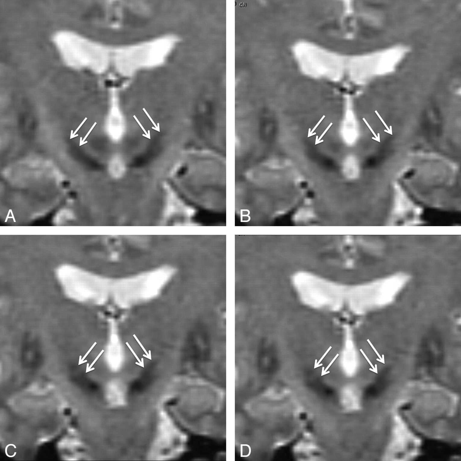
  • Fig 2.
    • Download figure
    • Open in new tab
    • Download powerpoint
    Fig 2.

    Coronal T2-weighted images showing the STN in a patient with advanced PD (spin-echo acquisition; TR/TE/NEX, 2200 ms/90 ms/2). Section thickness is 2 mm, located every 1 mm. A, The section is located 1 mm behind the anterior limit of the red nuclei. Both STNs are seen as almond-shaped hypointense structures above the locus niger (arrows). B, This section is located 1 mm in front of A, at the level of the anterior limit of the red nuclei. Both STNs are clearly seen (arrows). C and D, These sections are located 1 and 2 mm in front of B, showing the anterior extension of the STN (arrows).

  • Fig 3.
    • Download figure
    • Open in new tab
    • Download powerpoint
    Fig 3.

    Postoperative study of the position of electrode contacts in a patient with bilateral implantation of electrodes in the STN for the treatment of PD. Postoperative 3D MR imaging acquisition with fusion of anatomic data by using a histologically based deformable 3D atlas of the basal ganglia (Yelnik et al55). A, 3D posterior oblique view shows the caudate nuclei (blue), the STN (pink), the locus niger (black), and the electrode contacts (yellow, active contacts; blue, inactive contacts; contacts inside or behind the STN are seen in the transparency). B, Frontal view through the active contact. The exact position of the contacts inside the metallic artifact on the MR imaging acquisition is indicated on each side by a yellow dot. Both contacts are located inside the STN (pink). Anatomic limits of the putamen (blue), caudate (blue), locus niger (black), thalamus (green), and optical tracts (orange) on the basis of the 3D atlas are shown. C, Axial view through the right active contact (yellow dot; the left active contact is above and is not seen in this view). The right active contact inside the STN (pink), caudate, putamen, and red nuclei (orange) can be seen. D, Right sagittal view through the right active contact (yellow dot). The same color code is used for caudate, thalamus, locus niger, and optical tract (orange).

PreviousNext
Back to top

In this issue

American Journal of Neuroradiology: 31 (1)
American Journal of Neuroradiology
Vol. 31, Issue 1
1 Jan 2010
  • Table of Contents
  • Index by author
Advertisement
Print
Download PDF
Email Article

Thank you for your interest in spreading the word on American Journal of Neuroradiology.

NOTE: We only request your email address so that the person you are recommending the page to knows that you wanted them to see it, and that it is not junk mail. We do not capture any email address.

Enter multiple addresses on separate lines or separate them with commas.
Neuroimaging and Deep Brain Stimulation
(Your Name) has sent you a message from American Journal of Neuroradiology
(Your Name) thought you would like to see the American Journal of Neuroradiology web site.
CAPTCHA
This question is for testing whether or not you are a human visitor and to prevent automated spam submissions.
Cite this article
D. Dormont, D. Seidenwurm, D. Galanaud, P. Cornu, J. Yelnik, E. Bardinet
Neuroimaging and Deep Brain Stimulation
American Journal of Neuroradiology Jan 2010, 31 (1) 15-23; DOI: 10.3174/ajnr.A1644

Citation Manager Formats

  • BibTeX
  • Bookends
  • EasyBib
  • EndNote (tagged)
  • EndNote 8 (xml)
  • Medlars
  • Mendeley
  • Papers
  • RefWorks Tagged
  • Ref Manager
  • RIS
  • Zotero
0 Responses
Respond to this article
Share
Bookmark this article
Neuroimaging and Deep Brain Stimulation
D. Dormont, D. Seidenwurm, D. Galanaud, P. Cornu, J. Yelnik, E. Bardinet
American Journal of Neuroradiology Jan 2010, 31 (1) 15-23; DOI: 10.3174/ajnr.A1644
del.icio.us logo Twitter logo Facebook logo Mendeley logo
  • Tweet Widget
  • Facebook Like
  • Google Plus One
Purchase

Jump to section

  • Article
    • Abstract
    • Imaging of Patients before Electrode Implantation
    • Targeting
    • Intraoperative Confirmation of Electrode Placement
    • Postoperative Imaging
    • Conclusions
    • References
  • Figures & Data
  • Info & Metrics
  • Responses
  • References
  • PDF

Related Articles

  • No related articles found.
  • PubMed
  • Google Scholar

Cited By...

  • An experimental evaluation of the relationship between the induced radiofrequency heating near an implanted conductive medical device during MRI, scanner reported B1+rms, and scanner reported average transmit power
  • In Vivo Localization of Deep Brain Implants in Mice
  • MRI Powered and Triggered Current Stimulator for Concurrent Stimulation and MRI
  • Reducing RF-induced Heating near Implanted Leads through High-Dielectric Capacitive Bleeding of Current (CBLOC)
  • Lead-DBS v2: Towards a comprehensive pipeline for deep brain stimulation imaging
  • Automatic Localization of the Subthalamic Nucleus on Patient-Specific Clinical MRI by Incorporating 7T MRI and Machine Learning: Application in Deep Brain Stimulation
  • Crossref (48)
  • Google Scholar

This article has been cited by the following articles in journals that are participating in Crossref Cited-by Linking.

  • Lead-DBS v2: Towards a comprehensive pipeline for deep brain stimulation imaging
    Andreas Horn, Ningfei Li, Till A. Dembek, Ari Kappel, Chadwick Boulay, Siobhan Ewert, Anna Tietze, Andreas Husch, Thushara Perera, Wolf-Julian Neumann, Marco Reisert, Hang Si, Robert Oostenveld, Christopher Rorden, Fang-Cheng Yeh, Qianqian Fang, Todd M. Herrington, Johannes Vorwerk, Andrea A. Kühn
    NeuroImage 2019 184
  • A review of low-intensity focused ultrasound pulsation
    Alexander Bystritsky, Alex S. Korb, Pamela K. Douglas, Mark S. Cohen, William P. Melega, Amit P. Mulgaonkar, Antonio DeSalles, Byoung-Kyong Min, Seung-Schik Yoo
    Brain Stimulation 2011 4 3
  • Neurostimulation Devices for the Treatment of Neurologic Disorders
    Christine A. Edwards, Abbas Kouzani, Kendall H. Lee, Erika K. Ross
    Mayo Clinic Proceedings 2017 92 9
  • A Novel Brain Stimulation Technology Provides Compatibility with MRI
    Peter Serano, Leonardo M. Angelone, Husam Katnani, Emad Eskandar, Giorgio Bonmassar
    Scientific Reports 2015 5 1
  • Comparison of T2*-weighted and QSM contrasts in Parkinson's disease to visualize the STN with MRI
    Anneke Alkemade, Gilles de Hollander, Max C. Keuken, Andreas Schäfer, Derek V. M. Ott, Johannes Schwarz, David Weise, Sonja A. Kotz, Birte U. Forstmann, Quan Jiang
    PLOS ONE 2017 12 4
  • Improving Safety of MRI in Patients with Deep Brain Stimulation Devices
    Alexandre Boutet, Clement T. Chow, Keshav Narang, Gavin J. B. Elias, Clemens Neudorfer, Jürgen Germann, Manish Ranjan, Aaron Loh, Alastair J. Martin, Walter Kucharczyk, Christopher J. Steele, Ileana Hancu, Ali R. Rezai, Andres M. Lozano
    Radiology 2020 296 2
  • Reducing RF-Induced Heating Near Implanted Leads Through High-Dielectric Capacitive Bleeding of Current (CBLOC)
    Laleh Golestanirad, Leonardo M. Angelone, John Kirsch, Sean Downs, Boris Keil, Giorgio Bonmassar, Lawrence L. Wald
    IEEE Transactions on Microwave Theory and Techniques 2019 67 3
  • Cognitive and Psychiatric Effects of STN versus GPi Deep Brain Stimulation in Parkinson's Disease: A Meta-Analysis of Randomized Controlled Trials
    Jia-Wei Wang, Yu-Qing Zhang, Xiao-Hua Zhang, Yun-Peng Wang, Ji-Ping Li, Yong-Jie Li, Allan Siegel
    PLOS ONE 2016 11 6
  • PyDBS: an automated image processing workflow for deep brain stimulation surgery
    Tiziano D’Albis, Claire Haegelen, Caroline Essert, Sara Fernández-Vidal, Florent Lalys, Pierre Jannin
    International Journal of Computer Assisted Radiology and Surgery 2015 10 2
  • Automatic localization of the subthalamic nucleus on patient‐specific clinical MRI by incorporating 7 T MRI and machine learning: Application in deep brain stimulation
    Jinyoung Kim, Yuval Duchin, Reuben R. Shamir, Remi Patriat, Jerrold Vitek, Noam Harel, Guillermo Sapiro
    Human Brain Mapping 2019 40 2

More in this TOC Section

  • Mechanisms of Healing in Coiled Intracranial Aneurysms: A Review of the Literature
  • Ultra-High-Field MR Neuroimaging
  • 4D-CTA in Neurovascular Disease: A Review
Show more Review Articles

Similar Articles

Advertisement

Indexed Content

  • Current Issue
  • Accepted Manuscripts
  • Article Preview
  • Past Issues
  • Editorials
  • Editor's Choice
  • Fellows' Journal Club
  • Letters to the Editor
  • Video Articles

Cases

  • Case Collection
  • Archive - Case of the Week
  • Archive - Case of the Month
  • Archive - Classic Case

Special Collections

  • AJNR Awards
  • ASNR Foundation Special Collection
  • Most Impactful AJNR Articles
  • Photon-Counting CT
  • Spinal CSF Leak Articles (Jan 2020-June 2024)

More from AJNR

  • Trainee Corner
  • Imaging Protocols
  • MRI Safety Corner

Multimedia

  • AJNR Podcasts
  • AJNR Scantastics

Resources

  • Turnaround Time
  • Submit a Manuscript
  • Submit a Video Article
  • Submit an eLetter to the Editor/Response
  • Manuscript Submission Guidelines
  • Statistical Tips
  • Fast Publishing of Accepted Manuscripts
  • Graphical Abstract Preparation
  • Imaging Protocol Submission
  • Evidence-Based Medicine Level Guide
  • Publishing Checklists
  • Author Policies
  • Become a Reviewer/Academy of Reviewers
  • News and Updates

About Us

  • About AJNR
  • Editorial Board
  • Editorial Board Alumni
  • Alerts
  • Permissions
  • Not an AJNR Subscriber? Join Now
  • Advertise with Us
  • Librarian Resources
  • Feedback
  • Terms and Conditions
  • AJNR Editorial Board Alumni

American Society of Neuroradiology

  • Not an ASNR Member? Join Now

© 2025 by the American Society of Neuroradiology All rights, including for text and data mining, AI training, and similar technologies, are reserved.
Print ISSN: 0195-6108 Online ISSN: 1936-959X

Powered by HighWire