Advertisement

Main menu

  • Home
  • Content
    • Current Issue
    • Publication Preview--Ahead of Print
    • Past Issue Archive
    • Case of the Week Archive
    • Classic Case Archive
    • Case of the Month Archive
    • COVID-19 Content and Resources
  • For Authors
    • Author Policies
    • Manuscript Submission Guidelines
  • About Us
    • About AJNR
    • American Society of Neuroradiology
  • Submit a Manuscript
  • Podcasts
    • Podcasts
    • Subscribe on iTunes
  • More
    • Subscribers
    • Permissions
    • Advertisers
    • Alerts
    • Feedback

User menu

  • Subscribe
  • Alerts
  • Log in

Search

  • Advanced search
American Journal of Neuroradiology

American Journal of Neuroradiology

  • Subscribe
  • Alerts
  • Log in

Advanced Search

  • Home
  • Content
    • Current Issue
    • Publication Preview--Ahead of Print
    • Past Issue Archive
    • Case of the Week Archive
    • Classic Case Archive
    • Case of the Month Archive
    • COVID-19 Content and Resources
  • For Authors
    • Author Policies
    • Manuscript Submission Guidelines
  • About Us
    • About AJNR
    • American Society of Neuroradiology
  • Submit a Manuscript
  • Podcasts
    • Podcasts
    • Subscribe on iTunes
  • More
    • Subscribers
    • Permissions
    • Advertisers
    • Alerts
    • Feedback
  • Follow AJNR on Twitter
  • Visit AJNR on Facebook
  • Follow AJNR on Instagram
  • Join AJNR on LinkedIn
  • RSS Feeds
Case ReportHead and Neck

Hemifacial Lipomatosis, a Possible Subtype of Partial Hemifacial Hyperplasia: CT and MR Imaging Findings

P. Bou-Haidar, P. Taub and P. Som
American Journal of Neuroradiology May 2010, 31 (5) 891-893; DOI: https://doi.org/10.3174/ajnr.A1857
P. Bou-Haidar
  • Find this author on Google Scholar
  • Find this author on PubMed
  • Search for this author on this site
P. Taub
  • Find this author on Google Scholar
  • Find this author on PubMed
  • Search for this author on this site
P. Som
  • Find this author on Google Scholar
  • Find this author on PubMed
  • Search for this author on this site
  • Article
  • Figures & Data
  • Info & Metrics
  • References
  • PDF
Loading

Abstract

SUMMARY: We present a case of hemifacial hyperplasia in an infant manifesting predominantly as lipomatosis and hemihypertrophy of the maxilla. To our knowledge, there is only 1 other case report in the literature demonstrating the MR imaging features of this condition. Our case was manifest almost exclusively as lipomatosis, largely lacking muscular hypertrophy/hyperplasia.

Abbreviations

BWS
Beckwith-Wiedemann syndrome
HFH
hemifacial hypertrophy
MRA
MR angiography
PHFH
partial hemifacial hypertrophy
THFH
true hemifacial hypertrophy

Hemifacial hyperplasia is a rare developmental anomaly characterized by asymmetric growth of ≥1 part of the face. In this report of a female child born with facial asymmetry, CT and MR imaging revealed predominantly lipomatosis. The case suggests that hemifacial lipomatosis may be a subtype of partial hemifacial hyperplasia. We discuss the differential diagnosis of HFH and emphasize the importance of screening for embryonal malignancies.

Case Report

A 15-month-old female child presented to our department with right-sided facial fullness (Fig 1). The abnormality was noted at birth, and the child's parents stated that the fullness did not fluctuate in size. The right cheek appeared to grow at a rate similar to the left side. Physical examination was also remarkable for a slightly enlarged right auricle. There were no other significant medical problems. No biochemical or chromosomal abnormalities were detected. The child was otherwise healthy. A sonographic examination showed diffuse right facial adipose hypertrophy but no discrete vascular malformation.

Fig 1.
  • Download figure
  • Open in new tab
  • Download powerpoint
Fig 1.

Surface rendering from a CT study performed on the patient at 15 months of age.

CT examination at this time demonstrated predominantly increased fatty tissue on the right side of the face. The distribution of fat was largely in the malar region but also infiltrated the ipsilateral muscles of mastication and the parotid gland and extended into the ipsilateral parapharyngeal fat. The right parotid gland and muscles of mastication had a slightly marbled appearance, and there was enlargement of the right prestyloid parapharyngeal space (Fig 2A). These findings were demonstrated again on a follow-up MR imaging examination 4 months later (Fig 2B).

Fig 2.
  • Download figure
  • Open in new tab
  • Download powerpoint
Fig 2.

A, Axial CT scan of the patient at 15 months of age. B, T1-weighted MR image from a study of the patient at 2 years of age. Note the lipomatosis infiltration and corresponding marbling of the right parotid gland and muscles of mastication (arrow) and the expansion the right parapharyngeal space (asterisk)

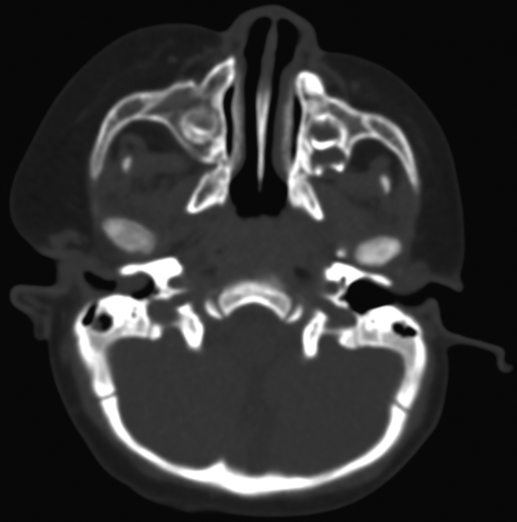
The bony structures of the right face showed slight asymmetry, with hypertrophy of the right maxilla and the right lateral pterygoid plate. There was also subtle enlargement of the right zygoma and the right mandible (Fig 3). Regarding dental development, there was slight early rupture of the right maxillary deciduous canine teeth, and enlargement of the premolar and molar teeth was also noted (Fig 4).

Fig 3.
  • Download figure
  • Open in new tab
  • Download powerpoint
Fig 3.

Axial CT scan through the maxilla of the patient at 15 months of age demonstrates mild hypertrophy of the right maxilla, lateral pterygoid plate zygoma, and mandibular head.

Fig 4.
  • Download figure
  • Open in new tab
  • Download powerpoint
Fig 4.

Frontal curved reformatted CT scan of the maxilla shows early development and enlargement of the right maxillary deciduous teeth.

MR imaging examination performed as a 6-month follow-up revealed stable and similar findings (Fig 5). No abnormal enhancement was identified. No discrete fibrous capsule to suggest an encapsulated lipoma was seen. The tongue appeared symmetric; however, accurate assessment was difficult because the patient was intubated for the study. Findings of an MRA examination were within normal limits, and no intracranial abnormalities were demonstrated. The child was otherwise developing normally and achieving normal milestones.

Fig 5.
  • Download figure
  • Open in new tab
  • Download powerpoint
Fig 5.

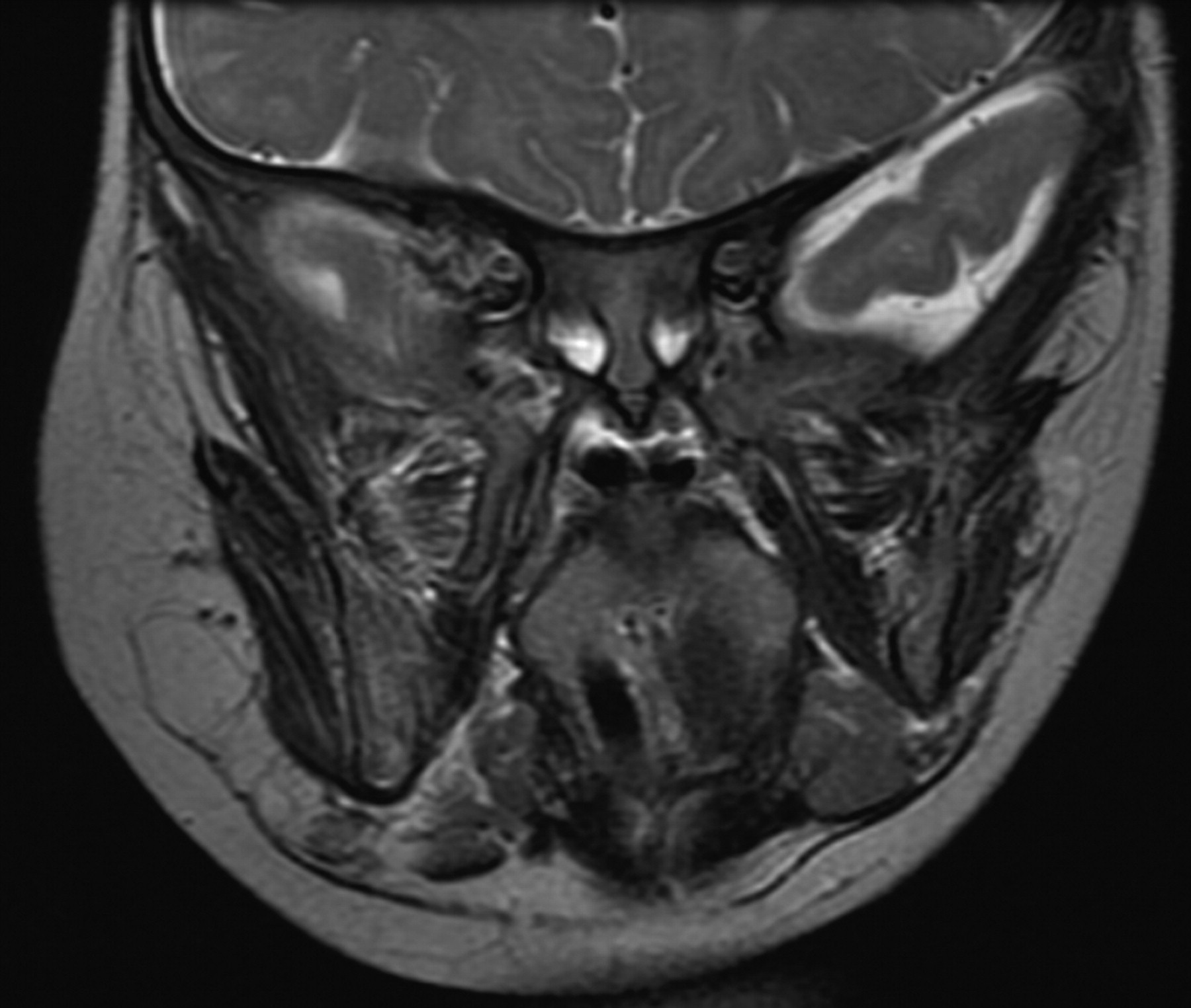
Coronal T2-weighted image of the patient at 2 years of age demonstrates stable findings similar to those in the earlier studies. Note the marbling of the right masticator muscles.

Discussion

HFH was first described by Meckel in 1822.1 It is a rare morphologic developmental anomaly characterized by asymmetric growth of ≥1 body part. Its true incidence is unknown and, to our knowledge, has not been published, and very little about this syndrome has been published in the radiology literature. Although it is often called HFH, it may be more accurately termed “hemifacial hyperplasia” to reflect the underlying pathology.2,3 Little is know about the etiology of this condition. Various theories, none clearly favored, have been proposed. These include vascular lymphatic, hormonal, and asymmetric development of the neural fold, with hyperplasia of neural crest cells.3 It is a condition that appears to affect the derivatives of the first pharyngeal arch,4 though the middle ear structures seem spared.

Isolated hemifacial hyperplasia should be a diagnosis of exclusion, because there are other more clinically burdensome conditions that can cause facial asymmetry. These conditions include the following: Proteus syndrome, BWS, neurofibromatosis type 1, and vascular malformations such as Klippel-Trenaunay syndrome and lymphaticovenous malformations.5 A full neurologic examination and brain imaging are prudent, and the radiologist may be the first to suggest screening for organomegaly and malignancies. Tumor surveillance, especially for malignancies of embryonal origin, is supported by a prospective study of children with isolated hemihyperplasia or hemihypertrophy. These tumors include Wilms' tumors, hepatoblastoma, and adrenal cell carcinoma.6

The extremely rare Proteus syndrome, suspected as the primary condition in the historic “elephant man,” causes a progressive overgrowth of all soft tissues and bones, including the skull, not limited to the region of isolated hemifacial hyperplasia. Patients with Proteus syndrome will often have involvement of the hands and feet and veracious epidermal nevi.7 BWS is a congenital cancer-predisposition syndrome associated with macroglossia, macrosomia, ear pits or ear creases, and midline abdominal wall defects. The associated hemihypertrophy seen in BWS may represent the mosaic form of macrosomia.8 The hemihypertrophy in neurofibromatosis type 1 can be due to even subtle plexiform neurofibromas and/or skeletal dysplasia. When vascular malformations result in facial asymmetry, it may be due to the bulk of abnormal vessels alone or combined with induced hypertrophy due to local increased vascular flow and local lymphedema.9 Occasionally, hemifacial atrophy (Parry-Romberg syndrome) can give the false appearance of hemifacial hyperplasia on the normal side, though these patients present later, between 5 and 15 years of age.

Rowe10 in 1962 classified HFH into THFH and PHFH. THFH manifests unilateral enlargement of all the viscerocranial structures bounded by the frontal bone, excluding the eye; inferiorly by the inferior border of the mandible and the midline; and laterally by the ear. THFH is characterized by enlargement of the teeth, bones, and soft tissues. Not all the included structures are enlarged in PHFH, as in our patient. Other findings in hemifacial hyperplasia include an enlarged tongue on the involved side, enlarged lips, and widening of the palate of the affected side.5

Our case, similar to a previous case report,11 demonstrated lipomatosis as the dominant feature and lacked significant facial musculature hyperplasia. Hemifacial myohyperplasia is a proposed newly described separate entity in which facial muscular hyperplasia is the dominant feature.3,4 This may explain the lack of the nasal deviation toward the affected side in our patient but rather deviation to the opposite (normal) side. Nasal deviation toward the affected side may be observed when the facial musculature around the nasolabial fold is involved and when the hypertrophied turbinates cause nasal septal deviation.4 The teeth, mandible, and maxilla should be carefully scrutinized for deformities to help one reach the correct diagnosis. This is especially important if only lipomatous overgrowth is seen initially.

Conclusions

Hemifacial hyperplasia is a rare developmental condition of asymmetric growth of ≥1 body part. MR imaging and CT illustrate hemifacial lipomatosis as the dominant feature, suggesting that it may represent a subtype of partial hemifacial hyperplasia.

References

  1. 1.↵
    1. Gorlin RJ,
    2. Meskin LH
    . Congenital hemihypertrophy: review of the literature and report of a case with special emphasis on oral manifestations. J Pediatr 1962;61:870–79
    CrossRefPubMed
  2. 2.↵
    1. Yoshimoto H,
    2. Yano H,
    3. Kobayashi K,
    4. et al
    . Increased proliferative activity of osteoblasts in congenital hemifacial hypertrophy. Plast Reconstr Surg 1998;102:1605–10
    CrossRefPubMed
  3. 3.↵
    1. Lee S,
    2. Sze R,
    3. Murakami C,
    4. et al
    . Hemifacial myohyperplasia: description of a new syndrome. Am J Med Genet 2001;103:326–33
    CrossRefPubMed
  4. 4.↵
    1. Urban PP,
    2. Roland B
    . Congenital isolated hemifacial hyperplasia. J Neurol 2009;256:1566–69. Epub 2009 May 8
    CrossRefPubMed
  5. 5.↵
    1. Lawoyin JO,
    2. Daramola JO,
    3. Lawoyin DO
    . Congenital hemifacial hypertrophy: report of two cases. Oral Surg Oral Med Oral Pathol 1989;68:27–30
    CrossRefPubMed
  6. 6.↵
    1. Hoyme HE,
    2. Seaver LH,
    3. Jones KL,
    4. et al
    . Isolated hemihyperplasia (hemihypertrophy): report of a prospective multicenter study of the incidence of neoplasia and review. Am J Med Genet 1998;79:274–78
    CrossRefPubMed
  7. 7.↵
    1. Zingade ND,
    2. Zingade NN
    . A rare case of Proteus syndrome. J Laryngol Otol 2008;122:e7
    CrossRefPubMed
  8. 8.↵
    1. DeBaun MR,
    2. Niemitz EL,
    3. McNeil DE,
    4. et al
    . Epigenetic alterations of H19 and LIT1 distinguish patients with Beckwith-Wiedemann syndrome with cancer and birth defects. Am J Hum Genet 2002;70:604–11
    CrossRefPubMed
  9. 9.↵
    1. Blei F
    . Vascular anomalies: from bedside to bench and back again. Curr Probl Pediatr Adolesc Health Care 2002;32:72–93
    CrossRefPubMed
  10. 10.↵
    1. Rowe NH
    . Hemifacial hypertrophy: review of the literature and addition of four cases. Oral Surg Oral Med Oral Pathol 1962;15:572–87
    CrossRefPubMed
  11. 11.↵
    1. Sugiyama M,
    2. Tanaka E,
    3. Ogawa I,
    4. et al
    . Magnetic resonance imaging in hemifacial hyperplasia. Dentomaxillofac Radiol 2001;30:235–38
    Abstract
  • Received July 8, 2009.
  • Accepted after revision July 15, 2009.
  • Copyright © American Society of Neuroradiology
View Abstract
PreviousNext
Back to top

In this issue

American Journal of Neuroradiology: 31 (5)
American Journal of Neuroradiology
Vol. 31, Issue 5
1 May 2010
  • Table of Contents
  • Index by author
Advertisement
Print
Download PDF
Email Article

Thank you for your interest in spreading the word on American Journal of Neuroradiology.

NOTE: We only request your email address so that the person you are recommending the page to knows that you wanted them to see it, and that it is not junk mail. We do not capture any email address.

Enter multiple addresses on separate lines or separate them with commas.
Hemifacial Lipomatosis, a Possible Subtype of Partial Hemifacial Hyperplasia: CT and MR Imaging Findings
(Your Name) has sent you a message from American Journal of Neuroradiology
(Your Name) thought you would like to see the American Journal of Neuroradiology web site.
CAPTCHA
This question is for testing whether or not you are a human visitor and to prevent automated spam submissions.
Citation Tools
Hemifacial Lipomatosis, a Possible Subtype of Partial Hemifacial Hyperplasia: CT and MR Imaging Findings
P. Bou-Haidar, P. Taub, P. Som
American Journal of Neuroradiology May 2010, 31 (5) 891-893; DOI: 10.3174/ajnr.A1857

Citation Manager Formats

  • BibTeX
  • Bookends
  • EasyBib
  • EndNote (tagged)
  • EndNote 8 (xml)
  • Medlars
  • Mendeley
  • Papers
  • RefWorks Tagged
  • Ref Manager
  • RIS
  • Zotero
Share
Hemifacial Lipomatosis, a Possible Subtype of Partial Hemifacial Hyperplasia: CT and MR Imaging Findings
P. Bou-Haidar, P. Taub, P. Som
American Journal of Neuroradiology May 2010, 31 (5) 891-893; DOI: 10.3174/ajnr.A1857
del.icio.us logo Digg logo Reddit logo Twitter logo CiteULike logo Facebook logo Google logo Mendeley logo
  • Tweet Widget
  • Facebook Like
  • Google Plus One
Purchase

Jump to section

  • Article
    • Abstract
    • Abbreviations
    • Case Report
    • Discussion
    • Conclusions
    • References
  • Figures & Data
  • Info & Metrics
  • References
  • PDF

Related Articles

  • No related articles found.
  • PubMed
  • Google Scholar

Cited By...

  • Parry Romberg Syndrome: 7 Cases and Literature Review
  • Crossref (9)
  • Google Scholar

This article has been cited by the following articles in journals that are participating in Crossref Cited-by Linking.

  • Parry Romberg Syndrome: 7 Cases and Literature Review
    M. Wong, C. D. Phillips, M. Hagiwara, D. R. Shatzkes
    American Journal of Neuroradiology 2015 36 7
  • Congenital infiltrating lipomatosis of the face: A subtype of hemifacial hyperplasia
    Ru Sun, Lisha Sun, Gang Li, Zhipeng Sun, Yanping Zhao, Xuchen Ma, Chongke Sun
    International Journal of Pediatric Otorhinolaryngology 2019 125
  • Lipomatosis: a diverse form of hemifacial hyperplasia
    Preeti Chawla Arora, Hemant R. Umarji, Aman Arora, Easwaran Ramaswami
    Imaging Science in Dentistry 2012 42 3
  • Clinical and Imaging Findings of True Hemifacial Hyperplasia
    Bansari A. Bhuta, Archana Yadav, Rajiv S. Desai, Shivani P. Bansal, Vipul V. Chemburkar, Prashant V. Dev
    Case Reports in Dentistry 2013 2013
  • Parry–Romberg syndrome: a mini review
    Tasleem Arif, Rafiya Fatima, Marwa Sami
    Acta Dermatovenerologica Alpina Pannonica et Adriatica 2020 29 4
  • Unilateral masticatory muscle hypertrophy with mandibular ramus hyperostosis
    Opeyemi O. Daramola, Mary Lou Sabino, Valerie A. Flanary
    International Journal of Pediatric Otorhinolaryngology Extra 2011 6 4
  • A rare cause of facial asymmetry: Recurrent hemifacıal lipomatosis
    YasinCelal Gunes, DenizSozmen Ciliz, Huseyin Cetin
    Turkish Journal of Plastic Surgery 2022 30 3
  • Partial facial hemihypertrophy: A case report and review of literature
    Adity Bansal, GoslaSrinivas Reddy, Ashi Chug
    Journal of Cleft Lip Palate and Craniofacial Anomalies 2020 7 2
  • Radiopaedia.org
    Daniel Bell, Dalia Ibrahim
    2017

More in this TOC Section

  • Correlation of Apparent Diffusion Coefficient at 3T with Prognostic Parameters of Retinoblastoma
  • Parathyroid Lesions: Characterization with Dual-Phase Arterial and Venous Enhanced CT of the Neck
  • MR Evaluation of Sinonasal Angiomatous Polyp
Show more HEAD AND NECK

Similar Articles

Advertisement

News and Updates

  • Lucien Levy Best Research Article Award
  • Thanks to our 2022 Distinguished Reviewers

Resources

  • Evidence-Based Medicine Level Guide
  • AJNR Podcast Archive
  • Librarian Resources
  • Terms and Conditions

Opportunities

  • Get Peer Review Credit from Publons

American Society of Neuroradiology

  • Neurographics
  • ASNR Annual Meeting
  • Fellowship Portal

© 2023 by the American Society of Neuroradiology | Print ISSN: 0195-6108 Online ISSN: 1936-959X

Powered by HighWire