Advertisement

Main menu

  • Home
  • Content
    • Current Issue
    • Accepted Manuscripts
    • Article Preview
    • Past Issue Archive
    • Video Articles
    • AJNR Case Collection
    • Case of the Week Archive
    • Case of the Month Archive
    • Classic Case Archive
  • Special Collections
    • AJNR Awards
    • ASNR Foundation Special Collection
    • Most Impactful AJNR Articles
    • Photon-Counting CT
    • Spinal CSF Leak Articles (Jan 2020-June 2024)
  • Multimedia
    • AJNR Podcasts
    • AJNR SCANtastic
    • Trainee Corner
    • MRI Safety Corner
    • Imaging Protocols
  • For Authors
    • Submit a Manuscript
    • Submit a Video Article
    • Submit an eLetter to the Editor/Response
    • Manuscript Submission Guidelines
    • Statistical Tips
    • Fast Publishing of Accepted Manuscripts
    • Graphical Abstract Preparation
    • Imaging Protocol Submission
    • Author Policies
  • About Us
    • About AJNR
    • Editorial Board
    • Editorial Board Alumni
  • More
    • Become a Reviewer/Academy of Reviewers
    • Subscribers
    • Permissions
    • Alerts
    • Feedback
    • Advertisers
    • ASNR Home

User menu

  • Alerts
  • Log in

Search

  • Advanced search
American Journal of Neuroradiology

American Journal of Neuroradiology

ASHNR American Society of Functional Neuroradiology ASHNR American Society of Pediatric Neuroradiology ASSR
  • Alerts
  • Log in

Advanced Search

  • Home
  • Content
    • Current Issue
    • Accepted Manuscripts
    • Article Preview
    • Past Issue Archive
    • Video Articles
    • AJNR Case Collection
    • Case of the Week Archive
    • Case of the Month Archive
    • Classic Case Archive
  • Special Collections
    • AJNR Awards
    • ASNR Foundation Special Collection
    • Most Impactful AJNR Articles
    • Photon-Counting CT
    • Spinal CSF Leak Articles (Jan 2020-June 2024)
  • Multimedia
    • AJNR Podcasts
    • AJNR SCANtastic
    • Trainee Corner
    • MRI Safety Corner
    • Imaging Protocols
  • For Authors
    • Submit a Manuscript
    • Submit a Video Article
    • Submit an eLetter to the Editor/Response
    • Manuscript Submission Guidelines
    • Statistical Tips
    • Fast Publishing of Accepted Manuscripts
    • Graphical Abstract Preparation
    • Imaging Protocol Submission
    • Author Policies
  • About Us
    • About AJNR
    • Editorial Board
    • Editorial Board Alumni
  • More
    • Become a Reviewer/Academy of Reviewers
    • Subscribers
    • Permissions
    • Alerts
    • Feedback
    • Advertisers
    • ASNR Home
  • Follow AJNR on Twitter
  • Visit AJNR on Facebook
  • Follow AJNR on Instagram
  • Join AJNR on LinkedIn
  • RSS Feeds

AJNR is seeking candidates for the AJNR Podcast Editor. Read the position description.

Research ArticleHead and Neck
Open Access

A Noninvasive Imaging Approach to Assess Plaque Severity: The Carotid Atherosclerosis Score

H.R. Underhill, T.S. Hatsukami, J. Cai, W. Yu, J.K. DeMarco, N.L. Polissar, H. Ota, X. Zhao, L. Dong, M. Oikawa and C. Yuan
American Journal of Neuroradiology June 2010, 31 (6) 1068-1075; DOI: https://doi.org/10.3174/ajnr.A2007
H.R. Underhill
  • Find this author on Google Scholar
  • Find this author on PubMed
  • Search for this author on this site
T.S. Hatsukami
  • Find this author on Google Scholar
  • Find this author on PubMed
  • Search for this author on this site
J. Cai
  • Find this author on Google Scholar
  • Find this author on PubMed
  • Search for this author on this site
W. Yu
  • Find this author on Google Scholar
  • Find this author on PubMed
  • Search for this author on this site
J.K. DeMarco
  • Find this author on Google Scholar
  • Find this author on PubMed
  • Search for this author on this site
N.L. Polissar
  • Find this author on Google Scholar
  • Find this author on PubMed
  • Search for this author on this site
H. Ota
  • Find this author on Google Scholar
  • Find this author on PubMed
  • Search for this author on this site
X. Zhao
  • Find this author on Google Scholar
  • Find this author on PubMed
  • Search for this author on this site
L. Dong
  • Find this author on Google Scholar
  • Find this author on PubMed
  • Search for this author on this site
M. Oikawa
  • Find this author on Google Scholar
  • Find this author on PubMed
  • Search for this author on this site
C. Yuan
  • Find this author on Google Scholar
  • Find this author on PubMed
  • Search for this author on this site
  • Article
  • Figures & Data
  • Supplemental
  • Info & Metrics
  • Responses
  • References
  • PDF
Loading

Article Figures & Data

Figures

  • Tables
  • Fig 1.
    • Download figure
    • Open in new tab
    • Download powerpoint
    Fig 1.

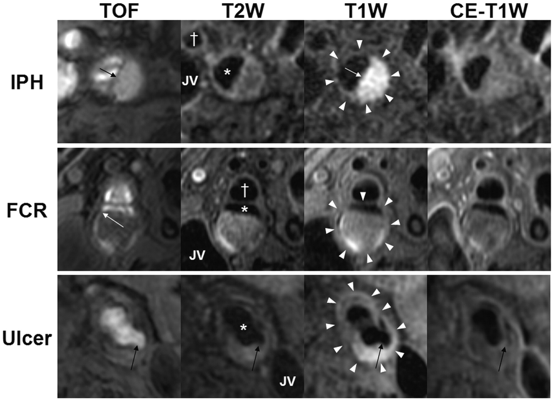
    In vivo identification of IPH, FCR, and ulceration. Each row contains multicontrast axial images from a single location in the carotid artery. The outer wall boundary (white arrowheads) of either the internal or common carotid artery, the lumen of the internal or common (asterisk) carotid artery, the external (cross, where applicable) carotid artery, and the JV are identified. The top row is of the right internal carotid artery from a 69-year-old man imaged at MSU. IPH (arrows) is characterized by a hyperintense signal intensity on TOF and T1WI. The second row is of the right internal carotid artery from an 80-year-old man imaged at PLA. An FCR (white arrow) is evident on TOF imaging by the hyperintense signal intensity extending from the lumen into the plaque and the absence of a fibrous cap on T2WI and CE-T1WI. The third row is of the left common carotid artery from a 74-year-old man imaged at PLA. An ulceration (black arrows) is present. Notably, the varying appearance of the ulcer between the different contrast weightings is due to reduced flow suppression caused by turbulent flow in the ulcerated region, particularly after contrast administration.

  • Fig 2.
    • Download figure
    • Open in new tab
    • Download powerpoint
    Fig 2.

    ROC curve analyses for the classification of IPH (A) and FCR (C) in the training set by maximum percentage LRNC. Adjacent to each ROC curve is a cumulative prevalence plot for IPH (B) and FCR (D) versus maximum percentage LRNC. The cumulative prevalence plots clearly depict changes in slope (arrows) for both IPH (B) and FCR (D), which were subsequently used to construct the CAS. E, ROC curve analysis for prediction of the presence of LRNC in the training set by maximum wall thickness, maximum total vessel area, and maximum NWI. F, In the cumulative prevalence plot for LRNC by maximum wall thickness, LRNC was absent in lesions with a maximum wall thickness <2 mm.

  • Fig 3.
    • Download figure
    • Open in new tab
    • Download powerpoint
    Fig 3.

    Flow diagram of CAS. Subjects with a maximum wall thickness >2 mm require additional evaluation. Further categorization of lesions can be determined by the size of the maximum percentage LRNC. Examples of matched cross-sectional images from 3 contrast weightings (TOF, T1WI, and CE-T1WI) for each category are provided. Images corresponding to CAS 1 (maximum wall thickness <2 mm) are from the left common carotid artery of a 69-year-old man imaged at MSU. The plaque in the CAS 2 (maximum percentage LRNC, ≤20%) example is from the right internal carotid artery of a 63-year-old man imaged at UW. There is a small LRNC (arrow) present on postcontrast imaging. Of note, there are also flow artifacts visible in the lumen—common artifacts in images distal to the bifurcation. The lesion in CAS 3 (maximum percentage LRNC, >20% and ≤40%) is from the left common carotid artery of a 65-year-old man imaged at AH. A noticeable LRNC (arrows) is present in both the anterior and posterior arterial wall. An example of a large LRNC (arrow) without IPH in the left common carotid artery of a 64-year-old man imaged at PLA is shown for CAS 4 (maximum percentage LRNC, >40%). Arrowheads indicate the outer wall boundary; asterisk, the lumen.

  • Fig 4.
    • Download figure
    • Open in new tab
    • Download powerpoint
    Fig 4.

    Prevalence of key features in the carotid artery is shown for each MR imaging location relative to the bifurcation. Distance from the bifurcation (0) is labeled on the x-axis, where positive and negative integers represent locations within the internal and common carotid arteries, respectively.

  • Fig 5.
    • Download figure
    • Open in new tab
    • Download powerpoint
    Fig 5.

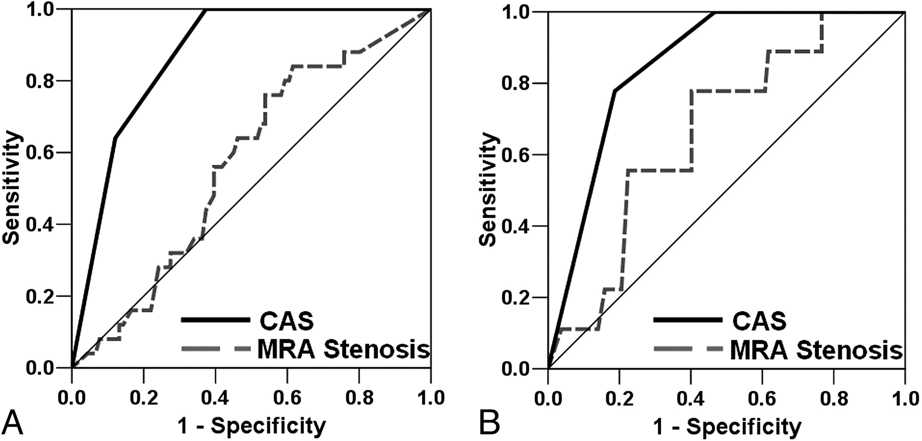
    ROC curve analyses for IPH (A) and FCR (B) by using either CAS or MRA stenosis.

Tables

  • Figures
    • View popup
    Table 1:

    Signal intensity of plaque features across different contrast weightingsa

    TOFT2WIPDT1WICE-T1WI
    Calcification–b–b–b–b–
    LRNCo/+–/o–/oo/+b–b
    IPH+b–/o–/o+b–/o
    • a Intensity relative to the sternocleidomastoid muscle: –indicates hypointense; o, isointense; +, hyperintense.

    • b Principal criteria for identification of a feature.

    • View popup
    Table 2:

    Demographic information (n = 334)

    Training Set (n= 196)Testing Set (n = 138)P Value
    Age (yr)69.0 ± 10.169.5 ± 9.5.61
    Male sex (%)74.076.1.70
    History of
        Hypertension (%)79.175.2.43
        Diabetes mellitus (%)20.427.5.15
        Coronary artery disease (%)34.339.3.41
    Smoking status
        Never (%)28.231.9.59
        Quit (%)49.744.2
        Current (%)22.123.9
    Imaging center
        AH (%)19.921.7.27
        MSU (%)21.413.0
        PLA (%)13.815.9
        UW (%)44.949.3
    Intraplaque hemorrhage (%)24.019.6.35
    Fibrous cap rupture (%)7.110.9.24
    Min lumen area (mm2)17.2 ± 10.417.9 ± 9.1.51
    Max wall area (mm2)61.1 ± 21.663.8 ± 24.5.29
    Max total vessel area (mm2)113.0 ± 35.7117.4 ± 38.3.29
    Max wall thickness (mm2)3.93 ± 1.794.00 ± 1.70.72
    Max normalized wall index0.696 ± 0.1390.674 ± 0.142.16
    Presence of calcification (%)72.473.9.80
    Max percentage calcification (%)a14.7 ± 10.416.1 ± 10.7.31
    Presence of LRNC (%)59.763.7.49
    Max percentage LRNCa (%)35.8 ± 18.331.3 ± 17.9.09
    MRA stenosis (%)47.6 ± 29.348.8 ± 32.4.83
    • a Only for arteries with calcification (n= 244) or LRNC (n = 206) present.

    • View popup
    Table 3:

    Prevalence of features for each level of CAS

    1234
    Training set (n = 196)
        No.16904248
        Calcification (%)12.580.078.672.9
        LRNC (%)0.030.0100.0100.0
        IPH (%)0.01.128.670.8
    Fibrous cap status
        Thick (%)0.021.1 (66.7)a23.812.5
        Thin (%)0.010.0 (33.3)a69.164.6
        Ruptured (%)0.00.07.122.9
    Testing Set (n = 138)
        No.17623425
        Calcification (%)17.683.979.480.0
        LRNC (%)11.843.5100.0100.0
        IPH (%)0.00.029.468.0
    Fibrous cap status
        Thick (%)11.8 (100.0)a30.7 (66.7)a29.44.0
        Thin (%)0.014.5 (33.3)a64.744.0
        Ruptured (%)0.00.05.952.0
    • a Lesions with an LRNC.

PreviousNext
Back to top

In this issue

American Journal of Neuroradiology: 31 (6)
American Journal of Neuroradiology
Vol. 31, Issue 6
1 Jun 2010
  • Table of Contents
  • Index by author
Advertisement
Print
Download PDF
Email Article

Thank you for your interest in spreading the word on American Journal of Neuroradiology.

NOTE: We only request your email address so that the person you are recommending the page to knows that you wanted them to see it, and that it is not junk mail. We do not capture any email address.

Enter multiple addresses on separate lines or separate them with commas.
A Noninvasive Imaging Approach to Assess Plaque Severity: The Carotid Atherosclerosis Score
(Your Name) has sent you a message from American Journal of Neuroradiology
(Your Name) thought you would like to see the American Journal of Neuroradiology web site.
CAPTCHA
This question is for testing whether or not you are a human visitor and to prevent automated spam submissions.
Cite this article
H.R. Underhill, T.S. Hatsukami, J. Cai, W. Yu, J.K. DeMarco, N.L. Polissar, H. Ota, X. Zhao, L. Dong, M. Oikawa, C. Yuan
A Noninvasive Imaging Approach to Assess Plaque Severity: The Carotid Atherosclerosis Score
American Journal of Neuroradiology Jun 2010, 31 (6) 1068-1075; DOI: 10.3174/ajnr.A2007

Citation Manager Formats

  • BibTeX
  • Bookends
  • EasyBib
  • EndNote (tagged)
  • EndNote 8 (xml)
  • Medlars
  • Mendeley
  • Papers
  • RefWorks Tagged
  • Ref Manager
  • RIS
  • Zotero
0 Responses
Respond to this article
Share
Bookmark this article
A Noninvasive Imaging Approach to Assess Plaque Severity: The Carotid Atherosclerosis Score
H.R. Underhill, T.S. Hatsukami, J. Cai, W. Yu, J.K. DeMarco, N.L. Polissar, H. Ota, X. Zhao, L. Dong, M. Oikawa, C. Yuan
American Journal of Neuroradiology Jun 2010, 31 (6) 1068-1075; DOI: 10.3174/ajnr.A2007
del.icio.us logo Twitter logo Facebook logo Mendeley logo
  • Tweet Widget
  • Facebook Like
  • Google Plus One
Purchase

Jump to section

  • Article
    • Abstract
    • Abbreviations
    • Materials and Methods
    • Results
    • Discussion
    • Conclusions
    • Footnotes
    • References
  • Figures & Data
  • Supplemental
  • Info & Metrics
  • Responses
  • References
  • PDF

Related Articles

  • PubMed
  • Google Scholar

Cited By...

  • Symptomatic and Asymptomatic Chronic Carotid Artery Occlusion on High-Resolution MR Vessel Wall Imaging
  • Association between coexisting intracranial artery and extracranial carotid artery atherosclerotic diseases and ipsilateral cerebral infarction: a Chinese Atherosclerosis Risk Evaluation (CARE-II) study
  • Comparison of carotid atherosclerotic plaques between subjects in Northern and Southern China: a Chinese atherosclerosis risk evaluation study
  • Signal of Carotid Intraplaque Hemorrhage on MR T1-Weighted Imaging: Association with Acute Cerebral Infarct
  • Prevalence and Characteristics of Carotid Artery High-Risk Atherosclerotic Plaques in Chinese Patients With Cerebrovascular Symptoms: A Chinese Atherosclerosis Risk Evaluation II Study
  • Prediction of High-Risk Plaque Development and Plaque Progression With the Carotid Atherosclerosis Score
  • Sex Differences of High-Risk Carotid Atherosclerotic Plaque with Less Than 50% Stenosis in Asymptomatic Patients: An In Vivo 3T MRI Study
  • Sustained Acceleration in Carotid Atherosclerotic Plaque Progression With Intraplaque Hemorrhage: A Long-Term Time Course Study
  • What Will Noninvasive Carotid Atherosclerosis Imaging Show Us About High-Risk Coronary Plaques?
  • Crossref (51)
  • Google Scholar

This article has been cited by the following articles in journals that are participating in Crossref Cited-by Linking.

  • Sustained Acceleration in Carotid Atherosclerotic Plaque Progression With Intraplaque Hemorrhage
    Jie Sun, Hunter R. Underhill, Daniel S. Hippe, Yunjing Xue, Chun Yuan, Thomas S. Hatsukami
    JACC: Cardiovascular Imaging 2012 5 8
  • Prevalence and Characteristics of Carotid Artery High‐Risk Atherosclerotic Plaques in Chinese Patients With Cerebrovascular Symptoms: A Chinese Atherosclerosis Risk Evaluation II Study
    Xihai Zhao, Daniel S. Hippe, Rui Li, Gador M. Canton, Binbin Sui, Yan Song, Feiyu Li, Yunjing Xue, Jie Sun, Kiyofumi Yamada, Thomas S. Hatsukami, Dongxiang Xu, Maoxue Wang, Chun Yuan
    Journal of the American Heart Association 2017 6 8
  • Ultrasound-Based Carotid Elastography for Detection of Vulnerable Atherosclerotic Plaques Validated by Magnetic Resonance Imaging
    Chengwu Huang, Xiaochang Pan, Qiong He, Manwei Huang, Lingyun Huang, Xihai Zhao, Chun Yuan, Jing Bai, Jianwen Luo
    Ultrasound in Medicine & Biology 2016 42 2
  • Prediction of High-Risk Plaque Development and Plaque Progression With the Carotid Atherosclerosis Score
    Dongxiang Xu, Daniel S. Hippe, Hunter R. Underhill, Minako Oikawa-Wakayama, Li Dong, Kiyofumi Yamada, Chun Yuan, Thomas S. Hatsukami
    JACC: Cardiovascular Imaging 2014 7 4
  • Clinical Factors Associated With High-Risk Carotid Plaque Features as Assessed by Magnetic Resonance Imaging in Patients With Established Vascular Disease (from the AIM-HIGH Study)
    Xue-Qiao Zhao, Thomas S. Hatsukami, Daniel S. Hippe, Jie Sun, Niranjan Balu, Daniel A. Isquith, John R. Crouse, Todd Anderson, John Huston, Nayak Polissar, Kevin O'Brien, Chun Yuan
    The American Journal of Cardiology 2014 114 9
  • Sex Differences of High-Risk Carotid Atherosclerotic Plaque with Less Than 50% Stenosis in Asymptomatic Patients: An In Vivo 3T MRI Study
    H. Ota, M.J. Reeves, D.C. Zhu, A. Majid, A. Collar, C. Yuan, J.K. DeMarco
    American Journal of Neuroradiology 2013 34 5
  • Association Between Carotid Bifurcation Geometry and Atherosclerotic Plaque Vulnerability
    Peirong Jiang, Zhensen Chen, Daniel S. Hippe, Hiroko Watase, Bin Sun, Ruolan Lin, Zheting Yang, Yunjing Xue, Xihai Zhao, Chun Yuan
    Arteriosclerosis, Thrombosis, and Vascular Biology 2020 40 5
  • Pulse Wave Imaging in Carotid Artery Stenosis Human Patients in Vivo
    Ronny X. Li, Iason Z. Apostolakis, Paul Kemper, Matthew D.J. McGarry, Ada Ip, Edward S. Connolly, James F. McKinsey, Elisa E. Konofagou
    Ultrasound in Medicine & Biology 2019 45 2
  • Non-Invasive Identification of Vulnerable Atherosclerotic Plaques Using Texture Analysis in Ultrasound Carotid Elastography: An In Vivo Feasibility Study Validated by Magnetic Resonance Imaging
    Chengwu Huang, Qiong He, Manwei Huang, Lingyun Huang, Xihai Zhao, Chun Yuan, Jianwen Luo
    Ultrasound in Medicine & Biology 2017 43 4
  • Signal of Carotid Intraplaque Hemorrhage on MR T1-Weighted Imaging: Association with Acute Cerebral Infarct
    D. Yang, Y. Liu, Y. Han, D. Li, W. Wang, R. Li, C. Yuan, X. Zhao
    American Journal of Neuroradiology 2020 41 5

More in this TOC Section

  • Correlation of Apparent Diffusion Coefficient at 3T with Prognostic Parameters of Retinoblastoma
  • Parathyroid Lesions: Characterization with Dual-Phase Arterial and Venous Enhanced CT of the Neck
  • Efficacy of Diffusion-Weighted Imaging for the Differentiation between Lymphomas and Carcinomas of the Nasopharynx and Oropharynx: Correlations of Apparent Diffusion Coefficients and Histologic Features
Show more HEAD AND NECK

Similar Articles

Advertisement

Indexed Content

  • Current Issue
  • Accepted Manuscripts
  • Article Preview
  • Past Issues
  • Editorials
  • Editor's Choice
  • Fellows' Journal Club
  • Letters to the Editor
  • Video Articles

Cases

  • Case Collection
  • Archive - Case of the Week
  • Archive - Case of the Month
  • Archive - Classic Case

Special Collections

  • AJNR Awards
  • ASNR Foundation Special Collection
  • Most Impactful AJNR Articles
  • Photon-Counting CT
  • Spinal CSF Leak Articles (Jan 2020-June 2024)

More from AJNR

  • Trainee Corner
  • Imaging Protocols
  • MRI Safety Corner

Multimedia

  • AJNR Podcasts
  • AJNR Scantastics

Resources

  • Turnaround Time
  • Submit a Manuscript
  • Submit a Video Article
  • Submit an eLetter to the Editor/Response
  • Manuscript Submission Guidelines
  • Statistical Tips
  • Fast Publishing of Accepted Manuscripts
  • Graphical Abstract Preparation
  • Imaging Protocol Submission
  • Evidence-Based Medicine Level Guide
  • Publishing Checklists
  • Author Policies
  • Become a Reviewer/Academy of Reviewers
  • News and Updates

About Us

  • About AJNR
  • Editorial Board
  • Editorial Board Alumni
  • Alerts
  • Permissions
  • Not an AJNR Subscriber? Join Now
  • Advertise with Us
  • Librarian Resources
  • Feedback
  • Terms and Conditions
  • AJNR Editorial Board Alumni

American Society of Neuroradiology

  • Not an ASNR Member? Join Now

© 2025 by the American Society of Neuroradiology All rights, including for text and data mining, AI training, and similar technologies, are reserved.
Print ISSN: 0195-6108 Online ISSN: 1936-959X

Powered by HighWire