
Article Figures & Data
Figures
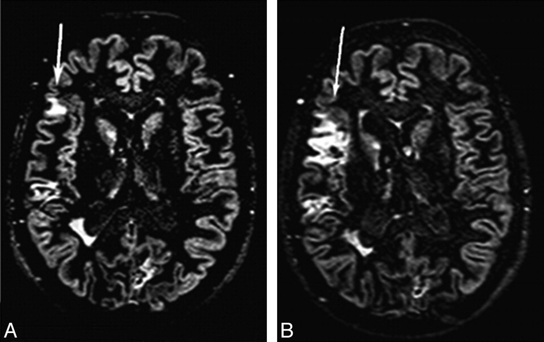
- Fig 1.
Brain axial DIR images from a patient with RRMS at baseline (A) and after 6 months (B). Multiple cortical lesions are visible. The size of 1 cortical lesion in the right parietal lobe (arrows) is increased after 6 months. Reprinted from Neuroimage, 42, Calabrese M et al, Morphology and evolution of cortical lesions in multiple sclerosis: a longitudinal MRI study, 1324–28, 2008, with permission from Elsevier.
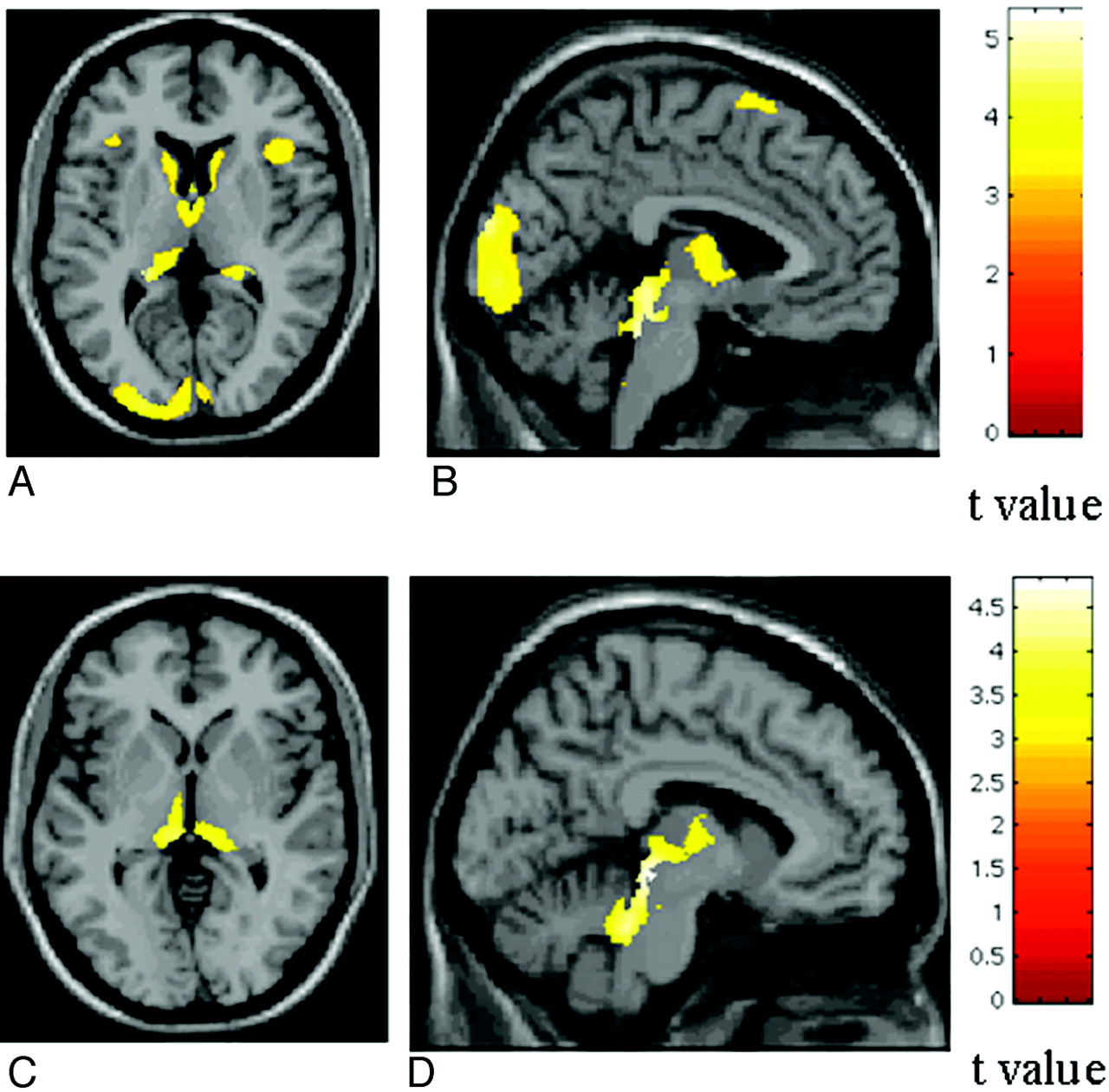
- Fig 2.
SPM regions with decreased GM concentration, overlaid on a high-resolution T1-weighted image, contrasting patients with different MS phenotypes. A and B, SPM regions with GM loss in patients with SPMS compared with those with RRMS. C and D, SPM regions with GM loss in patients with SPMS compared with those with PPMS. Reprinted from Neuroimage, 42, Ceccarelli A et al, A voxel-based morphometry study of grey matter loss in MS patients with different clinical phenotypes, 315–22, 2008, with permission from Elsevier.
- Fig 3.
SPM regions with anatomic correspondence between GM atrophy (blue) and GM mean diffusivity (red) changes in patients with PPMS compared with controls. A and B, An overlap is visible in the thalami. Reprinted with permission from Ceccarelli et al, 2009.66
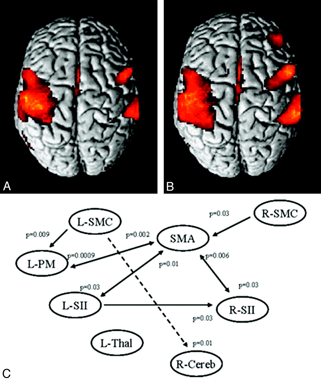
- Fig 4.
A and B, Cortical activations on a rendered brain from healthy controls (A) and patients with MS (B) from 8 European centers during the performance of a simple motor task with the right hand. Compared with controls, patients with MS had more significant activations bilaterally in several regions of the sensorimotor network. C, A dynamic causal model shows the results of the between-group analysis of effective connectivity. Increased strength of the connections in patients versus controls is reported as continuous black lines, whereas reduced strength of the connections in patients versus controls is reported as dotted black lines. Reprinted with permission from Rocca et al, 2009.81


In this issue


Jump to section
Related Articles
Cited By...
- Cortical Thickness and Serum NfL Explain Cognitive Dysfunction in Newly Diagnosed Patients With Multiple Sclerosis
- Increased total sodium concentration in gray matter better explains cognition than atrophy in MS
- Establishing pathological cut-offs of brain atrophy rates in multiple sclerosis
- Assessing Abnormal Iron Content in the Deep Gray Matter of Patients with Multiple Sclerosis versus Healthy Controls