Advertisement

Main menu

  • Home
  • Content
    • Current Issue
    • Accepted Manuscripts
    • Article Preview
    • Past Issue Archive
    • Video Articles
    • AJNR Case Collection
    • Case of the Week Archive
    • Case of the Month Archive
    • Classic Case Archive
  • Special Collections
    • AJNR Awards
    • ASNR Foundation Special Collection
    • Most Impactful AJNR Articles
    • Photon-Counting CT
    • Spinal CSF Leak Articles (Jan 2020-June 2024)
  • Multimedia
    • AJNR Podcasts
    • AJNR SCANtastic
    • Trainee Corner
    • MRI Safety Corner
    • Imaging Protocols
  • For Authors
    • Submit a Manuscript
    • Submit a Video Article
    • Submit an eLetter to the Editor/Response
    • Manuscript Submission Guidelines
    • Statistical Tips
    • Fast Publishing of Accepted Manuscripts
    • Graphical Abstract Preparation
    • Imaging Protocol Submission
    • Author Policies
  • About Us
    • About AJNR
    • Editorial Board
    • Editorial Board Alumni
  • More
    • Become a Reviewer/Academy of Reviewers
    • Subscribers
    • Permissions
    • Alerts
    • Feedback
    • Advertisers
    • ASNR Home

User menu

  • Alerts
  • Log in

Search

  • Advanced search
American Journal of Neuroradiology

American Journal of Neuroradiology

ASHNR American Society of Functional Neuroradiology ASHNR American Society of Pediatric Neuroradiology ASSR
  • Alerts
  • Log in

Advanced Search

  • Home
  • Content
    • Current Issue
    • Accepted Manuscripts
    • Article Preview
    • Past Issue Archive
    • Video Articles
    • AJNR Case Collection
    • Case of the Week Archive
    • Case of the Month Archive
    • Classic Case Archive
  • Special Collections
    • AJNR Awards
    • ASNR Foundation Special Collection
    • Most Impactful AJNR Articles
    • Photon-Counting CT
    • Spinal CSF Leak Articles (Jan 2020-June 2024)
  • Multimedia
    • AJNR Podcasts
    • AJNR SCANtastic
    • Trainee Corner
    • MRI Safety Corner
    • Imaging Protocols
  • For Authors
    • Submit a Manuscript
    • Submit a Video Article
    • Submit an eLetter to the Editor/Response
    • Manuscript Submission Guidelines
    • Statistical Tips
    • Fast Publishing of Accepted Manuscripts
    • Graphical Abstract Preparation
    • Imaging Protocol Submission
    • Author Policies
  • About Us
    • About AJNR
    • Editorial Board
    • Editorial Board Alumni
  • More
    • Become a Reviewer/Academy of Reviewers
    • Subscribers
    • Permissions
    • Alerts
    • Feedback
    • Advertisers
    • ASNR Home
  • Follow AJNR on Twitter
  • Visit AJNR on Facebook
  • Follow AJNR on Instagram
  • Join AJNR on LinkedIn
  • RSS Feeds

AJNR is seeking candidates for the AJNR Podcast Editor. Read the position description.

Review ArticleReview Articles
Open Access

Isolated Acute Nontraumatic Cortical Subarachnoid Hemorrhage

V. Cuvinciuc, A. Viguier, L. Calviere, N. Raposo, V. Larrue, C. Cognard and F. Bonneville
American Journal of Neuroradiology September 2010, 31 (8) 1355-1362; DOI: https://doi.org/10.3174/ajnr.A1986
V. Cuvinciuc
  • Find this author on Google Scholar
  • Find this author on PubMed
  • Search for this author on this site
A. Viguier
  • Find this author on Google Scholar
  • Find this author on PubMed
  • Search for this author on this site
L. Calviere
  • Find this author on Google Scholar
  • Find this author on PubMed
  • Search for this author on this site
N. Raposo
  • Find this author on Google Scholar
  • Find this author on PubMed
  • Search for this author on this site
V. Larrue
  • Find this author on Google Scholar
  • Find this author on PubMed
  • Search for this author on this site
C. Cognard
  • Find this author on Google Scholar
  • Find this author on PubMed
  • Search for this author on this site
F. Bonneville
  • Find this author on Google Scholar
  • Find this author on PubMed
  • Search for this author on this site
  • Article
  • Figures & Data
  • Info & Metrics
  • Responses
  • References
  • PDF
Loading

Article Figures & Data

Figures

  • Tables
  • Fig 1.
    • Download figure
    • Open in new tab
    • Download powerpoint
    Fig 1.

    Imaging algorithm and main causes of cSAH.
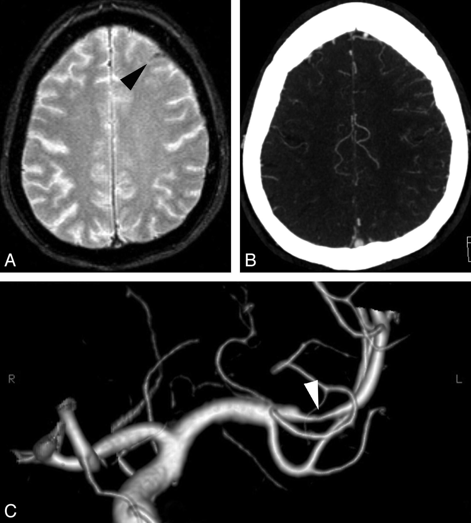
  • Fig 2.
    • Download figure
    • Open in new tab
    • Download powerpoint
    Fig 2.

    Cortical venous thrombosis. A, Axial GRE T2 image shows right central sulcus SAH (black arrowhead). B, Axial FLAIR image shows a thrombosed right cortical parietal vein (white arrowhead).

  • Fig 3.
    • Download figure
    • Open in new tab
    • Download powerpoint
    Fig 3.

    Dural sinus thrombosis. A, Axial brain CT shows left peri-rolandic SAH. B, Axial FLAIR image confirms the sulcal hemorrhage, which is also visible more anteriorly (arrowhead). C, Sagittal contrast-enhanced T1 shows a filling defect in the superior sagittal sinus.

  • Fig 4.
    • Download figure
    • Open in new tab
    • Download powerpoint
    Fig 4.

    RCVS. A, Axial brain CT scan shows bilateral frontoparietal sulcal SAH (white arrowheads). B, Axial FLAIR image confirms the cSAH (white arrowheads). C, Lateral projection of 3D TOF MRA shows multiple arterial stenoses and dilations, mainly on the anterior cerebral artery branches (white arrowheads). D, Lateral projection of a right internal carotid angiogram shows multiple stenoses and dilations on both anterior cerebral and middle cerebral arteries (black arrowheads). Note that DSA, even if it showed more clearly the arterial abnormalities, did not change the previously suspected diagnosis. Follow-up MRA performed at 3 months demonstrated disappearance of the arterial lesions (not shown).

  • Fig 5.
    • Download figure
    • Open in new tab
    • Download powerpoint
    Fig 5.

    Endocarditis. A, Axial brain CT scan shows an isolated slight right frontal subarachnoid hyperattenuation. B, Because of clinical aggravation the next day, another brain CT was performed and demonstrated a larger right Sylvian SAH. Subsequent MR imaging showed a right middle cerebral artery infarction due to M1 occlusion (not shown), which precluded the exact identification of this SAH origin.

  • Fig 6.
    • Download figure
    • Open in new tab
    • Download powerpoint
    Fig 6.

    Pial vasodilation. A, Axial GRE T2 image shows a left frontal sulcal SAH (black arrowhead), possibly located in the “watershed” territory between the anterior and the middle cerebral arteries. B, Axial maximum-intensity-projection reconstruction of CTA shows an asymmetry of the distal arteries, in favor of left pial vasodilation. C, Frontal projection of 3D angiography of the left carotid artery reveals a severe stenosis at the origin of the M2 branch (white arrowhead).

  • Fig 7.
    • Download figure
    • Open in new tab
    • Download powerpoint
    Fig 7.

    Eclampsia-related PRES. A, Axial FLAIR image shows a left frontal SAH and a left parietal subcortical hyperintensity. B, Axial FLAIR image shows bilateral cerebellar hyperintensities, very suggestive of PRES lesions.
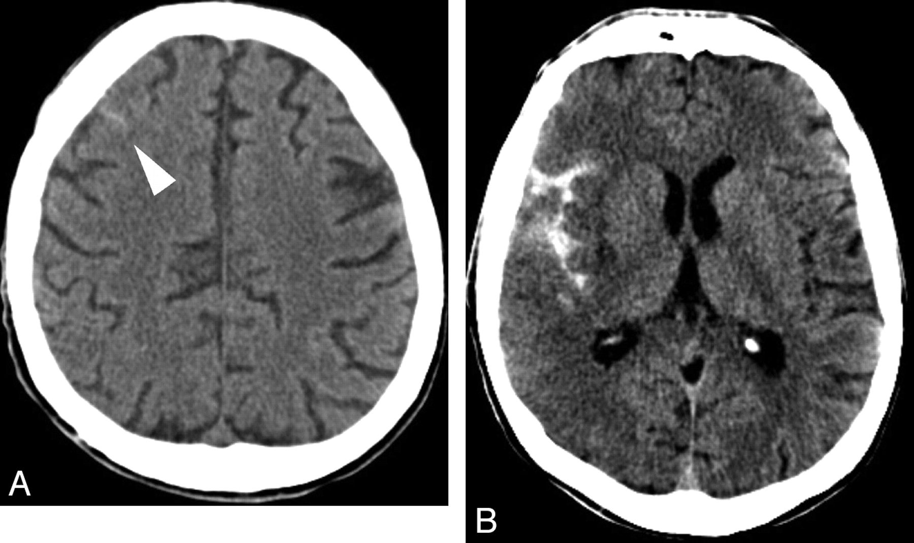
  • Fig 8.
    • Download figure
    • Open in new tab
    • Download powerpoint
    Fig 8.

    CAA. A, Axial brain CT scan shows a subtle left rolandic hyperattenuation favoring minimal SAH (white arrowhead). B and C, Axial GRE T2 images show left temporal lobar hemorrhage, multiple microbleeds, and cortical hemosiderosis. D, Axial FLAIR image obtained after 7 months shows a new asymptomatic SAH in a left parietal sulcus (white arrowhead), which was previously normal (see B).

Tables

  • Figures
  • Etiology of cSAH

    Etiology
    Pial arteriovenous malformations
    Dural arteriovenous fistulas
    Arterial dissection
    Dural/cortical venous thrombosis
    Vasculitides
    RCVS
    PRES
    High-grade stenosis
    Endocarditis
    CAA
    Coagulation disorders
    Abscess
    Cavernoma
    Primary and secondary brain tumors
PreviousNext
Back to top

In this issue

American Journal of Neuroradiology: 31 (8)
American Journal of Neuroradiology
Vol. 31, Issue 8
1 Sep 2010
  • Table of Contents
  • Index by author
Advertisement
Print
Download PDF
Email Article

Thank you for your interest in spreading the word on American Journal of Neuroradiology.

NOTE: We only request your email address so that the person you are recommending the page to knows that you wanted them to see it, and that it is not junk mail. We do not capture any email address.

Enter multiple addresses on separate lines or separate them with commas.
Isolated Acute Nontraumatic Cortical Subarachnoid Hemorrhage
(Your Name) has sent you a message from American Journal of Neuroradiology
(Your Name) thought you would like to see the American Journal of Neuroradiology web site.
CAPTCHA
This question is for testing whether or not you are a human visitor and to prevent automated spam submissions.
Cite this article
V. Cuvinciuc, A. Viguier, L. Calviere, N. Raposo, V. Larrue, C. Cognard, F. Bonneville
Isolated Acute Nontraumatic Cortical Subarachnoid Hemorrhage
American Journal of Neuroradiology Sep 2010, 31 (8) 1355-1362; DOI: 10.3174/ajnr.A1986

Citation Manager Formats

  • BibTeX
  • Bookends
  • EasyBib
  • EndNote (tagged)
  • EndNote 8 (xml)
  • Medlars
  • Mendeley
  • Papers
  • RefWorks Tagged
  • Ref Manager
  • RIS
  • Zotero
0 Responses
Respond to this article
Share
Bookmark this article
Isolated Acute Nontraumatic Cortical Subarachnoid Hemorrhage
V. Cuvinciuc, A. Viguier, L. Calviere, N. Raposo, V. Larrue, C. Cognard, F. Bonneville
American Journal of Neuroradiology Sep 2010, 31 (8) 1355-1362; DOI: 10.3174/ajnr.A1986
del.icio.us logo Twitter logo Facebook logo Mendeley logo
  • Tweet Widget
  • Facebook Like
  • Google Plus One
Purchase

Jump to section

  • Article
    • Abstract
    • Abbreviations
    • Imaging Protocol
    • Etiologies of cSAH
    • Conclusions
    • Acknowledgments
    • Footnotes
    • References
  • Figures & Data
  • Info & Metrics
  • Responses
  • References
  • PDF

Related Articles

  • No related articles found.
  • PubMed
  • Google Scholar

Cited By...

  • Synchronous subarachnoid haemorrhage and ischaemic stroke as a result of complete internal carotid artery occlusion
  • Recurrent migraine aura-like symptoms in an elderly woman: symptomatic cortical spreading depression?
  • Yield of diagnostic imaging in atraumatic convexity subarachnoid hemorrhage
  • Pearls & Oy-sters: Paraneoplastic cerebral vasculitis: Rare cause of spontaneous convexity subarachnoid hemorrhage
  • Centripetal Propagation of Vasoconstriction at the Time of Headache Resolution in Patients with Reversible Cerebral Vasoconstriction Syndrome
  • Cerebral Venous Thrombosis with Subarachnoid Hemorrhage: a Case Report
  • An Updated Definition of Stroke for the 21st Century: A Statement for Healthcare Professionals From the American Heart Association/American Stroke Association
  • Amyloid-{beta} Contributes to Blood-Brain Barrier Leakage in Transgenic Human Amyloid Precursor Protein Mice and in Humans With Cerebral Amyloid Angiopathy
  • Sporadic cerebral amyloid angiopathy revisited: recent insights into pathophysiology and clinical spectrum
  • Clinical Presentation, Etiology, and Long-Term Prognosis in Patients With Nontraumatic Convexal Subarachnoid Hemorrhage
  • Central Sulcus Focal Subarachnoid Hemorrhage in the Elderly: Cerebral Amyloid Angiopathy Is the Most Frequent Cause
  • Reply:
  • Investigating suspected subarachnoid haemorrhage in adults
  • Extracranial Internal Carotid Artery Stenosis as a Cause of Cortical Subarachnoid Hemorrhage
  • Crossref (115)
  • Google Scholar

This article has been cited by the following articles in journals that are participating in Crossref Cited-by Linking.

  • An Updated Definition of Stroke for the 21st Century
    Ralph L. Sacco, Scott E. Kasner, Joseph P. Broderick, Louis R. Caplan, J.J. (Buddy) Connors, Antonio Culebras, Mitchell S.V. Elkind, Mary G. George, Allen D. Hamdan, Randall T. Higashida, Brian L. Hoh, L. Scott Janis, Carlos S. Kase, Dawn O. Kleindorfer, Jin-Moo Lee, Michael E. Moseley, Eric D. Peterson, Tanya N. Turan, Amy L. Valderrama, Harry V. Vinters
    Stroke 2013 44 7
  • Controversies and evolving new mechanisms in subarachnoid hemorrhage
    Sheng Chen, Hua Feng, Prativa Sherchan, Damon Klebe, Gang Zhao, Xiaochuan Sun, Jianmin Zhang, Jiping Tang, John H. Zhang
    Progress in Neurobiology 2014 115
  • Cortical superficial siderosis: detection and clinical significance in cerebral amyloid angiopathy and related conditions
    Andreas Charidimou, Jennifer Linn, Meike W. Vernooij, Christian Opherk, Saloua Akoudad, Jean-Claude Baron, Steven M. Greenberg, Hans Rolf Jäger, David J. Werring
    Brain 2015 138 8
  • Amyloid-β Contributes to Blood–Brain Barrier Leakage in Transgenic Human Amyloid Precursor Protein Mice and in Humans With Cerebral Amyloid Angiopathy
    Anika M.S. Hartz, Björn Bauer, Emma L.B. Soldner, Andrea Wolf, Sandra Boy, Roland Backhaus, Ivan Mihaljevic, Ulrich Bogdahn, Hans H. Klünemann, Gerhard Schuierer, Felix Schlachetzki
    Stroke 2012 43 2
  • Subarachnoid Hemorrhage: Beyond Aneurysms
    Carrie P. Marder, Vinod Narla, James R. Fink, Kathleen R. Tozer Fink
    American Journal of Roentgenology 2014 202 1
  • Acute stroke differential diagnosis: Stroke mimics
    Pedro Vilela
    European Journal of Radiology 2017 96
  • Clinical Presentation, Etiology, and Long-Term Prognosis in Patients With Nontraumatic Convexal Subarachnoid Hemorrhage
    Markus Beitzke, Thomas Gattringer, Christian Enzinger, Gerit Wagner, Kurt Niederkorn, Franz Fazekas
    Stroke 2011 42 11
  • Magnetic resonance imaging of arterial stroke mimics: a pictorial review
    Gilles Adam, Marine Ferrier, Sofia Patsoura, Raluca Gramada, Zuzana Meluchova, Vanessa Cazzola, Jean Darcourt, Christophe Cognard, Alain Viguier, Fabrice Bonneville
    Insights into Imaging 2018 9 5
  • Subcortical vascular dementia
    Hidekazu Tomimoto
    Neuroscience Research 2011 71 3
  • Acute Convexity Subarachnoid Hemorrhage Related to Cerebral Amyloid Angiopathy: Clinicoradiological Features and Outcome
    Lionel Calviere, Victor Cuvinciuc, Nicolas Raposo, Alexandre Faury, Christophe Cognard, Vincent Larrue, Alain Viguier, Fabrice Bonneville
    Journal of Stroke and Cerebrovascular Diseases 2016 25 5

More in this TOC Section

  • Mechanisms of Healing in Coiled Intracranial Aneurysms: A Review of the Literature
  • Ultra-High-Field MR Neuroimaging
  • Armies of Pestilence: CNS Infections as Potential Weapons of Mass Destruction
Show more Review Articles

Similar Articles

Advertisement

Indexed Content

  • Current Issue
  • Accepted Manuscripts
  • Article Preview
  • Past Issues
  • Editorials
  • Editor's Choice
  • Fellows' Journal Club
  • Letters to the Editor
  • Video Articles

Cases

  • Case Collection
  • Archive - Case of the Week
  • Archive - Case of the Month
  • Archive - Classic Case

Special Collections

  • AJNR Awards
  • ASNR Foundation Special Collection
  • Most Impactful AJNR Articles
  • Photon-Counting CT
  • Spinal CSF Leak Articles (Jan 2020-June 2024)

More from AJNR

  • Trainee Corner
  • Imaging Protocols
  • MRI Safety Corner

Multimedia

  • AJNR Podcasts
  • AJNR Scantastics

Resources

  • Turnaround Time
  • Submit a Manuscript
  • Submit a Video Article
  • Submit an eLetter to the Editor/Response
  • Manuscript Submission Guidelines
  • Statistical Tips
  • Fast Publishing of Accepted Manuscripts
  • Graphical Abstract Preparation
  • Imaging Protocol Submission
  • Evidence-Based Medicine Level Guide
  • Publishing Checklists
  • Author Policies
  • Become a Reviewer/Academy of Reviewers
  • News and Updates

About Us

  • About AJNR
  • Editorial Board
  • Editorial Board Alumni
  • Alerts
  • Permissions
  • Not an AJNR Subscriber? Join Now
  • Advertise with Us
  • Librarian Resources
  • Feedback
  • Terms and Conditions
  • AJNR Editorial Board Alumni

American Society of Neuroradiology

  • Not an ASNR Member? Join Now

© 2025 by the American Society of Neuroradiology All rights, including for text and data mining, AI training, and similar technologies, are reserved.
Print ISSN: 0195-6108 Online ISSN: 1936-959X

Powered by HighWire